Comparative Study
. 2000 Oct;38(10):3689-95.
doi: 10.1128/JCM.38.10.3689-3695.2000.

Rapid quantification and differentiation of human polyomavirus DNA in undiluted urine from patients after bone marrow transplantation

Affiliations
Comparative Study

Rapid quantification and differentiation of human polyomavirus DNA in undiluted urine from patients after bone marrow transplantation

S S Biel et al. J Clin Microbiol. 2000 Oct.

Abstract

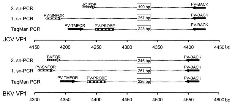
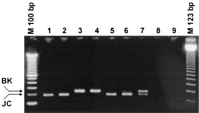
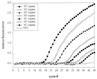
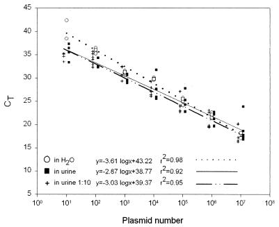
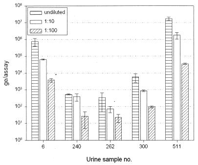
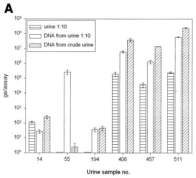
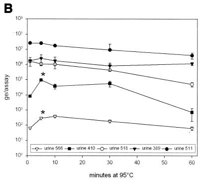
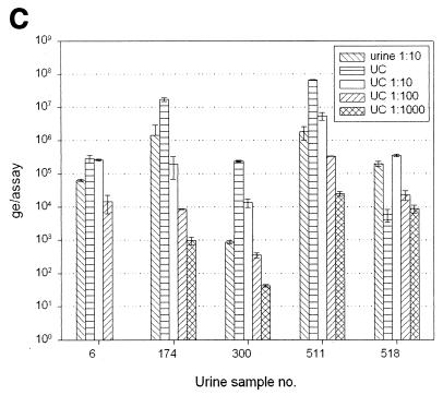
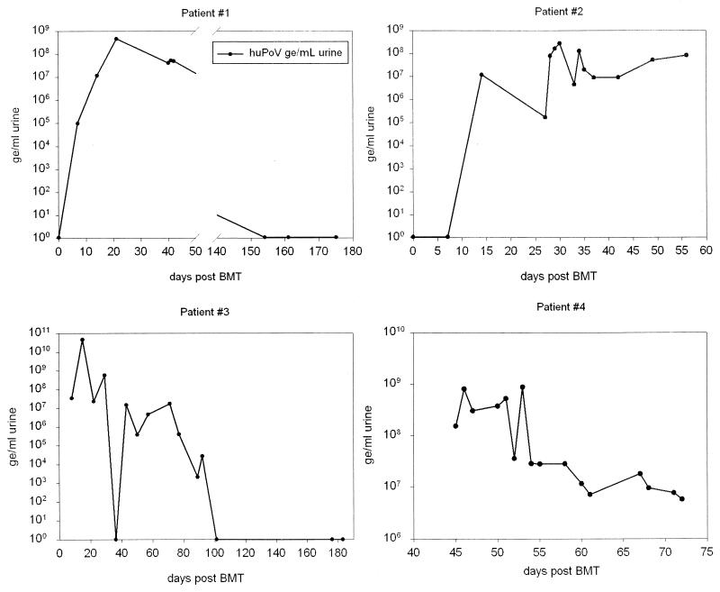
A combined PCR assay was developed for the detection and typing of human polyomavirus (huPoV) in clinical samples, consisting of (i) a qualitative seminested PCR assay (snPCR) to discriminate between huPoV BK and JC and (ii) a high-throughput, quantitative TaqMan PCR assay (TM-PCR) for the general detection of huPoV. The TM-PCR detects huPoV DNA in a linear range from 10(7) to 10(1) copies per assay. In reproducibility runs, the inter- and intra-assay variabilities were < or =60 and < or =50%, respectively. The snPCR assay uses a set of four primers for the same region of the BK and JC viral genomes. In the first round of amplification, two general primers were used; in the second round, one of these general primers and two additional, BK- or JC-specific primers were used simultaneously to produce amplicons of different sizes specific for BK virus (246 bp) and JC virus (199 bp), respectively. We tested different urine dilutions in order to determine the inhibitory effects of urine on PCR amplification. Furthermore, we compared the use of native urine with DNA purified by different preparation procedures. Our results show, that a 1:10 dilution of the urine led to complete reduction of the amplification inhibition found with 6% of undiluted urine samples. In a clinical study including 600 urine specimens, our assay turned out to be fast, cheap, and reliable in both qualitative and quantitative aspects.


Figures

FIG. 1
FIG. 1
Schematic representation indicating spacing, positions, and orientations of primers and the exonuclease probe. Whereas the TM-PCR utilizes the same primer set and exonuclease probe for quantification of both huPoVs, the snPCR is based on different primers for BKV and JCV in the second amplification round (three-primer PCR).
FIG. 2
FIG. 2
snPCR. The ethidium bromide-stained 2.5% agarose gel shows the PCR products of six urine samples (lanes 1 to 6), a mix of plasmids pPBK and pPJC as a positive control (lane 7), and two no-template controls (lanes 8 and 9). The different amplicon sizes for BKV (246 bp) and JCV (199 bp) are clearly visible.
FIG. 3
FIG. 3
TaqMan assay for the quantification of huPoV DNA. Amplification plots of serial dilutions from 107 to 101 copies of plasmid pPJC are shown. Relative fluorescence is plotted versus cycle number. Each amplification plot is the result of triplicate experiments.
FIG. 4
FIG. 4
Calibration curves were obtained by correlation of the CT value and the plasmid copy number. CT values were taken from amplifications of serial dilutions of plasmid pPJC in water (open circles), in six native urine specimens (solid squares), and in the same six urine specimens previously diluted 1:10 with water (crosses).
FIG. 5
FIG. 5
Urine dilution. HuPov DNA was quantified in native, 1:10-diluted, and 1:100-diluted urine. Each group of three bars represents one urine sample.
FIG. 6
FIG. 6
(A) Quantification of huPoV DNA in 1:10-diluted urine and in Qiagen DNA preparations from native urine and from 1:10-diluted urine. It is evident that DNA preparation from 1:10-diluted urine may be beneficial, whereas preparation from native urine can be affected by inhibitory factors, resulting in a decreased amount of detectable DNA (specimens 55 and 194). In most urine samples, DNA can be enriched at least by a factor of 10 by preparation (specimens 406, 457, and 511). (B) Urine was heated to 95°C for 1, 5, 10, 30, and 60 min immediately before use in PCR. Five different urine samples, covering a wide range of DNA contents, were investigated. There was no significant change in detectable DNA amounts as a result of heating longer than 5 min before PCR cycling. (C) Ultracentrifuge (UC) sedimentation of urine. The urine was sedimented and used for TM-PCR either pure or diluted with water. It is demonstrated that ultracentrifuge pretreatment of urine leads to enrichment of detectable huPoV DNA in some samples (samples 174, 300, and 511). In other samples PCR inhibitors seem to be enriched along with the virus (samples 5 and 518).
FIG. 6
FIG. 6
(A) Quantification of huPoV DNA in 1:10-diluted urine and in Qiagen DNA preparations from native urine and from 1:10-diluted urine. It is evident that DNA preparation from 1:10-diluted urine may be beneficial, whereas preparation from native urine can be affected by inhibitory factors, resulting in a decreased amount of detectable DNA (specimens 55 and 194). In most urine samples, DNA can be enriched at least by a factor of 10 by preparation (specimens 406, 457, and 511). (B) Urine was heated to 95°C for 1, 5, 10, 30, and 60 min immediately before use in PCR. Five different urine samples, covering a wide range of DNA contents, were investigated. There was no significant change in detectable DNA amounts as a result of heating longer than 5 min before PCR cycling. (C) Ultracentrifuge (UC) sedimentation of urine. The urine was sedimented and used for TM-PCR either pure or diluted with water. It is demonstrated that ultracentrifuge pretreatment of urine leads to enrichment of detectable huPoV DNA in some samples (samples 174, 300, and 511). In other samples PCR inhibitors seem to be enriched along with the virus (samples 5 and 518).
FIG. 7
FIG. 7
Inhibition of actin amplification caused by native urine. One nanogram of human genomic DNA was added to water and various native urine samples. Amplification plots are given for seven DNA-spiked urine samples and the DNA-spiked water control. The increased CT value for sample 381 reflects inhibition of actin amplification. The other samples display a nearly constant CT, indicating minor inhibitory influences.
FIG. 8
FIG. 8
Monitoring of four representative BMT patients. Lines indicate huPoV DNA loads as determined by TM-PCR. As determined by snPCR, all four patients were positive for BKV only during the time course.

References

    1. Arthur R R, Shah K V, Baust S J, Santos G W, Saral R. Association of BK viruria with hemorrhagic cystitis in recipients of bone marrow transplants. N Engl J Med. 1986;315:230–234. - PubMed
    1. Arthur R R, Shah K V. Occurrence and significance of papovaviruses BK and JC in the urine. Prog Med Virol. 1989;36:42–61. - PubMed
    1. Arthur R R, Dagostin S, Shah K V. Detection of BK virus and JC virus in urine and brain tissue by the polymerase chain reaction. J Clin Microbiol. 1989;27:1174–1179. - PMC - PubMed
    1. Azzi A, et al. Monitoring of polyomavirus BK viruria in bone marrow transplantation patients by DNA hybridization assay and by polymerase chain reaction: an approach to assess the relationship between BK viruria and hemorrhagic cystitis. Bone Marrow Transplant. 1994;14:235–240. - PubMed
    1. Azzi A, Zakrzewska K, Cesaro A, Fanci R, Bosi A. Monitoring of BKV viral load in urine of bone marrow transplantation patients and hemorrhagic cystitis. Acta Microbiol Immunol Hung. 1999;46:370–371.

Publication types