. 2000 Oct 2;192(7):965-76.
doi: 10.1084/jem.192.7.965.

CD1b-mediated T cell recognition of a glycolipid antigen generated from mycobacterial lipid and host carbohydrate during infection

Affiliations

CD1b-mediated T cell recognition of a glycolipid antigen generated from mycobacterial lipid and host carbohydrate during infection

D B Moody et al. J Exp Med. .

Abstract

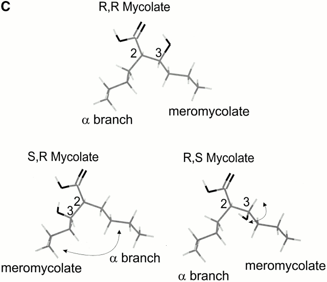
T cells recognize microbial glycolipids presented by CD1 proteins, but there is no information regarding the generation of natural glycolipid antigens within infected tissues. Therefore, we determined the molecular basis of CD1b-restricted T cell recognition of mycobacterial glycosylated mycolates, including those produced during tissue infection in vivo. Transfection of the T cell receptor (TCR) alpha and beta chains from a glucose monomycolate (GMM)-specific T cell line reconstituted GMM recognition in TCR-deficient T lymphoblastoma cells. This TCR-mediated response was highly specific for natural mycobacterial glucose-6-O-(2R, 3R) monomycolate, including the precise structure of the glucose moiety, the stereochemistry of the mycolate lipid, and the linkage between the carbohydrate and the lipid. Mycobacterial production of antigenic GMM absolutely required a nonmycobacterial source of glucose that could be supplied by adding glucose to media at concentrations found in mammalian tissues or by infecting tissue in vivo. These results indicate that mycobacteria synthesized antigenic GMM by coupling mycobacterial mycolates to host-derived glucose. Specific T cell recognition of an epitope formed by interaction of host and pathogen biosynthetic pathways provides a mechanism for immune response to those pathogenic mycobacteria that have productively infected tissues, as distinguished from ubiquitous, but innocuous, environmental mycobacteria.


Figures

Figure 1
Figure 1
Variable regions of the TCR α and β chains mediated CD1b-restricted recognition of GMM. (A) CD3 expression by untransfected J.RT3 cells and J.RT3 cells contransfected with the LDN5 TCR α (TCRAV3S1J9C1) and β (TCRBV7S1D2S1J2S1C2) chains was determined by FACS® with an anti-CD3ε (SPV-T3b; open histogram) compared with staining with a nonbinding isotype-matched control mAb (P3; filled histogram). MFI, mean fluorescence intensity. (B) J.RT3 cells cotransfected with LDN5 TCR α and β chains, the CD8-1 TCR α and β chains, or the LDN5 TCR β chain were assayed for IL-2 release in response to stimulation by OKT3 (20 μg/ml), M. phlei GMM (13 μg/ml), or the mycobacterial phospholipid containing the mannosyl phosphoisoprenoid antigen (PL; 1 μg/ml) presented by monocyte-derived dendritic cells. IL-2 release was quantified by measuring proliferative responses of IL-2–responsive HT-2 cells cultured with J.RT3 supernatants. (C) LDN5αβ/J.RT3 cells were assayed for IL-2 release when stimulated by GMM (5 μg/ml) presented by C1R B lymphoblastoid cells transfected with vector alone (mock) or cDNAs encoding either CD1a, CD1b, or CD1c.
Figure 1
Figure 1
Variable regions of the TCR α and β chains mediated CD1b-restricted recognition of GMM. (A) CD3 expression by untransfected J.RT3 cells and J.RT3 cells contransfected with the LDN5 TCR α (TCRAV3S1J9C1) and β (TCRBV7S1D2S1J2S1C2) chains was determined by FACS® with an anti-CD3ε (SPV-T3b; open histogram) compared with staining with a nonbinding isotype-matched control mAb (P3; filled histogram). MFI, mean fluorescence intensity. (B) J.RT3 cells cotransfected with LDN5 TCR α and β chains, the CD8-1 TCR α and β chains, or the LDN5 TCR β chain were assayed for IL-2 release in response to stimulation by OKT3 (20 μg/ml), M. phlei GMM (13 μg/ml), or the mycobacterial phospholipid containing the mannosyl phosphoisoprenoid antigen (PL; 1 μg/ml) presented by monocyte-derived dendritic cells. IL-2 release was quantified by measuring proliferative responses of IL-2–responsive HT-2 cells cultured with J.RT3 supernatants. (C) LDN5αβ/J.RT3 cells were assayed for IL-2 release when stimulated by GMM (5 μg/ml) presented by C1R B lymphoblastoid cells transfected with vector alone (mock) or cDNAs encoding either CD1a, CD1b, or CD1c.
Figure 2
Figure 2
LDN5 and LDN5αβ/J.RT3 cells recognized GMM but not other naturally occurring mycobacterial glycosylated mycolates. Glucose-6-O-monomycolate, free mycolate, glycerol mycolate, arabinose-5-O-monomycolate, trehalose-6-O-monomycolate, Myc PL, and trehalose dimycolate (cord factor) were purified from mycobacteria and cultured with monocyte-derived dendritic cells for presentation to LDN5 and LDN5αβ/J.RT-3. R indicates the meromycolate R group which, if present, may be a keto, wax ester, or methoxy derivative. M. tb, M. tuberculosis.
Figure 3
Figure 3
LDN5 and LDN5αβ/J.RT3 recognition of synthetic GMM was specific for the 6-linkage. G-3-MM and G-6-MM were synthesized and the structures were confirmed by electrospray ionization mass spectroscopy and NMR analysis. Antigens (ag) were presented by monocyte-derived dendritic cells and responses were determined by proliferation for LDN5 and by HT-2 assay of IL-2 release for LDN5αβ/J.RT3.
Figure 4
Figure 4
LDN5 and LDN5αβ/J.RT-3 cells specifically recognized the 2R, 3R stereochemical structure of the mycolic acid moiety of GMM. (A) Free C32 mycolates produced synthetically by condensation of palmitate were methylated and resolved with preparative silica TLC in chloroform into two pairs of enantiomers, 2R, 3R plus 2S, 3S (R f 0.42) and 2R, 3S plus 2S, 3R (R f 0.52), and then separately glucosylated to yield GMM. To check the purity of the fraction containing 2R, 3S plus 2S, 3R GMMs and to rule out racemization during the glucosylation reaction, GMMs were cleaved with acid to yield free mycolates, methylated to yield mycolic acid methyl esters (MAMEs) and resolved on silica TLC plates in chloroform. Left lane, MAMEs from the mixture of all four synthetic GMM stereoisomers; middle lane, MAME from 2R, 3R R. equi GMM; right lane, MAMEs from preparative TLC-purified mixture of 2R, 3S plus 2S, 3R GMMs. The lack of MAMEs from the mixture of 2R, 3S and 2S, 3R GMMs migrating at R f 0.42 indicated that this preparation was pure and that mycolic acids were not racemized during the glucosylation reaction. (B) The proliferative response of LDN5 and the IL-2 secretion of LDN5αβ/J.RT3 in response to stereoisomers of GMM containing C32 mycolates presented by monocyte-derived dendritic cells is shown. These results were typical of three experiments. (C) Structural relationship of natural (2R, 3R) mycolate composed of C (gray), H (light gray), and O (black) with synthetic analogues that are not known to occur naturally is depicted with truncated meromycolate and α branches. The synthetic 2S, 3R mycolate form differed from natural 2R, 3R mycolate in the absolute orientation of the α and meromycolate branches at C2 as indicated by the arrow. The 2R, 3S form differed from the natural mycolate in the absolute orientation of the 3-hydroxyl with regard to the meromycolate chain.
Figure 4
Figure 4
LDN5 and LDN5αβ/J.RT-3 cells specifically recognized the 2R, 3R stereochemical structure of the mycolic acid moiety of GMM. (A) Free C32 mycolates produced synthetically by condensation of palmitate were methylated and resolved with preparative silica TLC in chloroform into two pairs of enantiomers, 2R, 3R plus 2S, 3S (R f 0.42) and 2R, 3S plus 2S, 3R (R f 0.52), and then separately glucosylated to yield GMM. To check the purity of the fraction containing 2R, 3S plus 2S, 3R GMMs and to rule out racemization during the glucosylation reaction, GMMs were cleaved with acid to yield free mycolates, methylated to yield mycolic acid methyl esters (MAMEs) and resolved on silica TLC plates in chloroform. Left lane, MAMEs from the mixture of all four synthetic GMM stereoisomers; middle lane, MAME from 2R, 3R R. equi GMM; right lane, MAMEs from preparative TLC-purified mixture of 2R, 3S plus 2S, 3R GMMs. The lack of MAMEs from the mixture of 2R, 3S and 2S, 3R GMMs migrating at R f 0.42 indicated that this preparation was pure and that mycolic acids were not racemized during the glucosylation reaction. (B) The proliferative response of LDN5 and the IL-2 secretion of LDN5αβ/J.RT3 in response to stereoisomers of GMM containing C32 mycolates presented by monocyte-derived dendritic cells is shown. These results were typical of three experiments. (C) Structural relationship of natural (2R, 3R) mycolate composed of C (gray), H (light gray), and O (black) with synthetic analogues that are not known to occur naturally is depicted with truncated meromycolate and α branches. The synthetic 2S, 3R mycolate form differed from natural 2R, 3R mycolate in the absolute orientation of the α and meromycolate branches at C2 as indicated by the arrow. The 2R, 3S form differed from the natural mycolate in the absolute orientation of the 3-hydroxyl with regard to the meromycolate chain.
Figure 5
Figure 5
Mycobacterial production of antigenic GMM required exogenous glucose. (A) M. phlei and M. smegmatis were grown in standard (7H9; GIBCO BRL) medium that was supplemented with 1 g/dl d-glucose or the indicated carbohydrate. Mycobacteria were washed, lyophilized, and then extracted at 7.5 mg/ml with chloroform/methanol to give the total lipid fraction (dilution = 1). LDN5 proliferation in response to total lipid fractions presented by monocyte-derived dendritic cells is shown as the mean of duplicate samples with error bars indicating the range. (B) M. avium was cultured in medium supplemented with glucose at the indicated concentration for 24 h. Total extractable lipids were weighed and tested for their ability to stimulate IL-2 secretion by LDN5αβ/J.RT3 cells. The relative yield of mycobacteria after culture for 24 h with each of the indicated glucoses was estimated by the optical density of the culture fluid at 600 nm (OD600). Computer-assisted, densitometric charring of total lipids resolved by TLC indicated that GMM comprised ∼1 μg/mg of total lipid from mycobacteria grown at 100 mg/dl (data not shown).
Figure 6
Figure 6
LDN5 recognized a GMM-comigrating lipid from M. leprae isolated directly from infected tissue. M. leprae was harvested directly from armadillo liver and total lipid was extracted with chloroform/methanol (2:1), loaded on a preparative silica TLC plate, and developed in chloroform/methanol/water (60:16:2) compared with a GMM standard (10 μg) purified from in vitro cultures of M. phlei. The TLC plate was scored at 1-cm intervals as indicated, and the lipids were separately extracted from each 1-cm fraction using chloroform/methanol (2:1). Extracts from each fraction were dried under nitrogen, resuspended in T cell medium (1:5 dilution) with monocyte-derived dendritic cells, and tested for stimulation of LDN5. The margin of the preparative plate was reserved and developed by charring with a cupric acetate solution and shown in comparison with the M. phlei GMM standard.
Figure 7
Figure 7
The map of the antigenic determinants of the TCR-mediated response to natural glucose-6-O-(2R, 3R) monomycolate. The hydrophilic cap of GMM is formed from host-derived glucose esterified via the sixth carbon to mycobacterial mycolate. Recognition of GMM required TCR α and β chains with the variable regions corresponding to LDN5, CD1b expression on the APC, and all tested elements of the structure of the hydrophilic cap of natural GMM, including components supplied by the host and by the mycobacteria. Natural or synthetic analogues lacking the indicated chemical element (red) or proper stereoconfiguration (green) of glucose-6-O-(2R, 3R) monomycolate were not recognized by cells bearing the LDN5 TCR α and β chains, but analogues lacking the distal elements of the mycolate alkyl chains (gray) were recognized. Substitution of the anomeric carbon (blue) with α-linked glucose (trehalose mycolate) or α-linked glucose monomycolate (trehalose dimycolate) abolished recognition.

References

    1. Sturgill-Koszycki S., Schlesinger P.H., Chakraborty P., Haddix P.L., Collins H.L., Fok A.K., Allen R.D., Gluck S.L., Heuser J., Russell D.G. Lack of acidification in Mycobacterium phagosomes produced by exclusion of the vesicular proton-ATPase. Science. 1994;263:678–681. - PubMed
    1. Ferrari G., Langen H., Naito M., Pieters J. A coat protein on phagosomes involved in the intracellular survival of mycobacteria. Cell. 1999;97:435–447. - PubMed
    1. Kaufmann S.H. Immunity to intracellular microbial pathogens. Immunol. Today. 1995;16:338–342. - PubMed
    1. Flynn J.L., Chan J., Triebold K.J., Dalton D.K., Stewart T.A., Bloom B.R. An essential role for interferon gamma in resistance to Mycobacterium tuberculosis infection. J. Exp. Med. 1993;178:2249–2254. - PMC - PubMed
    1. North R.J. Importance of thymus-derived lymphocytes in cell-mediated immunity to infection. Cell. Immunol. 1973;7:166–176. - PubMed

Publication types