. 2000 Oct 2;192(7):1015-26.
doi: 10.1084/jem.192.7.1015.

Carbon monoxide generated by heme oxygenase 1 suppresses endothelial cell apoptosis

Affiliations

Carbon monoxide generated by heme oxygenase 1 suppresses endothelial cell apoptosis

S Brouard et al. J Exp Med. .

Abstract

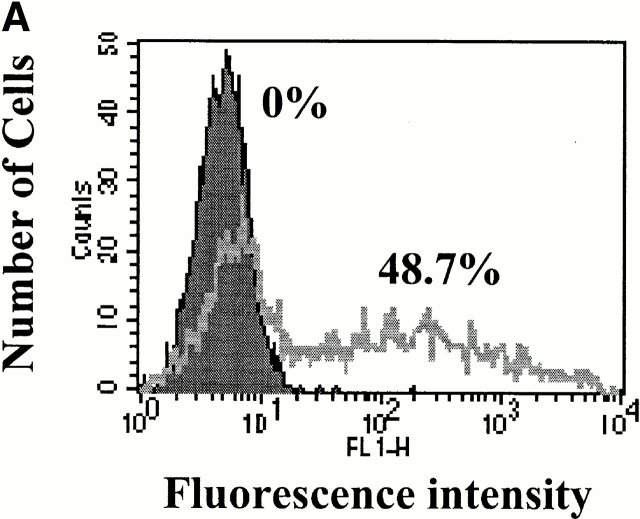
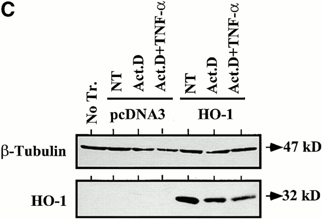
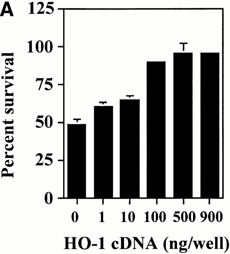
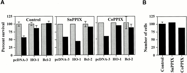
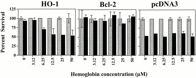
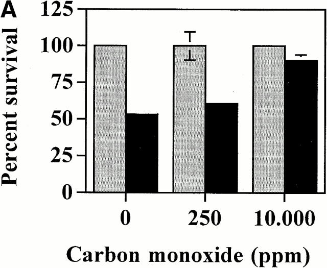
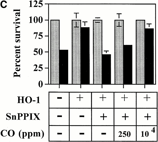
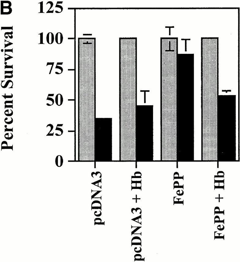
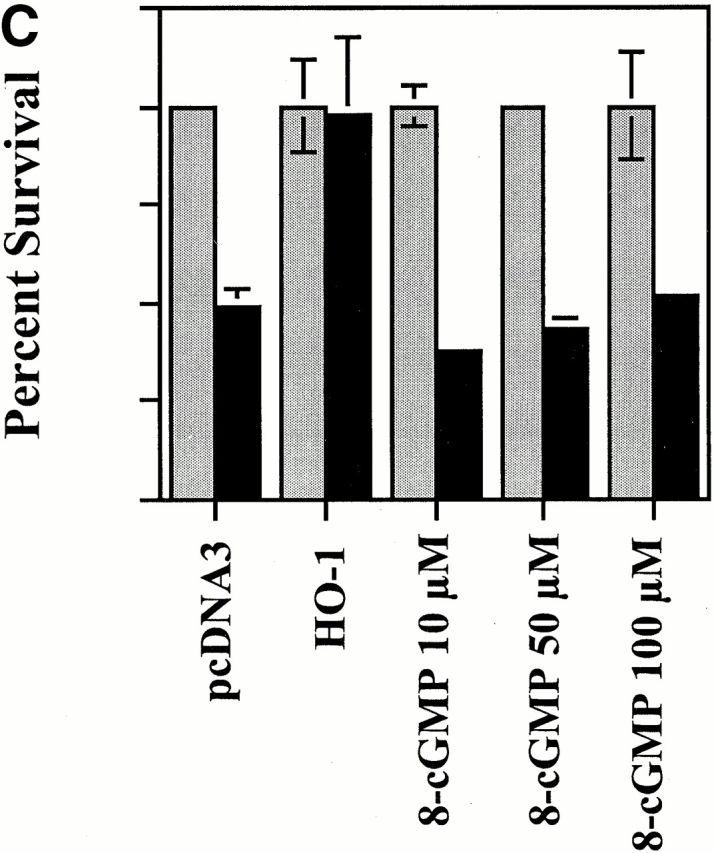
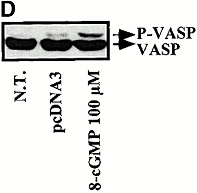
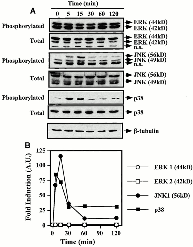
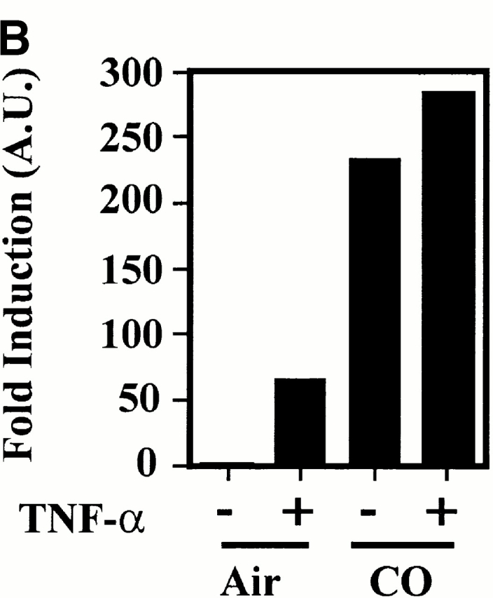
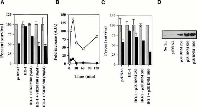
Heme oxygenase 1 (HO-1) inhibits apoptosis by regulating cellular prooxidant iron. We now show that there is an additional mechanism by which HO-1 inhibits apoptosis, namely by generating the gaseous molecule carbon monoxide (CO). Overexpression of HO-1, or induction of HO-1 expression by heme, protects endothelial cells (ECs) from apoptosis. When HO-1 enzymatic activity is blocked by tin protoporphyrin (SnPPIX) or the action of CO is inhibited by hemoglobin (Hb), HO-1 no longer prevents EC apoptosis while these reagents do not affect the antiapoptotic action of bcl-2. Exposure of ECs to exogenous CO, under inhibition of HO-1 activity by SnPPIX, substitutes HO-1 in preventing EC apoptosis. The mechanism of action of HO-1/CO is dependent on the activation of the p38 mitogen-activated protein kinase (MAPK) signaling transduction pathway. Expression of HO-1 or exposure of ECs to exogenous CO enhanced p38 MAPK activation by TNF-alpha. Specific inhibition of p38 MAPK activation by the pyridinyl imidazol SB203580 or through overexpression of a p38 MAPK dominant negative mutant abrogated the antiapoptotic effect of HO-1. Taken together, these data demonstrate that the antiapoptotic effect of HO-1 in ECs is mediated by CO and more specifically via the activation of p38 MAPK by CO.


Figures

Figure 1
Figure 1
HO-1 suppresses EC apoptosis. (A) 2F-2B ECs were transfected with a GFP-expressing plasmid and monitored for GFP expression by flow cytometry. The percentage of transfected ECs was assessed by measuring fluorescence intensity in ECs transfected with control (pcDNA3; filled histogram) versus GFP (open histogram) expression plasmids. (B) ECs were cotransfected with β-galactosidase plus control (pcDNA3) or HO-1 (β-actin/HO-1) expression vectors. EC apoptosis was induced by TNF-α plus Act.D and apoptosis of β-galactosidase–transfected ECs was quantified. Gray bars represent ECs treated with Act.D and black bars represent ECs treated with TNF-α plus Act.D. Results shown are the mean ± SD from duplicate wells taken from 1 representative experiment out of 10. (C) HO-1 expression was detected in BAECs by Western blot. No Tr, nontransfected. NT, nontreated. (D) 2F-2B ECs were cotransfected with β-galactosidase plus control (pcDNA3) or HO-1 (β-actin/HO-1) expression vectors. Gray bars represent untreated ECs and black bars represent ECs treated with etoposide (200 μM, 8 h) or subjected to serum deprivation (0.1% FCS for 24 h). Results shown are the mean ± SD from duplicate wells taken from one representative experiment out of three independent experiments. Similar results were obtained using BAECs.
Figure 1
Figure 1
HO-1 suppresses EC apoptosis. (A) 2F-2B ECs were transfected with a GFP-expressing plasmid and monitored for GFP expression by flow cytometry. The percentage of transfected ECs was assessed by measuring fluorescence intensity in ECs transfected with control (pcDNA3; filled histogram) versus GFP (open histogram) expression plasmids. (B) ECs were cotransfected with β-galactosidase plus control (pcDNA3) or HO-1 (β-actin/HO-1) expression vectors. EC apoptosis was induced by TNF-α plus Act.D and apoptosis of β-galactosidase–transfected ECs was quantified. Gray bars represent ECs treated with Act.D and black bars represent ECs treated with TNF-α plus Act.D. Results shown are the mean ± SD from duplicate wells taken from 1 representative experiment out of 10. (C) HO-1 expression was detected in BAECs by Western blot. No Tr, nontransfected. NT, nontreated. (D) 2F-2B ECs were cotransfected with β-galactosidase plus control (pcDNA3) or HO-1 (β-actin/HO-1) expression vectors. Gray bars represent untreated ECs and black bars represent ECs treated with etoposide (200 μM, 8 h) or subjected to serum deprivation (0.1% FCS for 24 h). Results shown are the mean ± SD from duplicate wells taken from one representative experiment out of three independent experiments. Similar results were obtained using BAECs.
Figure 2
Figure 2
The antiapoptotic effect of HO-1 is dose dependent. (A) 2F-2B ECs were cotransfected with increasing doses of HO-1 expression vector (β-actin/HO-1). EC apoptosis was induced by TNF-α plus Act.D. Results shown are the mean ± SD from duplicate wells taken from one representative experiment out of three. Similar results were obtained using BAECs. (B) The expression of HO-1 was detected in BAECs by Western blot. Values indicate the amount of HO-1 vector (pcDNA3/HO-1) used in each transfection (ng of DNA per 3 × 10⁵ cells). No Tr., nontransfected.
Figure 2
Figure 2
The antiapoptotic effect of HO-1 is dose dependent. (A) 2F-2B ECs were cotransfected with increasing doses of HO-1 expression vector (β-actin/HO-1). EC apoptosis was induced by TNF-α plus Act.D. Results shown are the mean ± SD from duplicate wells taken from one representative experiment out of three. Similar results were obtained using BAECs. (B) The expression of HO-1 was detected in BAECs by Western blot. Values indicate the amount of HO-1 vector (pcDNA3/HO-1) used in each transfection (ng of DNA per 3 × 10⁵ cells). No Tr., nontransfected.
Figure 3
Figure 3
The antiapoptotic effect of HO-1 is dependent on HO enzymatic activity. (A) 2F-2B ECs were cotransfected with β-galactosidase plus pcDNA3, HO-1 (β-actin/HO-1), or bcl-2 expression vectors. Cells were either left untreated (Control) or treated with the inhibitor of HO enzymatic activity SnPPIX. CoPPIX, a protoporphyrin that does not inhibit HO enzymatic activity, was used as a control treatment. Gray bars represent ECs treated with Act.D and black bars represent ECs treated with TNF-α plus Act.D. Results shown are the mean ± SD from duplicate wells taken from one representative experiment out of three. (B) 2F-2B ECs were transfected with β-galactosidase plus pcDNA3 expression vectors. ECs were either left untreated (Control) or were treated with SnPPIX and CoPPIX as in A. The results shown are the mean ± SD from duplicate wells taken from one representative experiment out of three.
Figure 4
Figure 4
Scavenging of CO by Hb suppresses the antiapoptotic effect of HO-1. 2F-2B ECs were cotransfected with β-galactosidase plus control (pcDNA3), HO-1 (β-actin/HO-1), or bcl-2 expression vectors. ECs were either left untreated (0) or were treated with increasing concentrations of Hb. Gray bars represent ECs treated with Act.D and black bars represent ECs treated with TNF-α plus Act.D. The results shown are the mean ± SD from duplicate wells taken from one representative experiment out of four.
Figure 5
Figure 5
Exogenous CO suppresses EC apoptosis in the absence of HO-1. (A) 2F-2B ECs were transfected with a β-galactosidase expression vector and exposed to exogenous CO. Gray bars represent ECs treated with Act.D alone and black bars represent ECs treated with TNF-α plus Act.D. (B) 2F-2B ECs were transfected with a β-galactosidase expression vector and exposed to exogenous CO (10,000 ppm) with or without Hb. Gray bars represent ECs treated with Act.D and black bars represent ECs treated with TNF-α plus Act.D. (C) 2F-2B ECs were cotransfected with β-galactosidase and HO-1 (β-actin/HO-1) expression vectors. Where indicated (+), HO-1 enzymatic activity was inhibited by SnPPIX and/or exposed to exogenous CO. Gray bars represent ECs treated with Act.D and black bars represent ECs treated with TNF-α plus Act.D. Results shown (A, B, and C) are the mean ± SD from duplicate wells taken from one representative experiment out of three.
Figure 5
Figure 5
Exogenous CO suppresses EC apoptosis in the absence of HO-1. (A) 2F-2B ECs were transfected with a β-galactosidase expression vector and exposed to exogenous CO. Gray bars represent ECs treated with Act.D alone and black bars represent ECs treated with TNF-α plus Act.D. (B) 2F-2B ECs were transfected with a β-galactosidase expression vector and exposed to exogenous CO (10,000 ppm) with or without Hb. Gray bars represent ECs treated with Act.D and black bars represent ECs treated with TNF-α plus Act.D. (C) 2F-2B ECs were cotransfected with β-galactosidase and HO-1 (β-actin/HO-1) expression vectors. Where indicated (+), HO-1 enzymatic activity was inhibited by SnPPIX and/or exposed to exogenous CO. Gray bars represent ECs treated with Act.D and black bars represent ECs treated with TNF-α plus Act.D. Results shown (A, B, and C) are the mean ± SD from duplicate wells taken from one representative experiment out of three.
Figure 6
Figure 6
ECs that express HO-1 suppress apoptosis of ECs that do not express HO-1. 2F-2B ECs were transfected with control (pcDNA3; I and II) or HO-1 (III) expression vectors. 16 h after transfection, ECs were harvested, washed, and cocultured at a ratio of 1:1 with ECs transfected with β-galactosidase (I and II) or with β-galactosidase plus HO-1 (III). Cocultures were maintained for an additional 24 h before induction of apoptosis by TNF-α plus Act.D. The percentage of survival was evaluated by counting the number of β-galactosidase–positive cells that retained normal morphology. Gray bars represent ECs treated with Act.D and black bars represent ECs treated with TNF-α plus Act.D. Results shown are the mean ± SD from duplicates taken from one representative experiment out of three.
Figure 7
Figure 7
Upregulation of endogenous HO-1 expression inhibits EC apoptosis via CO. (A) 2F-2B ECs were cotransfected with a β-galactosidase expression vector and exposed to FePP. Apoptosis was induced by TNF-α and Act.D. Gray bars represent ECs treated with Act.D and black bars represent ECs treated with TNF-α plus Act.D. The results shown are the mean ± SD from duplicate wells taken from one representative experiment out of three. (B) 2F-2B ECs were cotransfected with a β-galactosidase expression vector and exposed to FePP (6.25 μM). Where indicated, ECs were treated with Hb (50 μM). The results shown are the mean ± SD from duplicate wells taken from one representative experiment out of three.
Figure 7
Figure 7
Upregulation of endogenous HO-1 expression inhibits EC apoptosis via CO. (A) 2F-2B ECs were cotransfected with a β-galactosidase expression vector and exposed to FePP. Apoptosis was induced by TNF-α and Act.D. Gray bars represent ECs treated with Act.D and black bars represent ECs treated with TNF-α plus Act.D. The results shown are the mean ± SD from duplicate wells taken from one representative experiment out of three. (B) 2F-2B ECs were cotransfected with a β-galactosidase expression vector and exposed to FePP (6.25 μM). Where indicated, ECs were treated with Hb (50 μM). The results shown are the mean ± SD from duplicate wells taken from one representative experiment out of three.
Figure 8
Figure 8
Iron chelation by DFO suppresses EC apoptosis. (A) 2F-2B ECs were transfected with a β-galactosidase expression vector and exposed to DFO. Gray bars represent ECs treated with Act.D alone and black bars represent ECs treated with TNF-α plus Act.D. (B) 2F-2B ECs were cotransfected as described above in A. Where indicated (+), HO-1 enzymatic activity was inhibited by SnPPIX and iron was chelated by DFO, as described above in A. Gray bars represent ECs treated with Act.D and black bars represent ECs treated with TNF-α plus Act.D. (C) 2F-2B ECs were cotransfected as described above in A. Where indicated (+), CO was removed from the culture medium by Hb and/or iron was chelated by DFO as described above in A and B. Gray bars represent ECs treated with Act.D and black bars represent ECs treated with TNF-α plus Act.D. Results shown (A, B, and C) are the mean ± SD from duplicate wells taken from one representative experiment out of three.
Figure 10
Figure 10
The antiapoptotic effect of HO-1 does not act via a cGMP-dependent pathway. (A) 2F-2B ECs were cotransfected with β-galactosidase and HO-1 (β-actin/HO-1) expression vectors. HO-1–transfected cells were exposed to increasing doses of the guanylcyclase inhibitor ODQ. Gray bars represent ECs treated with Act.D alone and black bars represent ECs treated with TNF-α plus Act.D. The results shown are the mean ± SD from duplicate wells taken from one representative experiment out of three. (B) Activation of guanylcyclase was monitored in BAECs by analyzing the phosphorylation of VASP. P-VASP (50 kD) and VASP (46 kD) are the phosphorylated and nonphosphorylated forms of VASP, respectively. (C) 2F-2B ECs were with β-galactosidase or with β-galactosidase plus HO-1 (β-actin/HO-1) expression vectors as described above in A. Where indicated, ECs were exposed to the cGMP analogue 8-Br-cGMP, as described in Materials and Methods. Gray bars represent ECs treated with Act.D and black bars represent ECs treated with TNF-α plus Act.D. The results shown are the mean ± SD from duplicate wells taken from one representative experiment out of three. (D) Phosphorylation of VASP was analyzed by Western blot as described above in B. N.T., nontreated.
Figure 10
Figure 10
The antiapoptotic effect of HO-1 does not act via a cGMP-dependent pathway. (A) 2F-2B ECs were cotransfected with β-galactosidase and HO-1 (β-actin/HO-1) expression vectors. HO-1–transfected cells were exposed to increasing doses of the guanylcyclase inhibitor ODQ. Gray bars represent ECs treated with Act.D alone and black bars represent ECs treated with TNF-α plus Act.D. The results shown are the mean ± SD from duplicate wells taken from one representative experiment out of three. (B) Activation of guanylcyclase was monitored in BAECs by analyzing the phosphorylation of VASP. P-VASP (50 kD) and VASP (46 kD) are the phosphorylated and nonphosphorylated forms of VASP, respectively. (C) 2F-2B ECs were with β-galactosidase or with β-galactosidase plus HO-1 (β-actin/HO-1) expression vectors as described above in A. Where indicated, ECs were exposed to the cGMP analogue 8-Br-cGMP, as described in Materials and Methods. Gray bars represent ECs treated with Act.D and black bars represent ECs treated with TNF-α plus Act.D. The results shown are the mean ± SD from duplicate wells taken from one representative experiment out of three. (D) Phosphorylation of VASP was analyzed by Western blot as described above in B. N.T., nontreated.
Figure 9
Figure 9
Iron chelation and CO have additive effects in suppressing EC apoptosis. 2F-2B ECs were cotransfected with β-galactosidase and HO-1 (β-actin/HO-1) expression vectors. Where indicated (+), cells were treated with the inhibitor of HO enzymatic activity SnPPIX. ECs were exposed to CO (250 ppm) and to the iron chelator DFO. Gray bars represent ECs treated with Act.D and black bars represent ECs treated with TNF-α plus Act.D. The results shown are the mean ± SD from duplicate wells taken from one representative experiment out of three.
Figure 11
Figure 11
Activation of MAPK by TNF-α. (A) BAECs were stimulated with TNF-α (10 ng/ml; time 0) and MAPK phosphorylation was monitored by Western blot (0, 5, 15, 30, 60, and 120 min after TNF-α stimulation) using antibodies directed against the phosphorylated forms of each MAPK. One single membrane was used for all the stainings shown. Experiments were repeated three times with virtually identical results. n.s., nonspecific band. (B) Phosphorylation of different MAPKs was quantified. The results were presented as fold induction in arbitrary units (A.U.), compared with time 0, before TNF-α stimulation. The results in B correspond to the membranes shown in A.
Figure 12
Figure 12
HO-1 and CO modulate p38 MAPK activation in ECs. (A) BAECs were either nontransduced (NT), transduced with a β-galactosidase (βgal.), or HO-1 recombinant adenovirus, and were left untreated (−) or treated (+) with TNF-α (10 ng/ml for 15 min). p38 MAPK phosphorylation was monitored by Western blot using antibodies directed against the phosphorylated forms of each MAPK. Results are presented as fold induction of MAPK activation by TNF-α in arbitrary units (A.U.), compared with time 0, before TNF-α stimulation. (B) BAECs were stimulated (+) or not (−) by TNF-α (10 ng/ml, 30 min) in the presence or absence of CO (10,000 ppm). Phosphorylation of p38 MAPK was quantified as in A. The results are presented as fold induction of MAPK activation by TNF-α in arbitrary units (A.U.).
Figure 12
Figure 12
HO-1 and CO modulate p38 MAPK activation in ECs. (A) BAECs were either nontransduced (NT), transduced with a β-galactosidase (βgal.), or HO-1 recombinant adenovirus, and were left untreated (−) or treated (+) with TNF-α (10 ng/ml for 15 min). p38 MAPK phosphorylation was monitored by Western blot using antibodies directed against the phosphorylated forms of each MAPK. Results are presented as fold induction of MAPK activation by TNF-α in arbitrary units (A.U.), compared with time 0, before TNF-α stimulation. (B) BAECs were stimulated (+) or not (−) by TNF-α (10 ng/ml, 30 min) in the presence or absence of CO (10,000 ppm). Phosphorylation of p38 MAPK was quantified as in A. The results are presented as fold induction of MAPK activation by TNF-α in arbitrary units (A.U.).
Figure 13
Figure 13
The antiapoptotic effect of HO-1 acts via the activation of p38 MAPK. (A) 2F-2B ECs were cotransfected with β-galactosidase, control (pcDNA3), or HO-1 (β-actin/HO-1) expression vectors. Where indicated, ECs were treated with the p38 kinase inhibitor SB203580. Gray bars represent ECs treated with Act.D alone and black bars represent ECs treated with TNF-α plus Act.D. The results shown are the mean ± SD from duplicate wells taken from one representative experiment out of three. (B) BAECs were transfected with a control (pcDNA3) vector and stimulated with TNF-α in the presence (•) or absence (○) of the p38 kinase inhibitor SB203580 (20 μM). MAPK phosphorylation was monitored by Western blot (0, 5, 15, 30, 60, and 120 min after TNF-α stimulation) using antibodies directed against the phosphorylated forms of each MAPK. (C) 2F-2B ECs were cotransfected with β-galactosidase, control, HO-1 (β-actin/HO-1), and where indicated with a phosphorylation-deficient p38/CSBP1 dominant negative mutant (DNM) expression vector. The values indicate the amount of vector used, in nanograms of DNA per 300 × 103 cells. Apoptosis was induced as in A. Gray bars represent ECs treated with Act.D and black bars represent ECs treated with TNF-α plus Act.D. The results shown are the mean ± SD from duplicate wells taken from one representative experiment out of three. (D) The expression of the p38/CSBP1 dominant negative mutant was confirmed by Western blot using an anti-p38 specific antibody. No Tr., nontransfected ECs.

References

    1. Cines D.B., Pollak E.S., Buck C.A., Loscalzo J., Zimmerman G.A., McEver R.P., Pober J.S., Wick T.M., Konkle B.A., Schwartz B.S. Endothelial cells in physiology and in the pathophysiology of vascular disorders. Blood. 1998;91:3527–3561. - PubMed
    1. Hughes C.C., Savage C.O., Pober J.S. The endothelial cell as a regulator of T-cell function. Immunol. Rev. 1990;117:85–102. - PubMed
    1. Mantovani A., Bussolino F., Introna M. Cytokine regulation of endothelial cell functionfrom molecular level to the bedside. Immunol. Today. 1997;18:231–240. - PubMed
    1. Springer T.A. Adhesion receptors of the immune system. Nature. 1990;346:425–434. - PubMed
    1. Soares M.P., Ferran C., Sato K., Takigami K., Anrather J., Lin Y., Bach F.H. Protective responses of endothelial cells. In: World Health Organization and Foundation IPSEN. V. Boulyjenkov, K. Berg, and Y. Christen,, editor. Genes and Resistance to Disease. Springer-Verlag; Heidelberg: 2000. pp. 91–104.

Publication types

MeSH terms