

Resistance to experimental autoimmune encephalomyelitis in mice lacking the CC chemokine receptor (CCR)2
- PMID: 11015448
- PMCID: PMC2193310
- DOI: 10.1084/jem.192.7.1075
Resistance to experimental autoimmune encephalomyelitis in mice lacking the CC chemokine receptor (CCR)2
Abstract
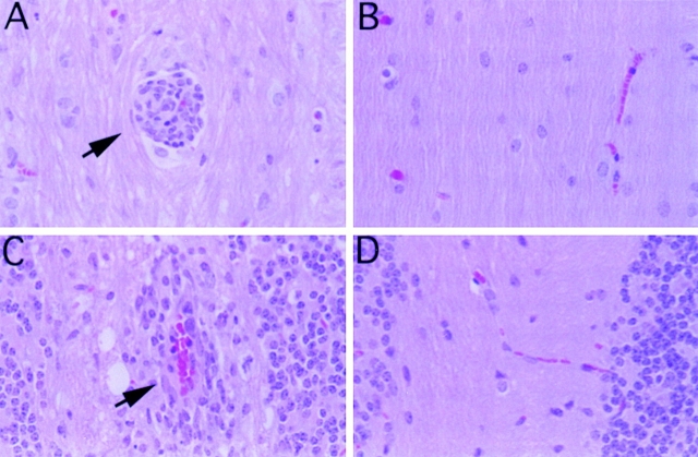
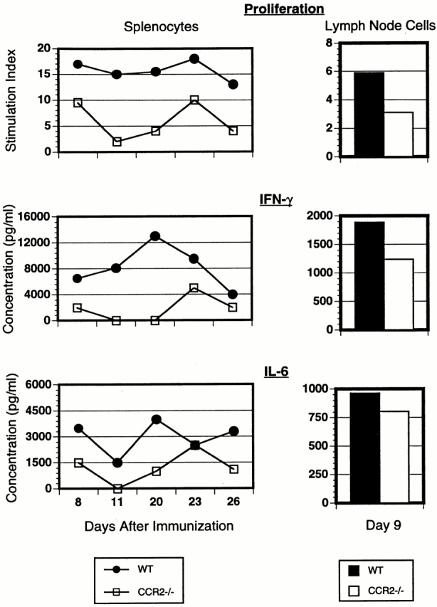
Monocyte recruitment to the central nervous system (CNS) is a necessary step in the development of pathologic inflammatory lesions in experimental autoimmune encephalomyelitis (EAE), a murine model of multiple sclerosis. Monocyte chemoattractant protein (MCP)-1, a potent agonist for directed monocyte migration, has been implicated in the pathogenesis of EAE. Here we report that deficiency in CC chemokine receptor (CCR)2, the receptor for MCP-1, confers resistance to EAE induced with a peptide derived from myelin oligodendrocyte glycoprotein peptide 35-55 (MOGp35-55). CCR2(-/)- mice immunized with MOGp35-55 failed to develop mononuclear cell inflammatory infiltrates in the CNS and failed to increase CNS levels of the chemokines RANTES (regulated on activation, normal T cell expressed and secreted), MCP-1, and interferon (IFN)-inducible protein 10 (IP-10) as well the chemokine receptors CCR1, CCR2, and CCR5. Additionally, T cells from CCR2(-/)- immunized mice showed decreased antigen-induced proliferation and production of IFN-gamma compared with wild-type immunized controls, suggesting that CCR2 enhances the T helper cell type 1 immune response in EAE. These data indicate that CCR2 plays a necessary and nonredundant role in the pathogenesis of EAE.
Figures


References
-
- Luster A.D. Chemokineschemotactic cytokines that mediate inflammation. N. Engl. J. Med. 1998;388:436–445. - PubMed
-
- Gu L., Tseng S.C., Rollins B.J. Monocyte chemoattractant protein-1. Chem. Immunol. 1999;72:7–29. - PubMed
-
- Charo I.F. CCR2from cloning to the creation of knockout mice. Chem. Immunol. 1999;72:30–41. - PubMed
-
- Frade J.M., Mellado M., del Real G., Gutierrez-Ramos J.C., Lind P., Martinez A.C. Characterization of the CCR2 chemokine receptorfunctional CCR2 receptor expression in B cells. J. Immunol. 1997;159:5576–5584. - PubMed
-
- Simpson J.E., Newcombe J., Cuzner M.L., Woodroofe M.N. Expression of monocyte chemoattractant protein-1 and other beta-chemokines by resident glia and inflammatory cells in multiple sclerosis lesions. J. Neuroimmunol. 1998;84:238–249. - PubMed
Publication types
MeSH terms
Substances
Grants and funding
LinkOut - more resources
Full Text Sources
Other Literature Sources
Molecular Biology Databases
Miscellaneous
