. 2000 Oct 10;97(21):11551-6.
doi: 10.1073/pnas.200238297.

The endocytic receptor protein LRP also mediates neuronal calcium signaling via N-methyl-D-aspartate receptors

Affiliations

The endocytic receptor protein LRP also mediates neuronal calcium signaling via N-methyl-D-aspartate receptors

B J Bacskai et al. Proc Natl Acad Sci U S A. .

Abstract

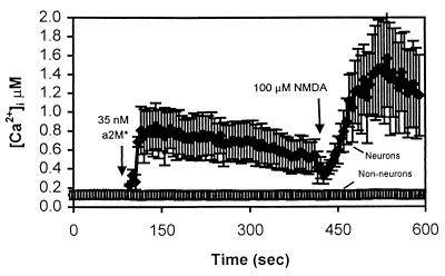
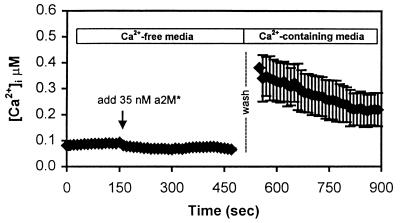
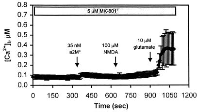
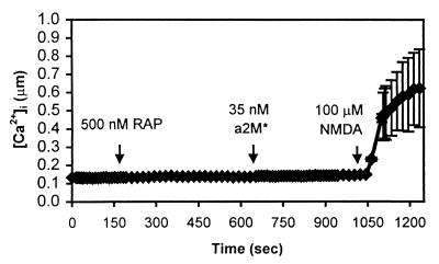
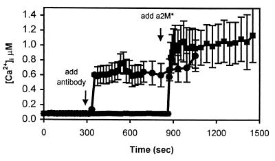
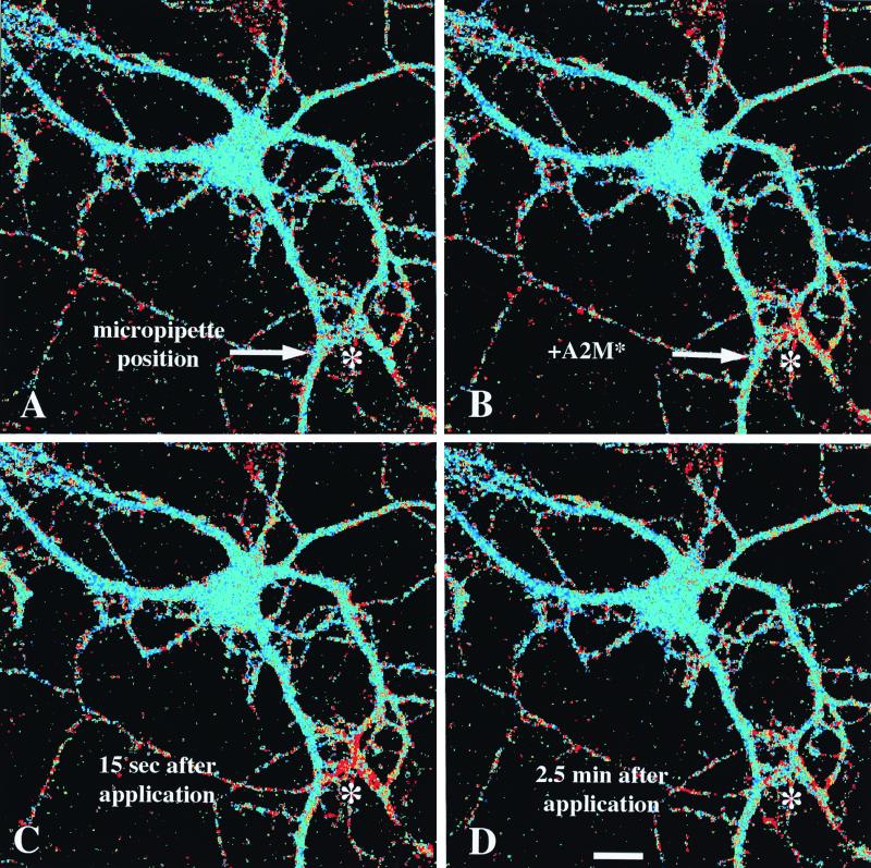
The low density lipoprotein receptor-related protein (LRP) is an endocytic receptor that is a member of the low density lipoprotein receptor family. We report that the LRP ligand, activated alpha(2)-macroglobulin (alpha(2)M*), induces robust calcium influx in cultured primary neurons, but not in nonneuronal LRP-containing cells in the same culture. The calcium influx is mediated through N-methyl-d-aspartate receptor channels, which explains the neuron specificity of the response. Microapplication of alpha(2)M* leads to a localized response at the site of application that dissipates rapidly, suggesting that the calcium signal is temporally and spatially discrete. Calcium influx to alpha(2)M* is blocked by the physiological LRP inhibitor, receptor-associated protein. Bivalent antibodies to the extracellular domain of LRP, but not Fab fragments of the same antibody, cause calcium influx, indicating that the response is specific to LRP and may require dimerization of the receptor. Thus, LRP is an endocytic receptor with a novel signaling role.


Figures

Figure 1
Figure 1
α2M* increases [Ca2+]i specifically in neurons. Primary cultures of mouse cortex were loaded for 30 min with 1 μM indo-1/AM and imaged by using a Bio-Rad 1024 Multiphoton confocal microscope. The traces represent a time course of intracellular calcium concentration in a field of cells in a single, representative experiment. Each trace is the average of six cells within the field, ± SD. Not all cells in the mixed cultures responded to α2M* treatment. The cells that did respond resembled neurons morphologically and also responded to NMDA application. Nonresponders had the generally flat appearance of glia and/or fibroblasts and did not respond to NMDA addition.
Figure 2
Figure 2
The calcium increase requires extracellular calcium. Cells were placed in nominally calcium-free buffer (not containing EGTA), and α2M* was added at approximately t = 150 sec. No intracellular calcium increase was observed. In fact, a small decrease was indicated. The calcium-free buffer was washed and replaced with calcium-containing buffer (2 mM) at t = 500 sec. After replacement of calcium, [Ca2+]i levels increased to about 400 nM, similar to levels normally observed after stimulation by α2M* in calcium-containing buffers. This suggests that α2M* was able to bind to LRP in the absence of calcium and initiate a calcium-signaling event. The response requires extracellular calcium, but not release of calcium from intracellular stores. The trace is the average of n = 7 cells in a field.
Figure 3
Figure 3
Calcium entry occurs through NMDAR channels. In this experiment, the cells were pretreated with 5 μM MK-801 for 5 min, and the NMDAR antagonist remained in the bath throughout the procedure. At t = 350 sec, 35 nM α2M* was added to the bath, resulting in a small but insignificant increase in [Ca2+]i in this field of cells. At t = 700 sec, 100 μM NMDA was added, and no change in [Ca2+]i was observed. Glutamate (10 μM), however, was capable of eliciting a calcium response at t = 900 sec. This is a representative trace of n = 4 experiments and is the mean of n = 15 neurons in a field.
Figure 4
Figure 4
The calcium response is blocked by RAP. RAP (500 nM) was added to the bath at t = 200 sec. RAP had no discernible effect on intracellular calcium. At t = 650 sec, 35 nM α2M* was added to the bath, but calcium was unaffected. NMDA was able to elicit a normal response in the seven cells in this field.
Figure 5
Figure 5
An antibody to the ligand-binding domain of LRP increases [Ca2+]i, but an antibody to an intracellular domain of LRP does not. This figure illustrates two experiments using rabbit polyclonal antibodies directed against LRP. In the top trace (●), R777 was added, which recognizes the ligand-binding domain of LRP. The addition of R777 increases [Ca2+]i in a neuron-specific manner. In the bottom trace (■), the addition of R704, which recognizes an intracellular domain of LRP, is unable to elicit an increase in [Ca2+]i. However, the subsequent addition of α2M* is able to generate a calcium response in these cells. Each trace is the average of seven cells in a field.
Figure 6
Figure 6
The calcium response is spatially restricted. A single cell was imaged with a multiphoton confocal microscope. α2M* was loaded into a glass micropipette, which was positioned with a micromanipulator near dendritic processes of the cell in the lower right corner of each image, as indicated by the asterisk (*). (A) Pseudocolor image of the cell immediately before administering α2M*. Blue colors represent low [Ca2+]i, and greens to yellows then reds represent increasing [Ca2+]i. (B) An image immediately after a 100-msec pressure pulse through the pipette, emitting a restricted cloud of α2M* just above the dendrites. (C) The cell approximately 15 sec after the pressure pulse, when the calcium change reaches its peak within a radius of about 25 μm of the tip of the pipette. (D) An image of the cell 2.5 min after the pressure pulse. The calcium concentration has returned to baseline by this time point. This result is representative of n = 6 experiments.

References

    1. Rebeck G W, Reiter J S, Strickland D K, Hyman B T. Neuron. 1993;11:575–580. - PubMed
    1. Herz J, Hamann U, Rogne S, Myklebost O, Gausepohl H, Stanley K K. EMBO J. 1988;7:4119–4127. - PMC - PubMed
    1. Strickland D K, Ashcom J D, Williams S, Burgess W H, Migliorini M, Argraves W S. J Biol Chem. 1990;265:17401–17404. - PubMed
    1. Willnow T E, Rohlmann A, Horton J, Otani H, Braun J R, Hammer R E, Herz J. EMBO J. 1996;15:2632–2639. - PMC - PubMed
    1. Williams S E, Ashcom J D, Argraves W S, Strickland D K. J Biol Chem. 1992;267:9035–9040. - PubMed

Publication types

MeSH terms

Substances

LinkOut - more resources