. 2000 Oct 10;97(21):11445-50.
doi: 10.1073/pnas.200363097.

IL-2-induced activation-induced cell death is inhibited in IL-15 transgenic mice

Affiliations

IL-2-induced activation-induced cell death is inhibited in IL-15 transgenic mice

J Marks-Konczalik et al. Proc Natl Acad Sci U S A. .

Abstract

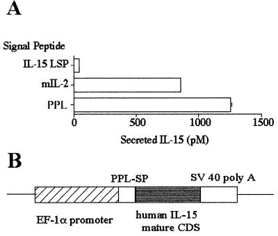
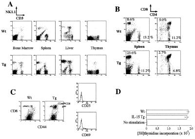
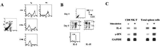
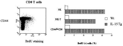
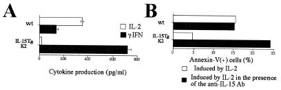
A transgenic (Tg) mouse expressing human IL-15 was generated to define the role of IL-15 in the normal immune response. Overexpression of IL-15 resulted in an increase of NK, CD44(hi)CD8 memory T cells, and gammadelta T cells. Additionally, we observed the emergence of a novel type of NK-T cells with CD8alphaalpha' expression. Due to the expansion and activation of NK cells, the IL-15Tg mouse showed enhanced innate immunity. In adaptive T cell immunity, the roles of IL-15 contrasted with those of IL-2. IL-15 inhibited IL-2-induced T cell death, which plays a role in the maintenance of peripheral self-tolerance. IL-15 thus seems to contribute to enhanced immune memory by selectively propagating memory T cells and by blocking T cell death mediated by IL-2.


Figures

Figure 1
Figure 1
Generation of the IL-15 transgenic mouse. (A) Ten micrograms of construct bearing the IL-15 SP (long signal peptide, LSP), IL-2 SP, or preprolactin (PPL) SP fused with human IL-15 cDNA (coding the mature peptide) under the human EF-1α promoter was transfected into 4 × 10⁶ COS cells by electroporation (250 V/950 μF). The supernatants were analyzed by a CTLL-2 assay at 48 h posttransfection. (B) Schematic design of the construct used to generate the IL-15Tg mouse.
Figure 2
Figure 2
Expansion of select lymphoid cells in the IL-15Tg mouse. (A) Analyses of cells from various tissues of wt and IL-15Tg mice. y axis, phosphatidylethanolamine-conjugated NK1.1 antibody; x axis, FITC-conjugated anti-CD3ɛ antibody. (B) Preferential increase of CD8 T cells in the IL-15Tg mouse. (C) Increase of CD44hiCD8 T cells in the IL-15Tg mouse. Memory CD8 T cells were defined by CD44/CD8 double staining (Left). Transgenic CD8 cells show high CD44 expression. CD44hiCD8 cells were further gated for the expression of the early T cell activation markers (FITC–anti-CD69 or anti-CD25) and demonstrated the lack of the expression of these antigens. (D) Normal response of T cells from the IL-15Tg mouse after anti-CD3 stimulation. Lymphocytes from wt and IL-15Tg spleens were incubated with a plate-coated anti-CD3 antibody for 48 h, then pulsed with 1 μCi per well of [3H]thymidine for 6 additional h.
Figure 3
Figure 3
(A) Properties of NK-T cells observed in the IL-15Tg mouse. Lymphocytes were isolated and purified from the liver. (Left) The NK-T population (FL1, FITC–anti-CD3ɛ; FL2, phycoerythrin–anti-NK1.1). Gated cells were further analyzed in the FL3 channel by the indicated antibodies (for CD8β, biotin–αCD8β/Cychrome–Streptavidin were used). As a control, preferential CD4 staining seen with the wt liver NK-T cells is shown. (B) Induction of CD8 NK-T cells from the bone marrow precursors of wt mice. Bone marrow cells were cultured with 10 nM IL-2 or IL-15. At day 6 and following, there was a propagation of NK and NK-T cells. The NK-T cells were further analyzed for their CD8 expression, which was >85% as shown as the histogram. (C) Production of γIFN but not IL-4 by the CD8 NK-T cells after stimulation examined by an RNase protection assay. NK-T cells were purified from a bone marrow culture using negative selection using the anti-CD8β antibody. NK-T cells and total spleen lymphocytes were stimulated by phorbol-myristate acetate (15 ng/ml) and ionomycin (0.75 μM) for 24 h before RNA extraction.
Figure 4
Figure 4
Enhanced BrdUrd incorporation into NK, NK-T, and CD44hiCD8 T cells from the IL-15Tg mouse. Cells were labeled with BrdUrd in vivo for 4 days. Lymphocytes were then stained by antibodies for NK and CD3 antigens, fixed, and stained by an FITC—anti-BrdUrd antibody. (Left) The selective incorporation of BrdUrd into the CD44hiCD8 T cells from the IL-15Tg(K2) mouse. Similar experiments were carried out with NK and NK-T cells, showing similar BrdUrd incorporation.
Figure 5
Figure 5
(A) Spontaneous lysis of YAC-1 cells by the IL-15Tg(K2) splenic cells. The effector/target ratio was compensated for based on the percentage of NK cells in the splenic lymphocytes. (B) Depletion of NK cells using the anti-asialo GM1 antibody abrogated the constitutive lytic activity to YAC-1 cells of the IL-15Tg(K2) splenic cells. The injection (i.p.) of the anti-asialo GM1 antibody resulted in rapid disappearance of NK cells from the spleen of the IL-15Tg mouse (8% to 0.3% within 24 h of injection). The NK-depleted splenic lymphocytes were then tested for cytotoxicity.
Figure 6
Figure 6
Augmented viral clearance in the IL-15Tg mouse against the vaccinia virus. 2 × 10⁸ plaque-forming units of the wt vaccinia virus were injected into five wt and IL-15Tg(K2) mice, and their blood was collected at the indicated time points after infection. After removing the plasma, blood cells were lysed by repeated freeze–thaw cycles, and the lysates were incubated with CV-1 cells for 36 h to monitor plaque formation.
Figure 7
Figure 7
(A) Reduced IL-2 production from T cells from the IL-15Tg mouse. CD3 T cells from either wt or IL-15Tg(K2) mouse were stimulated by CD3/CD28 antibodies, and cytokine production was measured by specific ELISA. (B) Reduced IL-2-induced AICD by the CD4 T cells purified from the IL-15Tg mouse and the restoration of AICD with the inclusion of an anti-IL-15 neutralizing antibody. CD4 cells (>95% pure) were first activated by CD3/28 antibodies for 48 h and then cultured with 1 nM IL-2 in the presence or absence of the M111 anti-IL-15 Ab (10 μg/ml, Genzyme) for 48 h. Cell death was induced in the third culture period by the treatment with anti-CD3/28 antibodies for 6 h. Dead cells were monitored by staining using the propidium iodide/Annexin V-GFP (CLONTECH). The data represent one of three independent experiments.

References

    1. Burton J D, Bamford R N, Peters C, Grant A J, Kurys G, Goldman C K, Brennan J, Roessler E, Waldmann T A. Proc Natl Acad Sci USA. 1994;91:4935–4939. - PMC - PubMed
    1. Bamford R N, Grant A J, Burton J D, Peters C, Kurys G, Goldman C K, Brennan J, Roessler E, Waldmann T A. Proc Natl Acad Sci USA. 1994;91:4940–4944. - PMC - PubMed
    1. Grabstein K H, Eisenman J, Shanebeck K, Rauch C, Srinivasan S, Fung V, Beers C, Richardson J, Schoenborn M A, Ahdieh M, et al. Science. 1994;264:965–968. - PubMed
    1. Giri J G, Kumaki S, Ahdieh M, Friend D J, Loomis A, Shanebeck K, DuBose R, Cosman D, Park L S, Anderson D M. EMBO J. 1995;14:3654–3663. - PMC - PubMed
    1. Waldmann T A, Tagaya Y. Annu Rev Immunol. 1999;17:19–49. - PubMed