. 2000 Oct 10;97(21):11238-43.
doi: 10.1073/pnas.200376897.

Three conformational states of scallop myosin S1

Affiliations

Three conformational states of scallop myosin S1

A Houdusse et al. Proc Natl Acad Sci U S A. .

Abstract

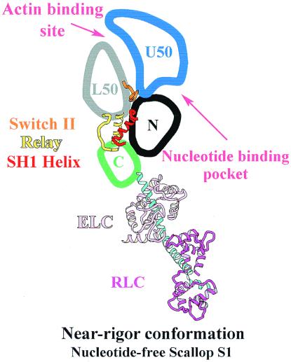
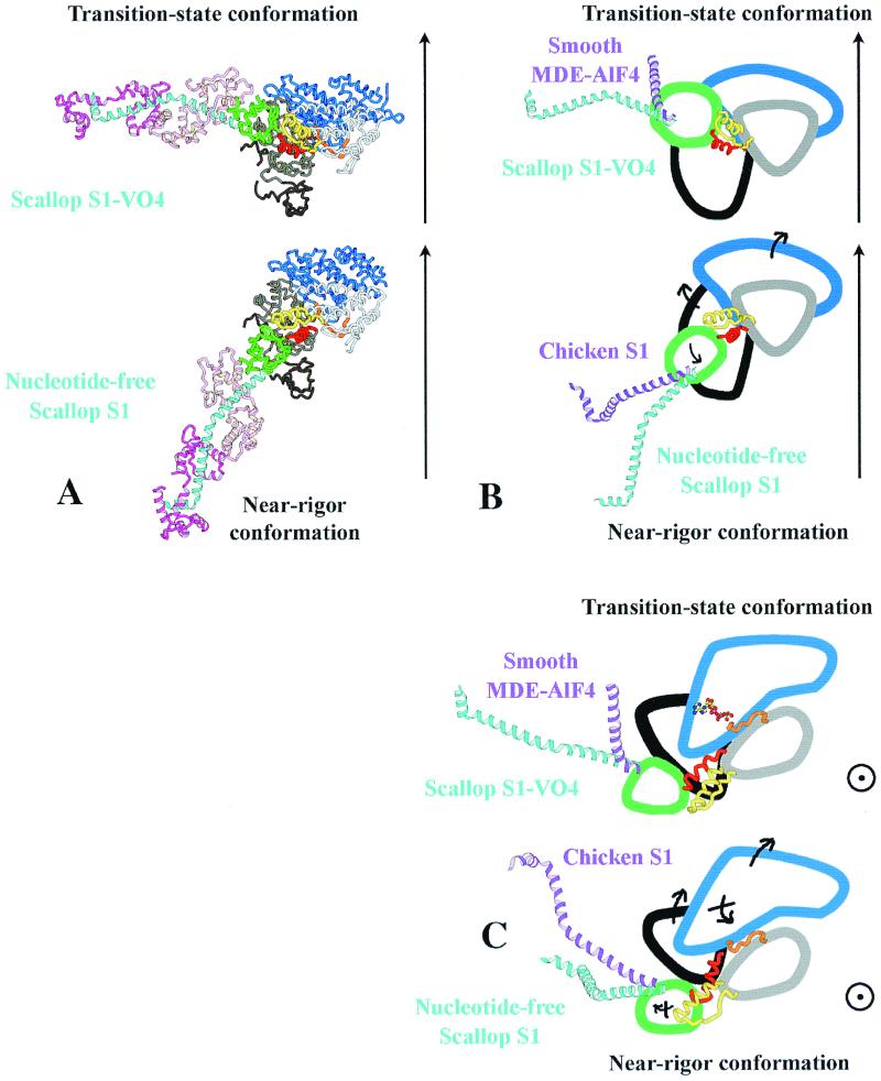
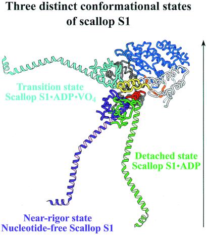
We have determined the structure of the intact scallop myosin head, containing both the motor domain and the lever arm, in the nucleotide-free state and in the presence of MgADP.V04, corresponding to the transition state. These two new structures, together with the previously determined structure of scallop S1 complexed with MgADP (which we interpret as a detached ATP state), reveal three conformations of an intact S1 obtained from a single isoform. These studies, together with new crystallization results, show how the conformation of the motor depends on the nucleotide content of the active site. The resolution of the two new structures ( approximately 4 A) is sufficient to establish the relative positions of the subdomains and the overall conformation of the joints within the motor domain as well as the position of the lever arm. Comparison of available crystal structures from different myosin isoforms and truncated constructs in either the nucleotide-free or transition states indicates that the major features within the motor domain are relatively invariant in both these states. In contrast, the position of the lever arm varies significantly between different isoforms. These results indicate that the heavy-chain helix is pliant at the junction between the converter and the lever arm and that factors other than the precise position of the converter can influence the position of the lever arm. It is possible that this pliant junction in the myosin head contributes to the compliance known to be present in the crossbridge.


Figures

Figure 1
Figure 1
Schematic drawing of the subdomains within the myosin head in the nucleotide-free scallop S1 structure. The four subdomains of the MD are labeled, as are three structural elements (joints) that articulate movements within the head: switch II (residues Ile-461 to Asn-470, orange); the relay (residues Asn-489 to Asp-515, yellow); and the SH1 helix (Cys-693 to Phe-707, red). Note that in all figures, the same colors have been kept for the four subdomains of the MD: the N-terminal (black), upper 50-kDa subdomain (blue), lower 50-kDa subdomain (gray), and the converter (green) as well as for the three joints. ELC, essential light chain; RLC, regulatory light chain.
Figure 2
Figure 2
(A) Ribbon diagrams of the nucleotide-free scallop S1 structure (Lower) and of scallop S1-VO4 (Upper) oriented such that the lower 50-kDa subdomains of these two structures superimpose. An arrow indicates the approximate direction of the actin filament axis relative to this subdomain, deduced from an electron microscope study of S1-decorated actin (16). The position of the ELC in the scallop nucleotide-free structure is very close to that found in the electron-microscope maps of actin decorated with vertebrate smooth muscle myosin S1 under rigor conditions (16). No data are available to indicate how S1 binds to actin in the prepower stroke state; for illustrative purposes only, we have chosen to orient this structure by assuming that the interactions with the lower 50-kDa subdomain would be conserved. The lever arm is positioned at ≈90° and 25° to the actin filament axis in the transition-state and near-rigor structures, respectively. (Note that for measuring angles, the lever-arm position is taken as a straight line drawn from the N-terminal side of the lever-arm helix to the sharp bend near the C terminus.) (B) Schematic drawings of the transition-state and the near-rigor conformations of scallop myosin from an interpretation of the structures seen in A. The rotation of the converter (green)/relay (yellow) module during the power stroke is amplified by the lever arm (scallop blue helix, light chains omitted for clarity). The direction of the movement of the subdomains in the transition between the two states is indicated with black arrows. Although the subdomains of the MD are similar in different isoforms, differences are seen in the lever-arm position. To illustrate this point, the position of the lever arm found in smooth muscle MDE (purple helix, Upper) and that of chicken striated muscle myosin S1 (purple helix, Lower) is compared with the positions found for scallop myosin in the transition state and near-rigor state, respectively. Differences in the bending of the heavy-chain helix at the junction between the converter and the lever arm result in markedly different orientations for the lever arm of these structures representing the same state. (C) Schematic drawing of an orthogonal view of the structures seen in A. In this orientation, the actin filament axis is approximately perpendicular to the page, and one can thus estimate the azimuthal component of the movement of the lever arm. This component is very small in the case of scallop. In contrast, bending of the heavy-chain helix at the pliant region in smooth MDE in the transition-state conformation could lead to a large azimuthal component during the power-stroke in this myosin. Comparison of the transition-state and near-rigor conformations in this view reveals changes in the position of the upper and lower 50-kDa subdomains related to differences in both the conformation of switch II and the actin-binding site.
Figure 3
Figure 3
Ribbon diagrams of the nucleotide-free scallop S1 structure in near-rigor, transition, and detached states, oriented such that the lower 50-kDa subdomains of these three structures superimpose. An arrow indicates the approximate direction of the actin filament axis relative to this subdomain, deduced from electron microscope studies (13, 16). The light chains bound to the heavy-chain helix of the lever arm in these three structures are omitted for clarity. Large differences are found in the position of the converter and result from relatively small rearrangements of the other three subdomains of the MD (not shown). In the three scallop S1 structures, the heavy-chain helix is straight at the junction between the converter and the lever arm, and the interactions at the interface between the converter and the ELC seem to be conserved.

References

    1. Rayment I, Rypniewski W R, Schmidt-Bäse K, Smith R, Tomchick D R, Benning M M, Winkelmann D A, Wesenberg G, Holden H M. Science. 1993;261:50–58. - PubMed
    1. Fisher A J, Smith C A, Thoden J B, Smith R, Sutoh K, Holden H M, Rayment I. Biochemistry. 1995;34:8960–8972. - PubMed
    1. Smith C A, Rayment I. Biochemistry. 1996;35:5404–5417. - PubMed
    1. Gulick A M, Bauer C B, Thoden J B, Rayment I. Biochemistry. 1997;36:11619–11628. - PubMed
    1. Dominguez R, Freyzon Y, Trybus K M, Cohen C. Cell. 1998;94:559–571. - PubMed

Publication types

LinkOut - more resources