. 2000 Oct 1;14(19):2501-14.
doi: 10.1101/gad.836800.

Multiple Ras-dependent phosphorylation pathways regulate Myc protein stability

Affiliations

Multiple Ras-dependent phosphorylation pathways regulate Myc protein stability

R Sears et al. Genes Dev. .

Abstract

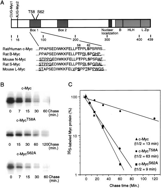
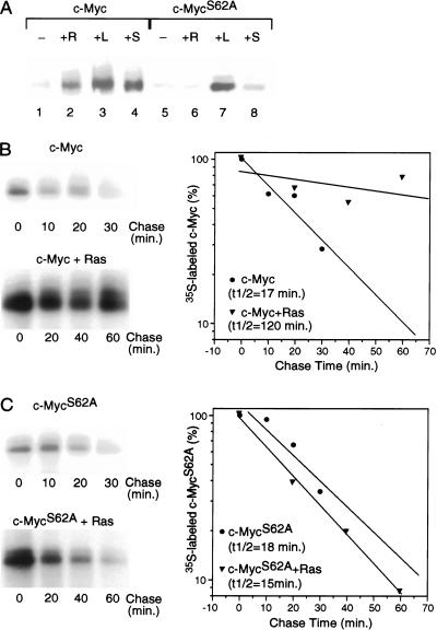
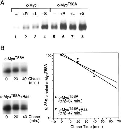
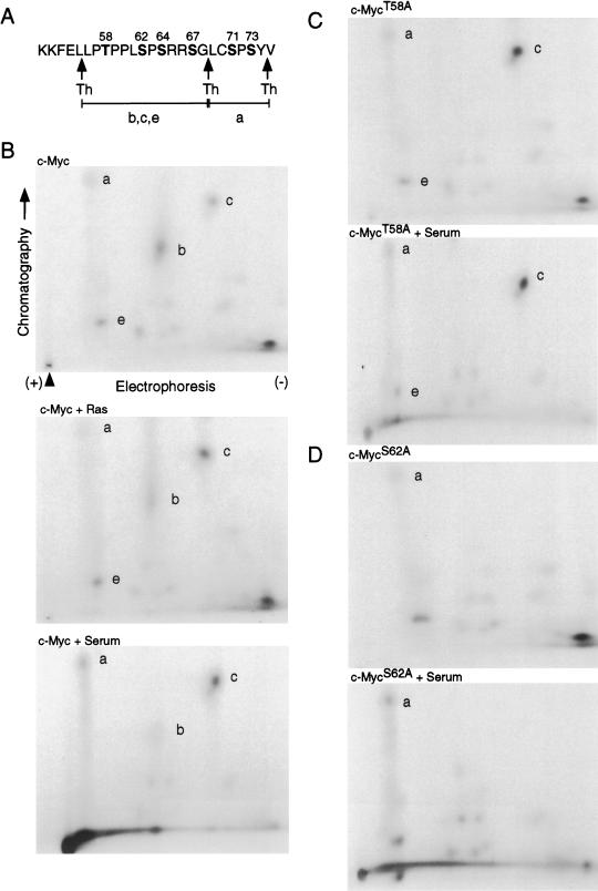
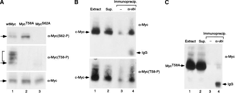
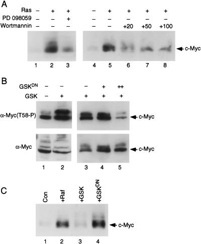
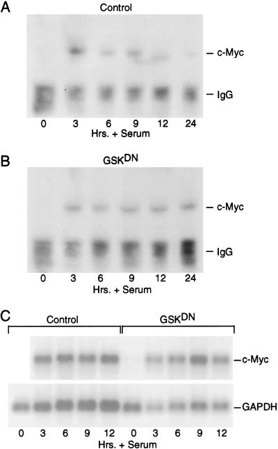
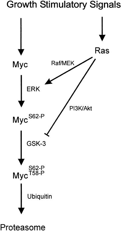
Our recent work has shown that activation of the Ras/Raf/ERK pathway extends the half-life of the Myc protein and thus enhances the accumulation of Myc activity. We have extended these observations by investigating two N-terminal phosphorylation sites in Myc, Thr 58 and Ser 62, which are known to be regulated by mitogen stimulation. We now show that the phosphorylation of these two residues is critical for determining the stability of Myc. Phosphorylation of Ser 62 is required for Ras-induced stabilization of Myc, likely mediated through the action of ERK. Conversely, phosphorylation of Thr 58, likely mediated by GSK-3 but dependent on the prior phosphorylation of Ser 62, is associated with degradation of Myc. Further analysis demonstrates that the Ras-dependent PI-3K pathway is also critical for controlling Myc protein accumulation, likely through the control of GSK-3 activity. These observations thus define a synergistic role for multiple Ras-mediated phosphorylation pathways in the control of Myc protein accumulation during the initial stage of cell proliferation.


Figures

Figure 1
Figure 1
Role of phosphorylation sites in determining the stability of Myc protein. (A) Schematic representation of the c-Myc protein. Translation start sites for Myc1 and Myc2 are shown. The helix–loop–helix (HLH)/leucine zipper (LZ) dimerization domains, the basic (B) DNA binding domain, the nuclear localization domain, and the two highly conserved Myc box 1 and 2 are shown. N-terminal phosphorylation sites Threonine 58 and Serine 62 are indicated. Depicted below is a comparison of the amino acid sequences surrounding the Thr 58 and Ser 62 residues in several related Myc proteins. Thr 58 and Ser 62 are in bold and divergent sequences are underlined. (B) Quiescent REF52 cells were infected with either Ad-Myc, Ad-MycT58A, or Ad-MycS62A, each at an MOI of 25. Infected cells were maintained in low serum (0.25%) medium. Eighteen hours postinfection, cells were labeled in vivo with 35S-methionine for 30 min and chased in low-serum medium containing excess unlabeled methionine for the indicated times. Labeled c-Myc was immunoprecipitated from equal cell numbers for each time point and analyzed by SDS-PAGE. (C) Immunoprecipitated 35S-labeled c-Myc from each sample shown in panel B was quantitated by PhosphorImager. Background was calculated from an equivalent area in each lane and subtracted from the value for labeled Myc in that lane. Time 0 was set at 100. Data is plotted on a semilog scale and best-fit lines were calculated using linear regression with the SigmaPlot program.
Figure 2
Figure 2
Role of Serine 62 phosphorylation in Ras-mediated stabilization of Myc. (A) Quiescent REF52 fibroblasts were infected with either Ad-Myc or Ad-MycS62A (MOI = 25) together with either Ad-Ras61L (+R) or a control virus (Ad-Con; all other samples; MOI = 200). Infected cells were maintained in low-serum medium (0.25%) for an additional 18 h and then harvested for Western blot analysis. Ad-Con-infected cells were either untreated (−), treated with 20% FCS for 4 h before harvesting (+S), or treated with 10 mM lactacystin for 4 h before harvesting (+L). Western blot analysis used the C-33 monoclonal c-Myc antibody. (B) Quiescent REF52 cells were infected with Ad-Myc (MOI = 25) together with either Ad-Con (upper panel) or Ad-Ras61L (lower panel; MOI = 200). Infected cells were maintained in low-serum (0.25%) medium. Eighteen hours postinfection, cells were labeled in vivo with 35S-methionine for 30 min and chased in low-serum medium containing excess unlabeled methionine for the indicated times. Labeled c-Myc was immunoprecipitated from equal cell numbers for each time point and analyzed by SDS-PAGE. 35S-labeled c-Myc from each sample was quantitated by PhosphorImager. Background was calculated from an equivalent area in each lane and subtracted from the value for labeled Myc in that lane. Time zero was set at 100. Data is plotted on a semi-log scale and best-fit lines were calculated using linear regression with the SigmaPlot program. (C) Quiescent REF52 cells were infected with Ad-MycS62A (MOI = 25) together with either Ad-Con (upper panel) or Ad-Ras61L (lower panel). Pulse/chase analyses were performed on the infected cells and immunoprecipitated 35S-labeled Myc was quantitated as described for panel B.
Figure 3
Figure 3
Role of Threonine 58 in controlling Myc stability. (A) Quiescent REF52 fibroblasts were infected with either Ad-Myc or Ad-MycT58A (MOI = 25) together with either Ad-Ras61L (+R) or Ad-Con (all other samples; MOI = 200). Infected cells were maintained in low-serum medium (0.25%) for an additional 18 h and then harvested for Western blot analysis. Ad-Con-infected cells were either untreated (−), treated with 20% FCS for 4 h before harvesting (+S), or treated with 10 mM lactacystin for 4 h before harvesting ( +L). Western blot analysis used the C-33 monoclonal c-Myc antibody. (B) Quiescent REF52 cells were infected with Ad-MycT58A (MOI = 25), together with either Ad-Con (upper panel) or Ad-Ras61L (lower panel; MOI = 200). Infected cells were maintained in low-serum (0.25%) medium. Eighteen hours postinfection, cells were labeled in vivo with 35S-methionine for 30 min and chased in low-serum medium containing excess unlabeled methionine for the indicated times. Labeled c-MycT58A was immunoprecipitated from equal cell numbers for each time point and analyzed by SDS-PAGE. 35S-labeled c-MycT58A from each sample was quantitated by PhosphorImager. Background was calculated from an equivalent area in each lane and subtracted from the value for labeled Myc in that lane. Time 0 was set at 100. Data is plotted on a semi-log scale and best-fit lines were calculated using linear regression with the SigmaPlot program.
Figure 4
Figure 4
Phosphopeptide mapping of c-Myc under various cell growth conditions. (A) Amino acid sequence of c-Myc between residues 51 and 75. Threonine and serine residues are in bold type and numbered. Thermolysin cleavage sites are indicated with arrows. Thermolytic peptides shown in the phosphopeptide maps are indicated (a,b,c,e). (B) Quiescent REF52 cells were infected with Ad-Myc (MOI = 25) together with either Ad-Ras61L (c-Myc + Ras) or Ad-Con (all other samples; MOI = 200). Infected cells were maintained in low-serum medium for 18 h and then in vivo labeled with 1 mCi/mL 32Pi for 2 h and harvested. Where indicated, Ad-Con-infected cells were stimulated with 20% FCS for 4 h before harvesting (c-Myc + Serum). 32P-labeled c-Myc was immunoprecipitated from cell lysates, fractionated by SDS-PAGE, transferred to Immobilon P membrane, and digested off the membrane with thermolysin. Thermolytic fragments were recovered and subject to two-dimensional separation. First-dimension electrophoresis was performed in buffer pH 1.9, and ascending chromatography was in phosphochromatography buffer. Chromatography and electrophoresis directions are shown, and the origin is indicated by an arrowhead. The same conditions were used for all two-dimensional maps shown. Thermolysin-digested phosphopeptides a, b, c, and e, described in panel A, are indicated. (C) Quiescent REF52 cells were infected and treated as described for panel B, except that Ad- Myc was replaced with Ad-MycT58A. Infected cells were in vivo labeled with 32Pi, and labeled MycT58A was subjected to thermolysin cleavage and phosphopeptide mapping as described for panel B. (D) Quiescent REF52 cells were infected and treated as described for panel B, except that Ad-Myc was replaced with Ad-MycS62A. Infected cells were in vivo labeled with 32Pi, and labeled MycS62A was subjected to thermolysin cleavage and phosphopeptide mapping as described for panel B.
Figure 5
Figure 5
Hierarchical phosphorylation of Serine 62 and Threonine 58 residues controls c-Myc ubiquitin-mediated degradation. (A) Quiescent REF52 cells were infected with either Ad-Myc, Ad-MycT58A, or Ad-MycS62A (MOI = 25). Infected cells were maintained in starvation medium for an additional 18 h. Four hours before harvesting, infected cells were treated with lactacystin. Each sample was analyzed by Western blotting with three separate antibodies: the monoclonal C-33 c-Myc antibody (lower panel), a polyclonal phosphospecific antibody that recognizes phosphorylated Thr 58 (middle panel), and a polyclonal phosphospecific antibody that recognizes Ser 62 (upper panel). Overlapping bands are indicated with arrows. Slower-mobility Myc bands are bracketed. (B) Quiescent REF52 cells were infected with Ad-Myc (MOI = 50). Infected cells were maintained in starvation medium for an additional 20 h. Six hours before harvesting, infected cells were treated with lactacystin. Ubiquitinated proteins were immunoprecipitated from harvested cell lysates with an anti-Ubiquitin antibody (Zymed; lane 4) or control extracts were mock treated and precipitated with proteinA/+G beads alone (lane 3). Immunoprecipitates along with one-tenth volume of the original extract (lane 1) or one-tenth volume of the supernatant following immunoprecipitation with anti-Ubi (lane 2) were all subjected to Western blot analysis with either the C-33 c-Myc antibody (upper panel) or the Thr 58 phosphospecific antibody (lower panel). C-Myc protein is indicated along with the IgG heavy chain from the anti-Ubi antibody. (C) Quiescent REF52 cells were infected with Ad-MycT58A (MOI = 50). Infected cells were treated as described in panel B. Ubiquitinated proteins were immunoprecipitated from harvested cell lysates, and Western blot analysis with the C-33 c-Myc antibody was performed on extract, supernatant, and immunoprecipitates as described for panel B.
Figure 6
Figure 6
Role of ERK and GSK-3 in Myc phosphorylation and stability. (A) Quiescent REF52 cells were infected with Ad-Myc (MOI = 25) together with either Ad-Ras61L (Ras, +) or Ad-Con (Ras, −; MOI = 200). Infected cells were maintained in low-serum medium for an additional 18 h. Where indicated, the MEK inhibitor PD 098059 (lane 3, 10 μM), or the PI-3K inhibitor Wortmanin (lanes 6–8, at the indicated mM concentrations), were added for 10 h before harvesting. Western blot analysis was performed on cell lysates with the C-33 c-Myc antibody. (B) Quiescent REF52 cells were infected with Ad-Myc (MOI = 25) together with Ad-Con (lane 1, MOI = 20; lane 3, MOI = 500), Ad-GSK (lanes 2–5, MOI = 20), and Ad-GSKDN (lanes 4,5, MOI = 100[+] or 500[++]). Infected cells were maintained in starvation medium for 20 h and then harvested for Western blot analysis with either the C-33 c-Myc antibody (lower panel) or the phosphospecific Thr 58-P antibody (upper panel). (C) Quiescent REF52 cells were infected with Ad-Myc (MOI = 25), together with either Ad-Con (lane 1), Ad-RafCAAX (lane 2), Ad-GSK (lane 3), or Ad-GSKDN (lane 4; MOI = 200). Infected cells were maintained in low-serum medium and harvested for Western blot analysis 18 h later with the C-33 c-Myc antibody.
Figure 7
Figure 7
GSK-3 regulates c-Myc accumulation following a growth response. (A) Quiescent REF52 cells were infected with Ad-Con (MOI = 200). Infected cells were maintained in starvation medium for 15 h and then stimulated with 20% FCS. Cells were harvested at the indicated times after serum stimulation, and endogenous c-Myc from cell lysates was concentrated by immunoprecipitation with the C-33 c-Myc antibody. Immunoprecipitated c-Myc was then visualized by Western blot analysis with the same C-33 Myc antibody. C-Myc protein and the IgG heavy chain from the immunoprecipitation are indicated. (B) Quiescent REF52 cells were infected with Ad-GSK-3DN (MOI = 200). Infected cells were treated as described for panel A. Endogenous c-Myc from each time point was immunoprecipitated and visualized as described for panel A. (C) Quiescent REF52 cells were infected with Ad-Con (control) or AGSKDN (MOI = 200). Infected cells were treated as described for panel A. Poly A+ RNA was isolated from approximately 300 μg of total RNA from each time point and subjected to Northern blot analysis. The blot was probed for c-Myc and GAPDH (loading control) as indicated.
Figure 8
Figure 8
Pathways controlling Myc phosphorylation and accumulation.

References

    1. Bhatia K, Huppi K, Spangler G, Siwarski D, Iyer R, Magrath I. Point mutations in the c-Myc transactivation domain are common in Burkitt's lymphoma and mouse plasmacytomas. Nat Genet. 1993;5:56–61. - PubMed
    1. Boyle WJ, van der Geer P, Hunter T. Phosphopeptide mapping and phosphoamino acid analysis by two-dimensional separation on thin-layer cellulose plates. Methods Enzymol. 1991;201:110–149. - PubMed
    1. Charron J, Malynn BA, Fisher P, Stewart V, Jeannotte L, Goff SP, Robertson EJ, Alt FW. Embryonic lethality in mice homozygous for a targeted disruption of the N-myc gene. Genes & Dev. 1992;6:2248–2257. - PubMed
    1. Ciechanover A, DiGiuseppe JA, Bercovich B, Orian A, Richter JD, Schwartz AL, Brodeur GM. Degradation of nuclear oncoproteins by the ubiquitin system in vitro. Proc Natl Acad Sci. 1991;88:139–143. - PMC - PubMed
    1. Clark HM, Yano T, Otsuki T, Jaffe ES, Shibata D, Raffeld M. Mutations in the coding region of c-MYC in AIDS-associated and other aggressive lymphomas. Cancer Res. 1994;54:3383–3386. - PubMed

MeSH terms

LinkOut - more resources