. 2000 Oct 1;14(19):2515-25.
doi: 10.1101/gad.177200.

Antagonism between C/EBPbeta and FOG in eosinophil lineage commitment of multipotent hematopoietic progenitors

Affiliations

Antagonism between C/EBPbeta and FOG in eosinophil lineage commitment of multipotent hematopoietic progenitors

E Querfurth et al. Genes Dev. .

Abstract

The commitment of multipotent cells to particular developmental pathways requires specific changes in their transcription factor complement to generate the patterns of gene expression characteristic of specialized cell types. We have studied the role of the GATA cofactor Friend of GATA (FOG) in the differentiation of avian multipotent hematopoietic progenitors. We found that multipotent cells express high levels of FOG mRNA, which were rapidly down-regulated upon their C/EBPbeta-mediated commitment to the eosinophil lineage. Expression of FOG in eosinophils led to a loss of eosinophil markers and the acquisition of a multipotent phenotype, and constitutive expression of FOG in multipotent progenitors blocked activation of eosinophil-specific gene expression by C/EBPbeta. Our results show that FOG is a repressor of the eosinophil lineage, and that C/EBP-mediated down-regulation of FOG is a critical step in eosinophil lineage commitment. Furthermore, our results indicate that maintenance of a multipotent state in hematopoiesis is achieved through cooperation between FOG and GATA-1. We present a model in which C/EBPbeta induces eosinophil differentiation by the coordinate direct activation of eosinophil-specific promoters and the removal of FOG, a promoter of multipotency as well as a repressor of eosinophil gene expression.


Figures
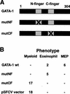
Figure 1
Figure 1
Effect of zinc finger mutations on myeloblast reprogramming by GATA-1. (A) Schematic drawing of GATA-1 and the two mutants with disrupted Zn-fingers. (B) Phenotypes of HD50M clones transfected with pNEO–GATA-1 or derivatives carrying mutations in either of the GATA-1 zinc fingers. The clones were picked, expanded, and characterized by IIF using monoclonal antibodies against lineage-specific surface antigens. The number of clones with MEP, eosinophil, and myeloblast phenotype is indicated.
Figure 2
Figure 2
Expression of FOG in chicken hematopoietic cell lines. A Northern blot was prepared from poly(A)+ RNA of the following chicken cell lines: HD3, HD37 (erythroid); HD50 (MEP); 1A1, HD50/4.8E (eosinophil); HD50/4.8M and HD57M (myeloid). The blot was probed sequentially with 32P-labeled chicken FOG cDNA (top) and chicken GAPDH cDNA (bottom) and signals detected by PhosphorImaging. The positions of the bands corresponding to the two mRNAs are indicated by arrows.
Figure 3
Figure 3
Ectopic expression of FOG in the myeloid cell line HD57M and the eosinophilic cell line 1A1. (A) Phenotype of FOG expressing myeloblast cell lines determined by IF/FC. The HD57M parental cell line is compared with the FOGexpressing clones HD57M FOG26 and HD57M FOG41. The graphs show the fluorescence intensity (log scale) obtained after staining with the indicated monoclonal antibodies plotted against the cell number (linear scale). (B) Western blot of lysates from the cell lines analyzed in A. Equal amounts of cellular protein were run on a 7.5% SDS–polyacrylamide gel and Western blotting was performed with the 12CA5 monoclonal antibody, detecting the HA tag on the FOG protein. The band corresponding to FOG is indicated. A nonspecific band (NS) served as a control for equal loading. (C) The surface antigen expression on the 1A1 eosinophil cell line, a 1A1 cell line stably transfected with the pEF–HA–PGKpuro vector (1A1 control), the HD50 MEP cell line, and two FOGexpressing 1A1 clones (1A1–FOG3 and 1A1–FOG20) was analyzed as in A. (D) Western blot of lysates from the cell lines analyzed in C. Proteins were separated by 10% SDS-PAGE and a Western blot sequentially probed with the following antibodies: 12CA5 (anti-HA-tag; a), anti-chicken C/EBPβ (b), anti-chicken Mim-1 (c), and anti-α-tubulin (d). The arrows indicate the positions of the bands corresponding to the antigens. (E) May-Gruenwald-Giemsa staining of 1A1 eosinophils, 1A1–FOG3 cells, and HD57 MEPs, as indicated. (F) Expression of endogenous FOG in MEPs derived by ectopic GATA-1 and FOG expression. A Northern blot of total RNA from HD50 MEPs, 1A1 eosinophils, 1A4 MEPs, and the 1A1–FOG3 and 1A1–FOG20 cell lines was sequentially probed with 32P-labeled cDNA for chicken FOG (a), chicken C/EBPβ (b), and chicken GAPDH (c), and signals detected by PhosphorImaging. The positions of the bands corresponding to the mRNAs are indicated by arrows.
Figure 4
Figure 4
FOG represses the eosinophil-specific EOS47 promoter through the NF of GATA-1. (A) Schematic representation of the positions of transcription factor binding sites present in the chicken EOS47 promoter (from McNagny et al. 1998). (B) Activation of the EOS47 promoter by GATA-1 and repression by FOG. The EOS47/-152-LUC reporter (1.0 μg) was cotransfected with pRSV–βGal (0.25 μg) into Q2bn fibroblasts. Expression vectors for Ets-1 (pCRNCM–cEts-1, 0.1 μg), c-Myb (pCRNCM–cMyb, 0.1μg), GATA-1 (pSPCMV–GATA-1, 0.3 μg), mutNF GATA-1 (pSPCMV–GATA-1mutNF, 0.3 μg), FOG (pEF–HAFOG–EFpuro, 0.5 μg), or equivalent amounts of the corresponding empty vectors (pCRNCM; pSPCMV; pEF–HA–PGKpuro, lanes indicated with ) were added as indicated. After 48 hr, luciferase and β-galactosidase activities were measured, and the luciferase activity normalized to the β-galactosidase activity. The basal promoter activity was arbitrarily assigned a value of one. Three independent transfections were carried out for each effector combination and standard deviations are indicated by the error bars. (C) The HD57–GATA–ER cell line was transfected with the pEF–HAFOG–PGKpuro expression vector and stable clones expressing FOG identified (HD57–GATA–ER FOG7, FOG8, FOG15, and FOG16). These, as well as a control nonexpressing clone (FOG2), were subjected to Western blot analysis as in Fig. 3B. Note that the expression level of clone 8 (lane 3) is lower than that of the other HA–FOG-expressing clones (the nonspecific band [NS] served as a control for equal loading). (D) HD57M GATA-1–ER clones not expressing (parental control and FOG2 clone) or expressing FOG (FOG7, FOG8, FOG15, and FOG16 clones) were induced with 0.1 μM βE for 36 hr (bottom) or left uninduced (top). Cells were analyzed for expression of the indicated antigens as in Fig. 3A. (E) The up-regulation of EOS47 antigen expression in D is given as a percent of that observed in the parental HD57M GATA–ER clone.
Figure 5
Figure 5
Down-regulation of FOG mRNA during eosinophil differentiation. (A) Cells from the C/EBPβ–ER-expressing HD57 clone 3, and the nonexpressing HD57 clone 12 were induced with 1.0 μM β-estradiol, and EOS47 expression determined by IIF after 1, 2, and 3 d of induction. Expression is given as percent of antigen-positive cells. No EOS47 expression was observed in the absence of βE in either clone (data not shown). (B) A Northern blot of total cellular RNA harvested at the timepoints in A was hybridized sequentially to probes for chicken FOG, chicken GATA-1, and chicken GAPDH. Results were analyzed by PhosphorImaging. (C) Primary MEPs were obtained by infection of yolk-sac blood island cells with the E26–WT and E26–βER viruses. After phenotyping to identify pure MEP populations (MEP21+, MEP26+, EOS47−, MYL51/2−, MHC II−), four to six clones for each construct were pooled. The pooled cells were left untreated or exposed to βE (1 μM final concentration). After 1, 2, and 3 d, one aliquot was removed for total RNA preparation (see D), and one aliquot washed free of βE and replated. A total of 6 d after induction, EOS47 antigen expression of the replated cells was determined by IIF, and plotted as a function of the time cells were exposed to βE (days). No change in antigen expression was observed in the absence of βE (data not shown). (D) A Northern blot of total RNA prepared as outlined in C was hybridized sequentially to probes for chicken FOG, chicken GATA-1, mim-1, and chicken GAPDH. Results were analyzed by PhosphorImaging.
Figure 6
Figure 6
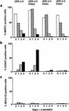
FOG inhibits C/EBPβ-mediated induction of eosinophil gene expression. HD57-C/EBPβ-ER clone 3 cells were transfected with the pEF–HAFOG–PGKpuro vector and the resulting puromycin-resistant clones analyzed for FOG expression by Western blot analysis (not shown). Two FOG-expressing (FOG1 and FOG7) and a nonexpressing control clone (FOG2) were further analyzed. These cell lines, along with the parental HD57-C/EBPβ-ER clone 3 cells, were induced with 1 μM βE or left untreated, and their expression of surface markers analyzed after 1, 2, and 4 d of exposure to βE. The results for MEP21 (a), EOS47 (b), and MYL51/2 (c) are shown, and are given as the percentage of cells staining positive. MEP26 and MHC II staining gave results similar to those for MEP21 and MYL51/2, respectively.
Figure 7
Figure 7
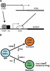
(A) Model for the induction of eosinophil-specific genes by C/EBP, as exemplified by the EOS47 promoter. In the MEPs, the EOS47 gene is inactive because of the absence of C/EBP, and the presence of FOG, which inhibits the activity of GATA-1 bound to the promoter (indicated by blunt-ended line). Upon induction of C/EBP, FOG is down-regulated, and the C/EBP site occupied (arrow), leading to the synergistic activation of the EOS47 promoter by GATA-1 and C/EBP. (B) Lineage determination by controlled collapse of the MEP phenotype. See text for explanation.

Similar articles

Cited by

References

    1. Beug H, Doederlein G, Freudenstein C, Graf T. Erythroblast cell lines transformed by a temperature sensitive mutant of avian erythroblastosis virus: A model system to study erythroid differentiation in vitro. J Cell Physiol Suppl. 1982;1:195–207. - PubMed
    1. Briegel K, Lim KC, Plank C, Beug H, Engel JD, Zenke M. Ectopic expression of a conditional GATA-2/estrogen receptor chimera arrests erythroid differentiation in a hormone-dependent manner. Genes & Dev. 1993;7:1097–1109. - PubMed
    1. Chomczynski P, Sacchi N. Single step isolation of RNA by acid guanidinium isothiocyanate method. Anal Biochem. 1987;162:156–159. - PubMed
    1. Crispino JD, Lodish MB, MacKay JP, Orkin SH. Use of altered specificity mutants to probe a specific protein-protein interaction in differentiation: The GATA-1:FOG complex. Mol Cell. 1999;3:219–228. - PubMed
    1. Deconinck AE, Mead PE, Tevosian SG, Crispino JD, Katz SG, Zon LI, Orkin SH. FOG acts as a repressor of red blood cell development in Xenopus. Development. 2000;127:2031–40. - PubMed

Publication types

MeSH terms