. 2000 Oct 1;14(19):2534-46.
doi: 10.1101/gad.831800.

A multifactor complex of eukaryotic initiation factors, eIF1, eIF2, eIF3, eIF5, and initiator tRNA(Met) is an important translation initiation intermediate in vivo

Affiliations

A multifactor complex of eukaryotic initiation factors, eIF1, eIF2, eIF3, eIF5, and initiator tRNA(Met) is an important translation initiation intermediate in vivo

K Asano et al. Genes Dev. .

Abstract

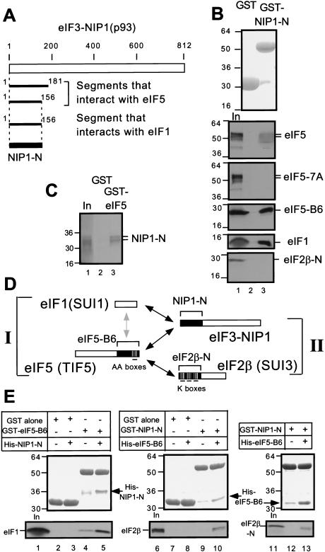
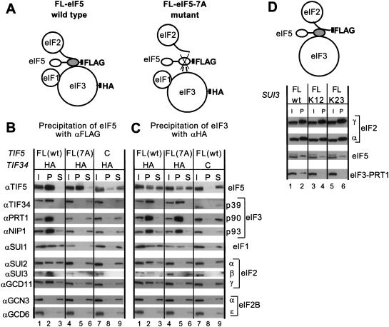
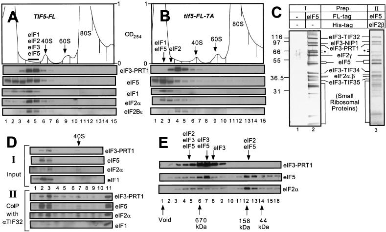
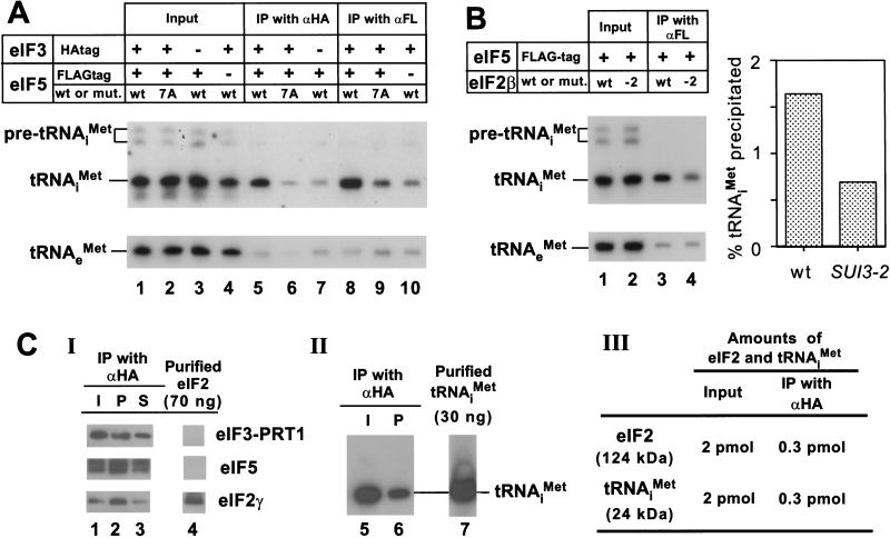
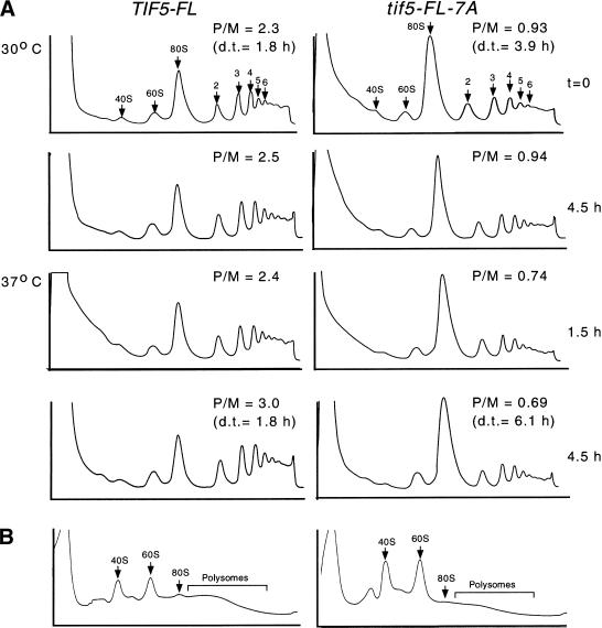
Translation initiation factor 2 (eIF2) bound to GTP transfers the initiator methionyl tRNA to the 40S ribosomal subunit. The eIF5 stimulates GTP hydrolysis by the eIF2/GTP/Met-tRNA(i)(Met) ternary complex on base-pairing between Met-tRNA(i)(Met) and the start codon. The eIF2, eIF5, and eIF1 all have been implicated in stringent selection of AUG as the start codon. The eIF3 binds to the 40S ribosome and promotes recruitment of the ternary complex; however, physical contact between eIF3 and eIF2 has not been observed. We show that yeast eIF5 can bridge interaction in vitro between eIF3 and eIF2 by binding simultaneously to the amino terminus of eIF3 subunit NIP1 and the amino-terminal half of eIF2beta, dependent on a conserved bipartite motif in the carboxyl terminus of eIF5. Additionally, the amino terminus of NIP1 can bind concurrently to eIF5 and eIF1. These findings suggest the occurrence of an eIF3/eIF1/eIF5/eIF2 multifactor complex, which was observed in cell extracts free of 40S ribosomes and found to contain stoichiometric amounts of tRNA(i)(Met). The multifactor complex was disrupted by the tif5-7A mutation in the bipartite motif of eIF5. Importantly, the tif5-7A mutant is temperature sensitive and displayed a substantial reduction in translation initiation at the restrictive temperature. We propose that the multifactor complex is an important intermediate in translation initiation in vivo.


Figures

Figure 1
Figure 1
Simultaneous interactions in vitro involving minimal binding segments of eIF3–NIP1, eIF2β, eIF5, and full-length eIF1. (A) Summary of yeast two-hybrid interactions detected between segments of NIP1 and full-length eIF5 and eIF1 (see Materials and Methods for details). The empty box indicates the 812-amino acid NIP1 polypeptide. Segments of NIP1 that interacted with eIF5 and eIF1 are listed below with locations in the sequence. (B) and (C) The amino-terminal segment of eIF3–NIP1 (NIP1-N) binds to eIF1 and eIF5. 20μg of GST–NIP1-N (B, lane 3), GST–eIF5 (C, lane 3), or GST alone (B and C, lanes 2), expressed in Escherichia coli and immobilized on glutathione sepharose beads, was incubated with the 35S-labeled proteins listed on the right. After extensive washing, the bound proteins were analyzed by SDS-PAGE followed by Coomassie staining (B, top panel) and autoradiography (B, lower five panels; C). Lanes 1 of B and C, 50% of the input amounts of labeled proteins. (D) Schematic illustration of interactions involving full-length eIF1 and segments of eIF2, eIF3, and eIF5 indicated with brackets and designated as in the text. Boxes indicate primary structures of the proteins involved and the filled regions denote the minimal binding domains. Black or gray arrows indicate direct interactions; the gray arrow denotes a relatively weak interaction. (E) Bridging experiments. 5 μg of GST–eIF5-B6, GST–NIP1-N, or GST alone immobilized on beads was mixed with 35S eIF1 (lanes 2–5), 35S eIF2β (lanes 7–10), or 35S eIF2β-N (lanes 12,13) in the presence or absence (as indicated) of 60 μg of His–NIP1-N or His–eIF5-B6 expressed in E. coli and purified by Ni2+ affinity chromatography. Bound proteins were detected by SDS-PAGE, followed by Coomassie staining (middle) and autoradiography (bottom). The GST-fusion or His-tagged proteins used in each reaction are listed above each of the middle panels. In lanes 1, 6, and 11, 50% of the input amounts of 35S-proteins were loaded.
Figure 2
Figure 2
Coimmunoprecipitation of eIF2 and eIF3 from cell extracts dependent on AA-box 2 in the carboxyl terminus of eIF5. (A) Schematic model of the structures of the multifactor complexes found in KAY50 and KAY51 (Table 1) containing wild-type eIF5-FL (left) or eIF5-FL-7A (right), as deduced from this study. The modular structure of eIF5 is represented by two ovals (amino-terminal and carboxy-terminal domains) connected with a thick line (less conserved region). The shaded oval in eIF5 corresponds to the B6 fragment, necessary and sufficient for binding to eIF2 and eIF3 (Asano et al. 1999). This oval is crossed in the mutant, indicating the 7A mutation in AA-box 2. The curved line attached to eIF2 represents the amino-terminal half of eIF2β containing the K-boxes which interact with the carboxy-terminal domain of eIF5. Filled squares indicate the epitope tags on eIF3-HA-TIF34 or FL-eIF5. Dotted lines indicate a weakened interaction. (B, C) Coimmunoprecipitation of eIFs with eIF5-FL or TIF34-HA. WCEs were prepared from strains KAY50, KAY51, KAY10, and KAY37 (Table 1), grown in YPD medium at 30°C. Aliquots of WCEs were incubated with anti-FLAG (B) or anti-HA (C) affinity resin and after extensive washing, the bound proteins were analyzed by SDS-PAGE and immunoblotting using the antibodies indicated on the left. Lanes 1, 4, and 7, 20% input (I) amounts of WCE; lanes 2, 5, and 8, the entire precipitated (P) fractions; lanes 3, 6, and 9, 10% of supernatant (S) fractions. (D) Coimmunoprecipitation of eIFs with eIF2β-FL. The schematic model is similar to that described in B and C except that eIF2β carries the FLAG epitope. WCEs prepared from strains KAY33 (SUI3), KAY25 (SUI3-FL), KAY29 (sui3-FL-K12) and KAY30 (sui3-FL-K23) (Asano et al. 1999) were immunoprecipitated with FLAG affinity resin and the precipitated proteins were analyzed by immunoblotting using the appropriate antibodies. (I) 20% input amount of WCE (lanes 1, 3, and 5); (P) the entire precipitated fractions (lanes 2, 4, and 6). K12 and K23 indicate the Ala substitutions present in K-boxes 1–2 or 2–3, respectively, in the mutant eIF2β-FL proteins.
Figure 3
Figure 3
The multifactor complex containing eIF1, eIF2, eIF3, and eIF5 can be isolated free of 40S ribosomes. (A,B) Twenty A260 units of WCEs prepared from KAY50 (TIF5-FL) (A) and KAY51 (tif5-FL-7A) (B) were resolved on 15%–40% sucrose gradients by centrifugation for 4.5 h at 39,000 rpm. Gradients were separated into 20 0.6 mL-fractions while scanning continuously at A254 as shown ( top panels). Portions of the fractions were precipitated with 10% TCA and analyzed by SDS-PAGE and immunoblotting using antibodies against the factors listed beside the five bottom panels. (C) Silver staining of purified preparations of the multifactor complex. The staining patterns of Type I (lane 2) and Type II (lane 3) preparations are shown along with a mock preparation obtained from a strain containing untagged eIF5 (lane 1) in parallel with the Type I preparation. (*) Degradation products of TIF32. (D) Size fractionation and coimmunoprecipitation analysis of a Type II preparation of the multifactor complex. A sample containing 0.5 mL (50 μg protein) of Type II preparation was resolved on 7.5%–30% sucrose gradient by centrifugation for 5 h at 41,000 rpm. The gradient was separated into 10 1.2 mL-fractions while scanning continuously at A254. One hundred μL of each fraction were precipitated with 10% TCA (top panels, input), 400 μL were immunoprecipitated with anti-TIF32 antibodies (bottom panels, CoIP with αTIF32), and the resulting samples were analyzed by immunoblotting using antibodies against the factors listed to the right. The position of the 40S ribosomes is indicated. (Lane 11) 1% of the Type II preparation separated on the sucrose gradient. (E) Size fractionation of Type I preparation of the multifactor complex. Two hundred microliters of Type I preparation was separated on a Superose 6 sizing column (Pharmacia), preequilibrated with buffer A without pepstatin, aprotinin, and leupeptin, using the FPLC system (Pharmacia), as described previously (Phan et al. 1998). Portions of the fractions were precipitated with 10 % TCA, and analyzed by SDS-PAGE and immunoblotting using antibodies against the factors listed on the right. Lane 1: sample from the void volume. Arrows below the panels indicate the elution positions of size standards (BioRad) determined in a parallel experiment. Arrows above the panels indicate the positions of different complexes deduced from the immunoblot analysis.
Figure 4
Figure 4
The multifactor complex contains initiator tRNAiMet. (A) The eIF5-FL/eIF3-HA complex contains tRNAiMet. Samples containing 1 mg of WCE in 1 mL of buffer A (Asano et al. 1999) prepared in the presence of 0.2 U/μL of RNasin (Promega) from strains used in Figure 2B,C were incubated for 2 h at 4°C with 100 μL of Protein A Sepharose (Pharmacia) that was preincubated with 15 μL of mouse monoclonal anti-HA antibodies (BAbCO) or M2 FLAG affinity resin (Sigma). After washing with 1 mL buffer A four times, the beads were suspended in water, extracted twice with phenol and once with phenol/chloroform, and precipitated with ethanol. Precipitated RNAs were separated by denaturing polyacrylamide gel electrophoresis, followed by Northern blot analysis using probes specific for tRNAiMet or tRNAeMet, as described previously (Anderson et al. 1998) (lanes 5–10). Lanes 1–4, RNA samples phenol-extracted and ethanol-precipitated from 2% of the input WCEs used for the immunoprecipitations. The top panel describes the antibodies used for immunoprecipitation, the presence of epitope tags on eIF5 or eIF3–TIF34, and the presence of wild-type (wt) or tif5-7A (7A) forms of eIF5 in the extracts. (B) Evidence that tRNAiMet is bound to eIF2 in the multifactor complex. One milligram aliquots of WCEs prepared from strains KAY56 (SUI3) and KAY57 (SUI3-2) were incubated at 37°C for 5 min and subjected to immunoprecipitation with FLAG affinity resin, followed by Northern blotting, as described above (lanes 3 and 4, respectively). Lanes 1 and 2, RNA samples phenol-extracted and ethanol-precipitated from 2% of the input WCEs used for the immunoprecipitations. The histogram on the right shows the percentages of the amounts of tRNAiMet present in the starting extracts that were immunoprecipitated based on phosphorimaging analysis of the Northern data. (C) Quantification of the amounts of eIF2 and tRNAiMet in the multifactor complex. A 200 μL aliquot (20 μg) of Type I preparation was immunoprecipitated for 2 h at 4°C with 20 μL Protein A Sepharose beads that had been preincubated with 3 μL of anti-HA antibodies. (Panel I) After washing the beads four times with buffer A, the immunoprecipitated proteins were subjected to immunoblot analysis as described in Figure 2C, along with 70 ng of purified eIF2 (lane 4). Lane 1, 20 μL starting Type I preparation; lane 3, 10 % of supernatant fraction from the immunoprecipitation. (Panel II) RNA was extracted from a duplicate immunoprecipitation conducted as in Panel I and subjected to Northern blot analysis as described above (A), along with 30 ng purified tRNAiMet (lane 7). Lane 5, RNA extracted from 100 μL of the starting Type I preparation. (Panel III) The results of quantification of data in panels I–II.
Figure 5
Figure 5
Analysis of polysome profiles in isogenic TIF5 and tif5-7A strains. (A) Yeast strains KAY50 and KAY51 grown exponentially in YPD medium at 30°C (t = 0) were shifted to 37°C (or kept at 30°C as a control) for the indicated times. Cycloheximide was added to the cultures for 5 min prior to harvesting the cells. WCEs were prepared and resolved by velocity-sedimentation on 5%–45% sucrose gradients. Fractions were collected while scanning continuously at A254. The positions of 40S and 60S subunits, 80S ribosomes and polysomes are indicated. (P/M) Ratio of A254 in the combined polysome fractions to that in the 80S peak; (d.t.) cell doubling time in hours. (B) WCEs prepared from yeast strains grown exactly as described for top panels in A were centrifuged on 5%–45% sucrose gradients containing 0.7 M NaCl for 2.5 h at 39,000 rpm and analyzed as in A.

References

    1. Anderson J, Phan L, Cuesta R, Carlson BA, Pak M, Asano K, Bjork GR, Tamame M, Hinnebusch AG. The essential Gcd10p–Gcd14p nuclear complex is required for 1-methyladenosine modification and maturation of initiator methionyl-tRNA. Genes & Dev. 1998;12:3650–3662. - PMC - PubMed
    1. Anderson J, Phan L, Hinnebusch AG. The Gcd10p/Gcd14p complex is the essential two-subunit tRNA(1-methyladenosine) methyltransferase of Saccharomyces cerevisiae. Proc Natl Acad Sci. 2000;97:5173–5178. - PMC - PubMed
    1. Asano K, Kinzy TZ, Merrick WC, Hershey JWB. Conservation and diversity of eukaryotic translation initiation factor eIF3. J Biol Chem. 1997;272:1101–1109. - PubMed
    1. Asano K, Phan L, Anderson J, Hinnebusch AG. Complex formation by all five homologues of mammalian translation initiation factor 3 subunits from yeast Saccharomyces cerevisiae. J Biol Chem. 1998;273:18573–18585. - PubMed
    1. Asano K, Krishnamoorthy T, Phan L, Pavitt GD, Hinnebusch AG. Conserved bipartite motifs in yeast eIF5 and eIF2Bε, GTPase-activating and GDP–GTP exchange factors in translation initiation, mediate binding to their common substrate eIF2. EMBO J. 1999;18:1673–1688. - PMC - PubMed

Publication types

MeSH terms