. 2000 Oct 2;151(1):1-14.
doi: 10.1083/jcb.151.1.1.

Protein ligands to HuR modulate its interaction with target mRNAs in vivo

Affiliations

Protein ligands to HuR modulate its interaction with target mRNAs in vivo

C M Brennan et al. J Cell Biol. .

Abstract

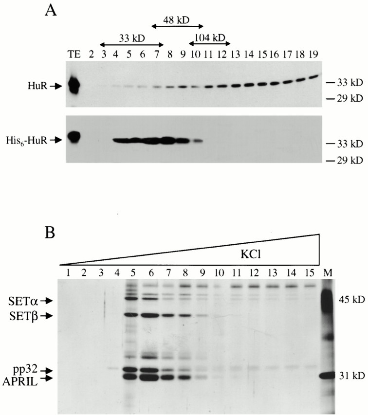
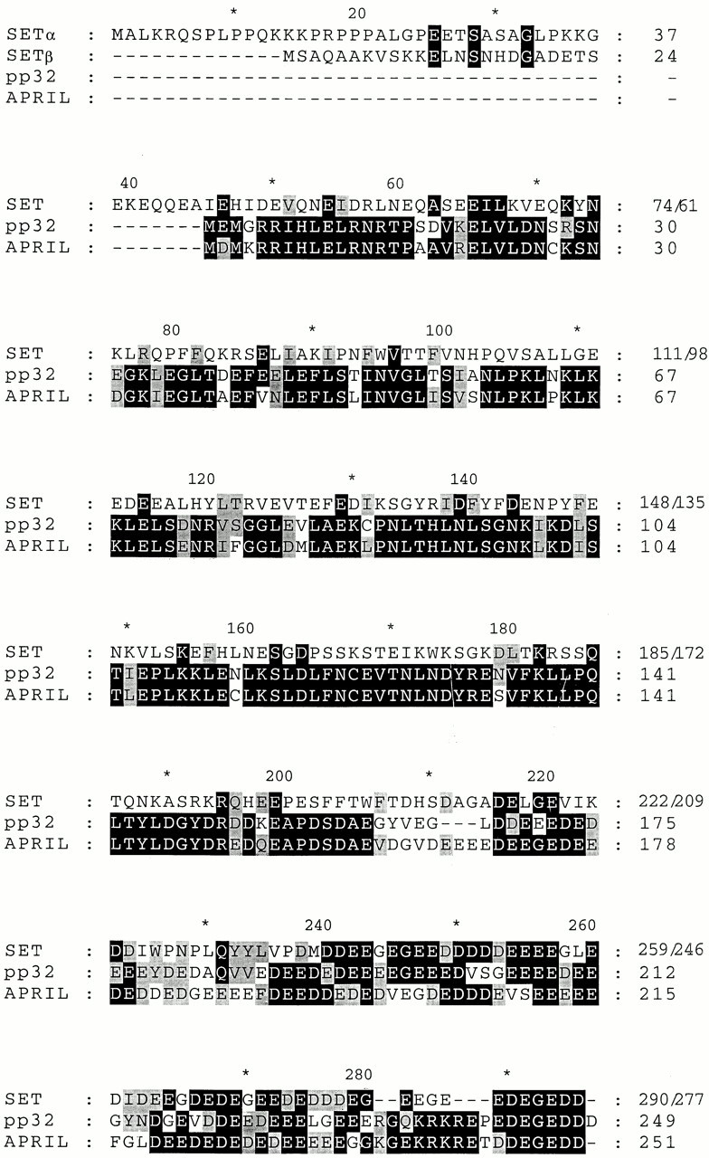
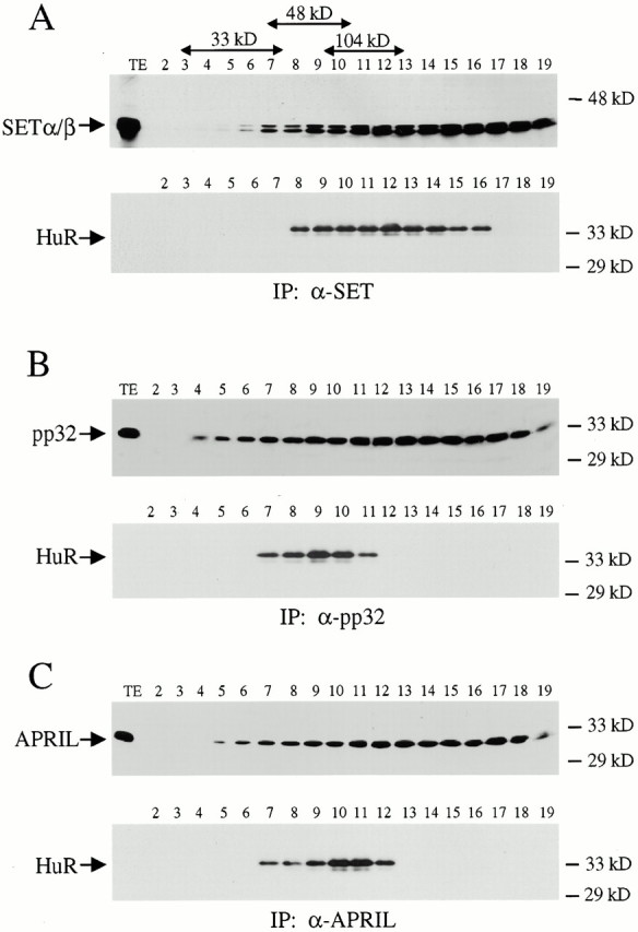
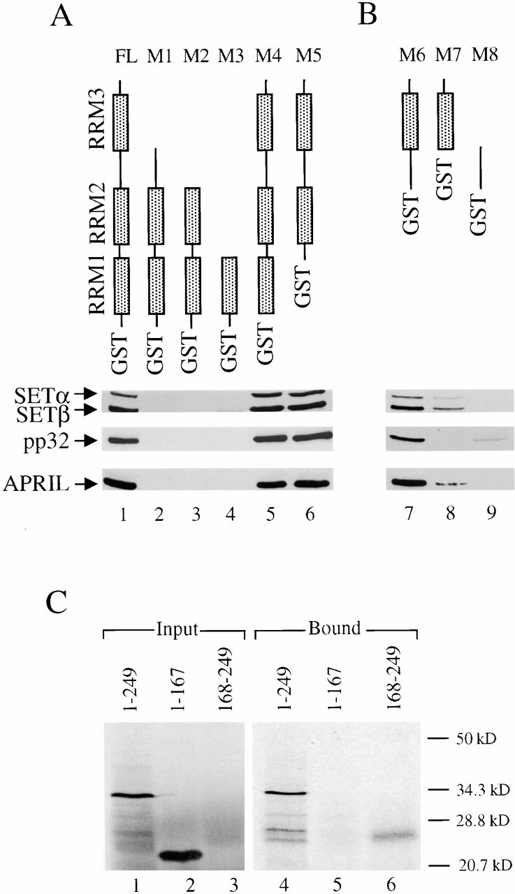
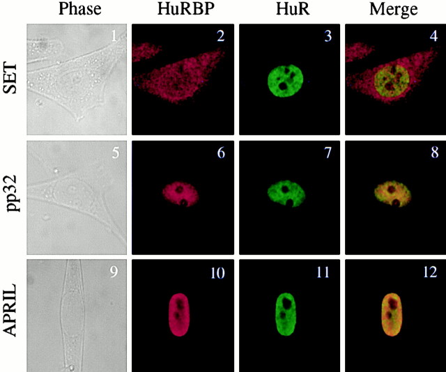
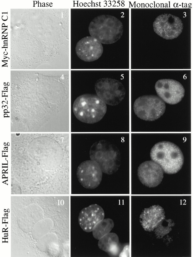
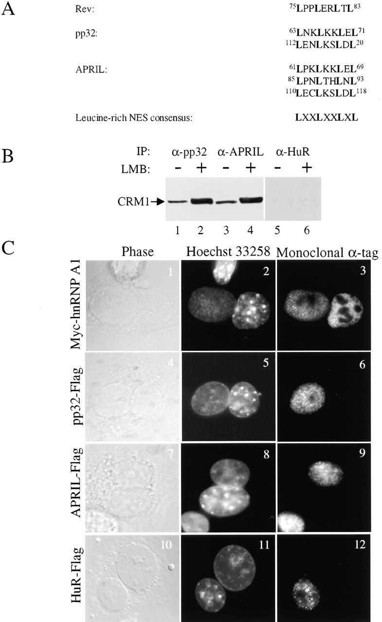
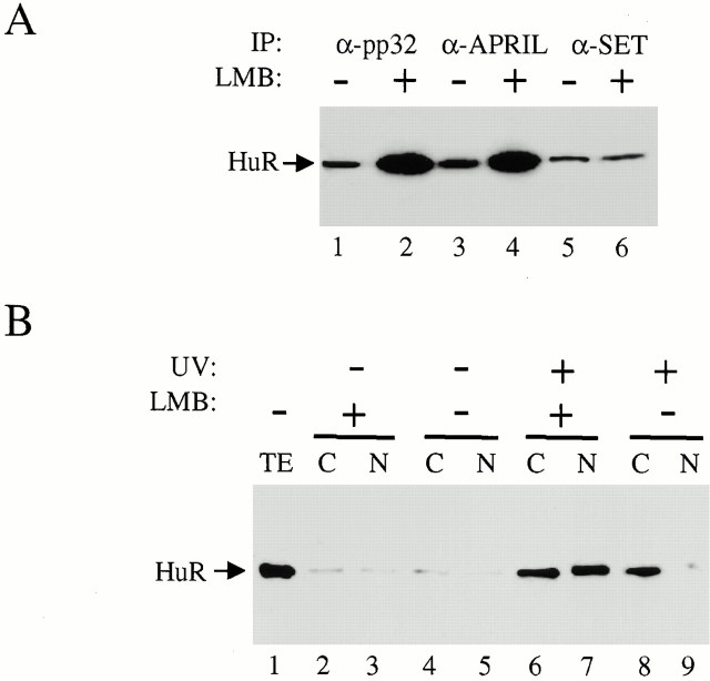
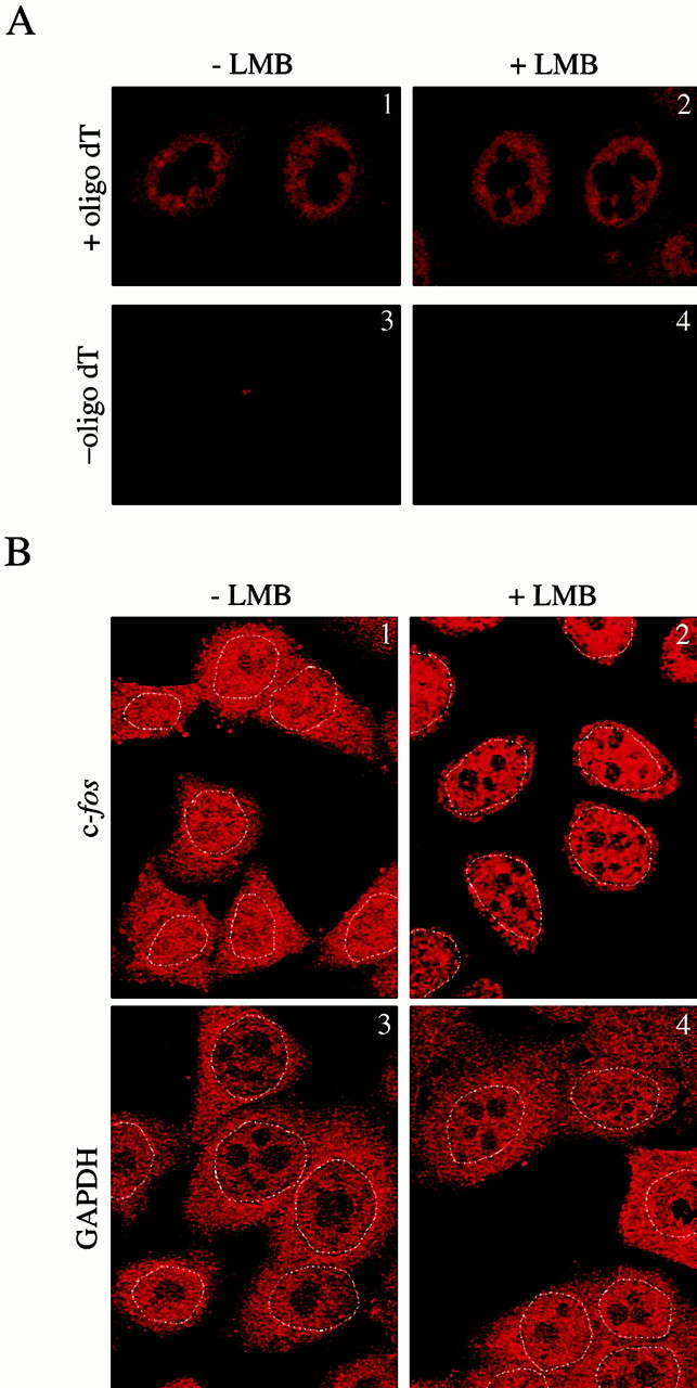
AU-rich elements (AREs) present in the 3' untranslated regions of many protooncogene, cytokine, and lymphokine messages target them for rapid degradation. HuR, a ubiquitously expressed member of the ELAV (embryonic lethal abnormal vision) family of RNA binding proteins, selectively binds AREs and stabilizes ARE-containing mRNAs in transiently transfected cells. Here, we identify four mammalian proteins that bind regions of HuR known to be essential for its ability to shuttle between the nucleus and the cytoplasm and to stabilize mRNA: SETalpha, SETbeta, pp32, and acidic protein rich in leucine (APRIL). Three have been reported to be protein phosphatase 2A inhibitors. All four ligands contain long, acidic COOH-terminal tails, while pp32 and APRIL share a second motif: rev-like leucine-rich repeats in their NH(2)-terminal regions. We show that pp32 and APRIL are nucleocytoplasmic shuttling proteins that interact with the nuclear export factor CRM1 (chromosomal region maintenance protein 1). The inhibition of CRM1 by leptomycin B leads to the nuclear retention of pp32 and APRIL, their increased association with HuR, and an increase in HuR's association with nuclear poly(A)+ RNA. Furthermore, transcripts from the ARE-containing c-fos gene are selectively retained in the nucleus, while the cytoplasmic distribution of total poly(A)+ RNA is not altered. These data provide evidence that interaction of its ligands with HuR modulate HuR's ability to bind its target mRNAs in vivo and suggest that CRM1 is instrumental in the export of at least some cellular mRNAs under certain conditions. We discuss the possible role of these ligands upstream of HuR in pathways that govern the stability of ARE-containing mRNAs.


Figures

Figure 1
Figure 1
HuR exists in complexes and interacts with four protein ligands in vitro. (A) HeLa whole-cell extract (Gu et al. 1997) was fractionated on a 5–20% glycerol gradient. Fractions were run on a 12% gel and immunoblots probed with the 3A2 anti–HuR antibody (Gallouzi et al. 2000). As markers, phosphorylase B (104 kD), ovalbumin (48 kD), and carbonic anhydrase (33 kD) were run on a parallel gradient. (B) To identify HuR binding proteins, GST-HuR was immobilized on glutathione Sepharose (Amersham Pharmacia Biotech) and incubated with HeLa nuclear extract pretreated with RNase A. After washing, bound proteins were eluted from the column with a KCl gradient (0.1–2 M), fractionated on a 12% denaturing gel, and visualized by silver staining. Fractions were pooled, run on a second gel, and the indicated proteins were identified (see Materials and Methods).
Figure 2
Figure 2
Sequences of the HuR binding proteins. Sequences shaded in black indicate identities, while gray denotes similarities. pp32 contains two rev-like leucine rich repeats (residues 63–71, and 112–120), whereas APRIL contains three (residues 61–69, 85–93, and 110–118). The calculated molecular weights of SETα, SETβ, pp32, and APRIL are: 33.5, 32.1, 28.6, and 28.8 kD, respectively. Note that APRIL migrates faster than pp32 in SDS-PAGE despite the fact that it is two amino acids longer. Residue three of our SETα cDNA clone is different from that reported (proline; Nagata et al. 1995).
Figure 3
Figure 3
Coimmunoprecipitation of HuR from a glycerol gradient of HeLa whole-cell extract using antiligand antibodies. HeLa whole-cell extract was prepared and fractionated on a glycerol gradient as described in Fig. 1. A portion of each fraction was electrophoresed on a 12% denaturing gel, transferred to nitroceullose, and probed with antiligand antibodies (see Materials and Methods). 10-fold larger portions were immunoprecipitated with anti–SET (A), anti–pp32 (B), or anti–APRIL (C). These precipitates were analyzed as above, but probed with the 3A2 anti–HuR monoclonal antibody. One gradient was used for all of the results in this figure. The molecular weight markers are described in Fig. 1.
Figure 4
Figure 4
RRM3 and the hinge region of HuR are required for ligand binding, while the acidic tail of pp32 binds HuR. (A and B) Eight deletion mutants of GST-HuR were overexpressed in Escherichia coli, purified on glutathione sepharose, and incubated with HeLa nuclear extract treated with RNase A. The beads were then washed and the ligands were eluted, run on a 12% denaturing gel, transferred to nitrocellulose, and probed with affinity purified antibodies to SETα/β, pp32, or APRIL. FL denotes full length HuR (326 amino acids) fused to GST. Those amino acids of HuR included in the mutants are as follows: M1, 2–242; M2, 2–189; M3, 2–100; M4, 19–326; M5, 101–326; M6, 190–326; M7, 243–326; M8, 190–242. (C) Plasmids encoding full-length pp32 (249 amino acids, lane 1), its NH2-terminal region (amino acids 1–167, lane 2), and its acidic tail (amino acids 168–249, lane 3) were transcribed and translated in vitro in the presence of 35S-methionine and incubated with GST-HuR on glutathione sepharose. Bound polypeptides were run on a denaturing gel and detected by autoradiography (lanes 4–6). While the migration of all pp32 polypeptides is retarded (presumably because of their acidic nature), that of the acidic tail (amino acids 168–249, lanes 3 and 6) is most retarded.
Figure 5
Figure 5
In vivo localization of HuR binding proteins. HeLa cells were fixed, permeabilized, incubated with affinity-purified polyclonal antiligand antibodies and the 3A2 monoclonal anti–HuR antibody, and visualized by confocal microscopy. The secondary antibodies were Texas red–conjugated anti–rabbit and Alexa 488 conjugated anti-mouse, respectively.
Figure 6
Figure 6
pp32- and APRIL-Flag shuttle. HeLa cells were transiently transfected with vectors encoding Myc-hnRNP C1 (Nakielny and Dreyfuss 1996), pp32-Flag, APRIL-Flag, or HuR-Flag (Fan and Steitz 1998a), cocultured with mouse L929 cells, and fused as described (Fan and Steitz 1998a). Both L929 cells (before fusion) and heterokaryons were treated with cycloheximide. Fixed cells were probed with either the M2 anti–Flag monoclonal or the 9E10 anti–Myc monoclonal (Sigma-Aldrich); the secondary antibody was a goat anti–mouse antibody conjugated to Alexa 488. Cells were also stained with Hoescht 33258 (Sigma-Aldrich) to distinguish the human and mouse nuclei; the mouse (but not human) nuclei display a speckled pattern.
Figure 7
Figure 7
pp32 and APRIL complex with CRM1, and their nuclear export is inhibited by leptomycin B. (A) pp32 and APRIL contain several rev-like leucine-rich repeats. (B) HeLa whole-cell extract was prepared from cells grown in either the presence or absence of 10 ng/ml leptomycin B (12-h treatment). Anti–pp32 or –APRIL antiserum or 3A2 anti–HuR antibody (in amounts determined to quantitatively precipitate the target protein) was used for immunoprecipitation and the precipitates were probed with a polyclonal anti–CRM1 antibody (Fornerod et al. 1997a). (C) HeLa cells transfected with Myc-hnRNP A1 (Michael et al. 1995), pp32-Flag, APRIL-Flag, or HuR-Flag (Fan and Steitz 1998a) were incubated with 10 ng/ml of leptomycin B for 12 h. They were then fused with L929 cells, permeabilized, and immunofluorescence was performed as reported (Fan and Steitz 1998a). The antibodies and dye used in this experiment are identical to those described in Fig. 6.
Figure 8
Figure 8
Leptomycin B treatment of HeLa cells results in increased ligand association with HuR, as well as a change in HuR's in vivo pattern of RNA binding. (A) HeLa whole-cell extract was prepared from cells grown either in the presence or absence of LMB, precipitated with anti–pp32, –APRIL, or –SET antiserum, and the precipitates were probed with the 3A2 anti–HuR monoclonal antibody. (B) HeLa cells grown in either the presence or absence of LMB were subjected to UV irradiation to induce HuR-RNA cross-links and fractionated into nucleus (N) and cytoplasm (C) (Pinol-Roma et al. 1989). The poly(A)+ RNA in each fraction was purified on oligo(dT)-sepharose and degraded with RNase (see Materials and Methods). Subsequently, cross-linked HuR was detected by immunoblotting with the 3A2 antibody. In lanes 2–5, the cells were treated exactly the same, but the UV treatment was omitted. Lane 1 shows the protein present in 1% the amount (compared with the other lanes) of UV-treated extract (total extract, TE) before fractionation.
Figure 9
Figure 9
LMB causes c-fos mRNA nuclear retention. (A) HeLa cells grown with or without LMB (see Materials and Methods) were fixed, permeabilized, probed with a digoxigenin-labeled oligo(dT35) probe, and visualized by confocal microscopy with comparable exposure times. A rhodamine-labeled antidigoxigenin antibody (Boehringer) was used for detection. (B) The distribution of c-fos and GAPDH mRNA was examined in HeLa cells with (2 and 4) and without (1 and 3) LMB treatment, as above. The 5′ digoxigenin-labeled antisense oligonucleotide probe complementary to nucleotides 288–328 of c-fos mRNA (Calbiochem) (1 and 2) was used at 5 ng/μl with a 1:200 dilution of sheep antidigoxigenin Fab-rhodamine antibody (Boehringer). Identical results were obtained with a c-fos 3′ untranslated region probe complementary to nucleotides 3363–3473 (a gift of J.-L. Veyrune). Cells in 3 and 4 were treated the same as those in 1 and 2, except that the hybridization was performed using a digoxigenin-labeled oligonucleotide complementary to nucleotides 46–85 of GAPDH mRNA (Calbiochem) as a probe. All antisense probes gave single bands on Northern blots (not shown). The dotted lines indicating the nuclear boundaries were constructed from the phase images of the same cells. Treatment of the fixed cells with RNase before hybridization with probes yielded no signal, as in A, 3 and 4. Likewise, no signal was detected with the c-fos probes without serum induction.

Similar articles

Cited by

References

    1. Adler H.T., Nallaseth F.S., Walter G., Tkachuk D.C. HRX leukemic fusion proteins form a heterocomplex with the leukemia-associated protein SET and protein phosphatase 2A. J. Biol. Chem. 1997;272:28407–28414. - PubMed
    1. Akamatsu W., Okano H.J., Osumi N., Inoue T., Nakamura S., Sakakibara S., Miura M., Matsuo N., Darnell R.B., Okano H. Mammalian ELAV-like neuronal RNA-binding proteins HuB and HuC promote neuronal development in both the central and the peripheral nervous systems. Proc. Natl. Acad. Sci. USA. 1999;96:9885–9890. - PMC - PubMed
    1. Anderson N.G., Maller J.L., Tonks N.K., Sturgill T.W. Requirement for integration of signals from two distinct phosphorylation pathways for activation of MAP kinase. Nature. 1990;343:651–653. - PubMed
    1. Andjelkovic M., Jakubowicz T., Cron P., Ming X.F., Han J.W., Hemmings B.A. Activation and phosphorylation of a pleckstrin homology domain containing protein kinase (RAC-PK/PKB) promoted by serum and protein phosphatase inhibitors. Proc. Natl. Acad. Sci. USA. 1996;93:5699–5704. - PMC - PubMed
    1. Andrews G.K., Harding M.A., Calvet J.P., Adamson E.D. The heat shock response in HeLa cells is accompanied by elevated expression of the c-fos proto-oncogene. Mol. Cell. Biol. 1987;7:3452–3458. - PMC - PubMed

Publication types

MeSH terms