Comparative Study
. 2000 Oct 2;151(1):41-52.
doi: 10.1083/jcb.151.1.41.

Agrin isoforms with distinct amino termini: differential expression, localization, and function

Affiliations
Comparative Study

Agrin isoforms with distinct amino termini: differential expression, localization, and function

R W Burgess et al. J Cell Biol. .

Abstract

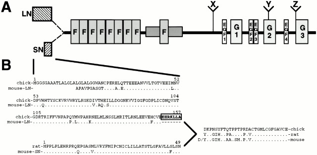
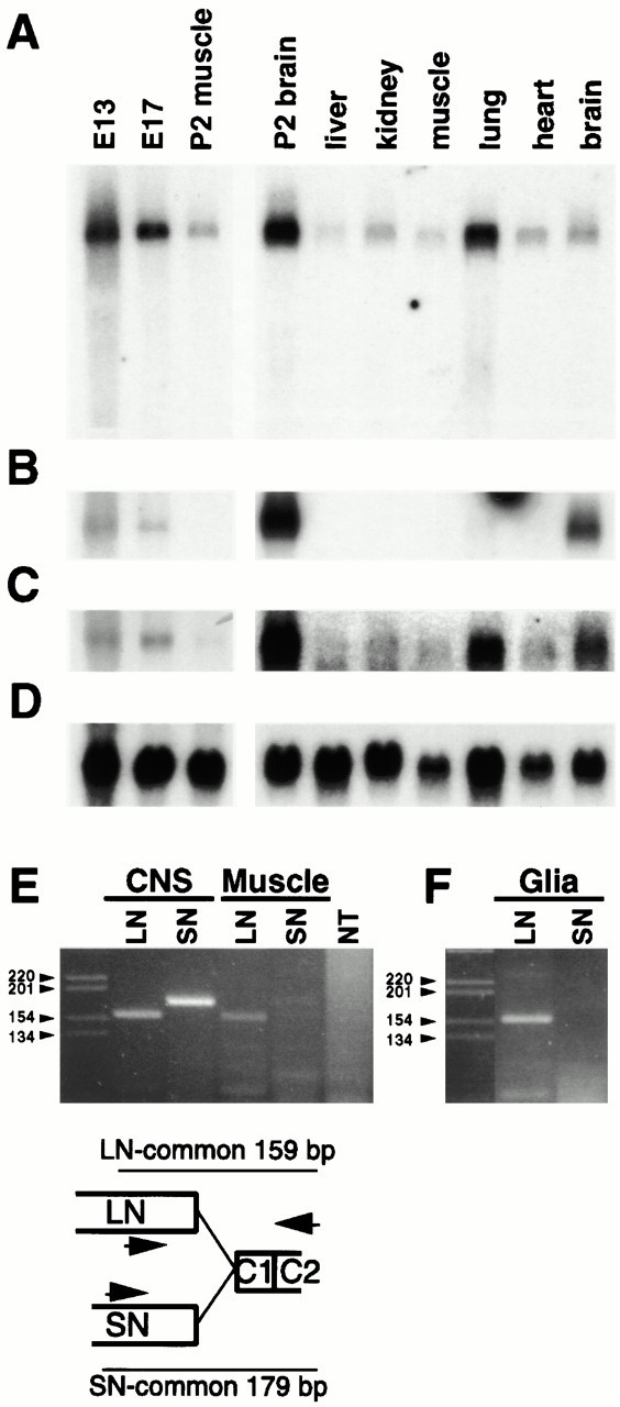
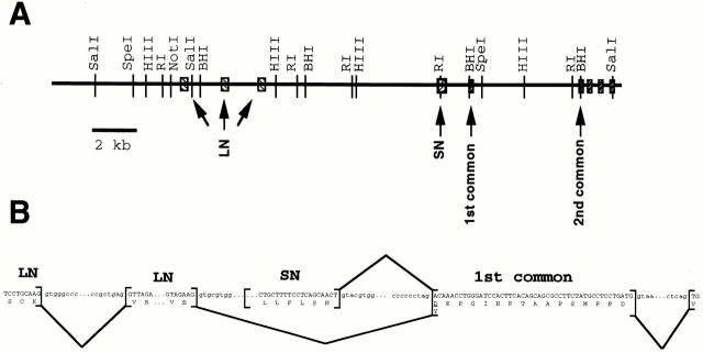
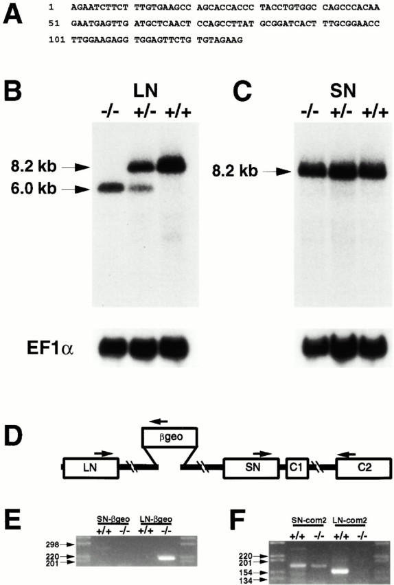
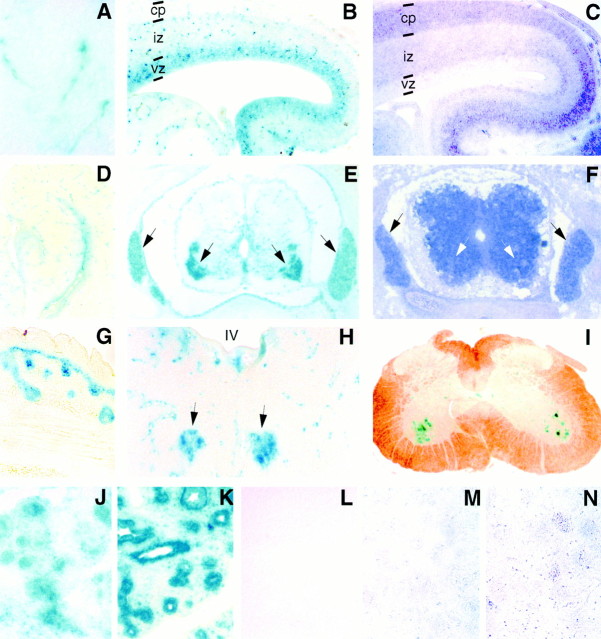
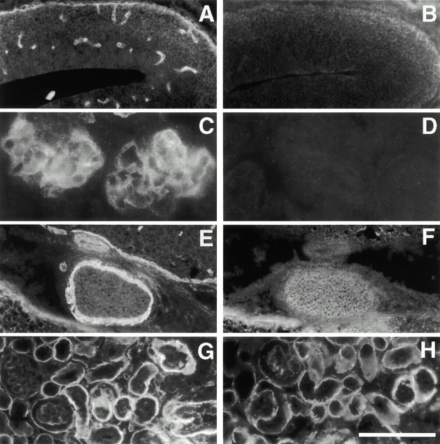
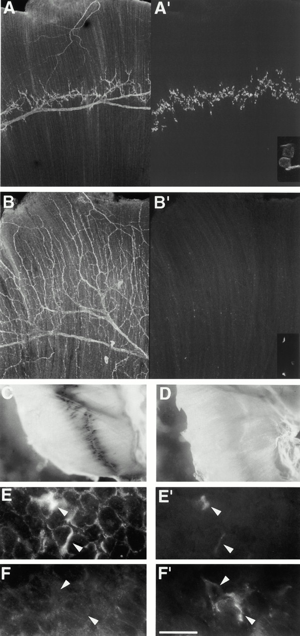
The proteoglycan agrin is required for postsynaptic differentiation at the skeletal neuromuscular junction, but is also associated with basal laminae in numerous other tissues, and with the surfaces of some neurons. Little is known about its roles at sites other than the neuromuscular junction, or about how its expression and subcellular localization are regulated in any tissue. Here we demonstrate that the murine agrin gene generates two proteins with different NH(2) termini, and present evidence that these isoforms differ in subcellular localization, tissue distribution, and function. The two isoforms share approximately 1,900 amino acids (aa) of common sequence following unique NH(2) termini of 49 or 150 aa; we therefore call them short NH(2)-terminal (SN) and long NH(2)-terminal (LN) isoforms. In the mouse genome, LN-specific exons are upstream of an SN-specific exon, which is in turn upstream of common exons. LN-agrin is expressed in both neural and nonneural tissues. In spinal cord it is expressed in discrete subsets of cells, including motoneurons. In contrast, SN-agrin is selectively expressed in the nervous system but is widely distributed in many neuronal cell types. Both isoforms are externalized from cells but LN-agrin assembles into basal laminae whereas SN-agrin remains cell associated. Differential expression of the two isoforms appears to be transcriptionally regulated, whereas the unique SN and LN sequences direct their distinct subcellular localizations. Insertion of a "gene trap" construct into the mouse genome between the LN and SN exons abolished expression of LN-agrin with no detectable effect on expression levels of SN-agrin or on SN-agrin bioactivity in vitro. Agrin protein was absent from all basal laminae in mice lacking LN-agrin transcripts. The formation of the neuromuscular junctions was as drastically impaired in these mutants as in mice lacking all forms of agrin. Thus, basal lamina-associated LN-agrin is required for neuromuscular synaptogenesis, whereas cell-associated SN-agrin may play distinct roles in the central nervous system.


Figures

Figure 1
Figure 1
Distinct NH2 termini of agrin. (A) Schematic of agrin protein structure, showing its major domains (laminin G domains, EGF repeats, and follistatin [F] repeats), sites of alternative splicing (X, Y, Z) and alternate NH2-terminal extensions (SN and LN). (B) The two alternative NH2 termini of mouse agrin, aligned with prototype LN and SN sequences from chick and rat, respectively. Dots indicate identity. The boxed heptapeptide in the chick sequence represents an alternatively spliced exon; a homologous segment has not yet been identified in murine LN-agrin.
Figure 2
Figure 2
The NH2 terminus of SN-agrin mediates its association with cells. CHO cells were transfected with vectors encoding the NH2 terminus of SN-agrin fused to FLAG (A and B), full-length SN-agrin (C and D), or the NH2 terminus of LN-agrin fused to FLAG (E and F). Cells were stained with anti-FLAG (A, B, E, and F) or anti-agrin (C and D), either live to detect cell surface proteins (A, C, and E) or after fixation and permeabilization (B, D, and F). SN-agrin reached the cell surface, but no staining of live cells was detctable with the LN–FLAG construct. Staining of fixed and permeabilized cells indicated expression levels were similar for each construct. Bar, 20 μm.
Figure 3
Figure 3
Tissue distribution of LN- and SN-agrin. (A–D) Northern analysis. Blots were hybridized sequentially with probes specific for LN-agrin (A), SN-agrin (B), all agrin isoforms (C), and the ubiquitously expressed RNA, EF1α (D). In adults, LN-agrin RNA is broadly distributed whereas SN-agrin is selectively expressed in brain. Both forms are present at E13 and E17 (RNA was from whole embryos). The sum of SN- and LN-agrins appear to account for the common signal. (E) Analysis of E18 CNS and muscle RNA by RT-PCR indicated that LN is present in both tissues, whereas SN was below the level of detection in muscle. NT, PCR performed with a mixture of LN and SN primers but no template. The PCR strategy and predicted sizes of products are shown in the sketch. (F) Analysis of cortical glial cultures by RT-PCR indicates that LN is expressed by glia, whereas SN is not. The strategy is the same as that in E.
Figure 4
Figure 4
Location of SN, LN, and common exons in the mouse agrin gene. (A) Map of the 5′ end of the agrin gene, as determined by restriction digestion, PCR, and Southern blotting. LN-specific sequences are encoded by at least three exons that lie 8-kb upstream of a single exon that encodes SN-specific sequences. The SN exon is separated by introns of 0.4 and 4 kb from first and second common exons. The second common exon corresponds to the first exon mapped by Rupp et al. 1992. (B) Predicted pattern of splicing to generate LN- and SN-agrin transcripts. The intron–exon boundaries all contain concensus splice donor (gt) and acceptor (ag) sequences and preserve the predicted agrin reading frame.
Figure 5
Figure 5
Characterization of a gene trap insertion that selectively intercepts LN-agrin transcripts. (A) Partial sequence of the transcript intercepted by the agrinLN gene trap insertion was determined by 5′ RACE. The sequence matches LN-agrin and terminates at the end of the LN-specific exon. (B and C) Northern analysis of poly A+ mRNA from the CNS of E18 agrinLN/LN, agrinLN/+, and agrin+/+ littermates, hybridized with LN- (B) and SN-specific (C) probes. The wild-type LN transcript is shifted from 8.2 to 6.0 kb by the β-geo insertion, whereas the size and abundance of the SN transcript are unaffected. Blots were standardized with EF1α. (D–F) RT-PCR analysis of RNA from CNS of mutants and controls, using strategy shown in D. Amplification from LN to β-geo yielded a band in mutants but not wild-type littermates, whereas primers in SN and β-geo gave no amplification product in either genotype (E). The wild-type transcript for SN-agrin was still present in mutant CNS, but the LN transcript is below the level of detection by RT-PCR (F).
Figure 6
Figure 6
Expression of LN- and SN-agrin. The β-geo insertion allowed LN-agrin expression to be analyzed by lacZ staining in heterozygotes (A, B, D, E, and G–L), whereas SN-agrin RNA was detected by in situ hybridization (C, F, M, and N). (A and B) In the brains of E14 mice, LN-agrin is expressed in cerebral blood vessels and in the ventricular (vz) and intermediate zone (iz) of the cortex, but not in the cortical plate (cp; B). (C) SN-agrin is expressed in a pattern that is complementary to the LN pattern, being most abundant in the postmitotic cells of the cortical plate. (D) In E18 embryos, LN-agrin continues to have very restricted expression in the hippocampus, being present only near the ventricles and in blood vessels. (E) In the spinal cord, LN-agrin is strongly expressed by motoneurons and sensory neurons of the DRG (arrows). (F) SN-agrin is widely distributed in many neuronal cells types in both the DRG and the spinal cord, although motoneurons are not more intensely positive than other cell types (white arrows). (H) LN-agrin is also expressed by cranial nerve motor nuclei in the hindbrain (E18 horizontal section; IV, forth ventricle; arrowheads, oculomotor nuclei). (I) Motoneuron expression of LN-agrin persists into adulthood. (G and J–N) Nonneuronal cells that abut basal laminae express LN-agrin but not SN-agrin. Epidermal cells in E18 skin (G), kidney glomeruli and tubules (J), and pulmonary epithelium (K) are all positive for LN-agrin. No lacZ activity was present in littermate controls in any tissue (E18 kidney shown, L). Consistent with northern blotting, SN-agrin is not expressed at levels detectable by in situ hybridization in nonneuronal tissues such as kidney (M) or lung (N).
Figure 7
Figure 7
LN-agrin accounts for all agrin in BLs. Immunohistochemistry with an anti-agrin antibody shows that agrin is present in BLs of cerebral microvasulature (A), renal glomeruli (C), and pia and sheath of the optic nerve (E) at E18. No immunoreactivity is detectable at any of these sites in agrinLN/LN mice (B, D, and F). Basal laminae remain intact as indicated by anti-laminin staining in wild-type (G) and agrinLN/LN mutant (H) kidney. Bar: (A, B, E, and F) 250 μm; (C and D) 50 μm; and (G and H) 125 μm.
Figure 8
Figure 8
Neuromuscular junctions fail to form in agrinLN/LN muscle. (A and B) Diaphragms from E18 mice were stained with antibodies against neurofilaments and synaptophysin to visualize axons (A and B) and with α-bungarotoxin to visualize AChRs (A′ and B′). In wild-type mice (A and A′), the synapses form a narrow endplate band in the center of the muscle. Plaques of AChRs are clear and bright (A′, inset). In agrinLN/LN muscle (B and B′), neurites fail to stop at synaptic sites and little postsynaptic specialization is seen. Small, dim clusters of AChRs are present (B′, inset), but they are usually not apposed to nerves. (C and D) Acetylcholinesterase accumulates in the end plate band of E18 control muscles (C) but not agrinLN/LN muscles (D). At E18, agrin is present in the extra-synaptic basal lamina of control myotubes, as shown in cross-sections of intercostal muscle (E). All muscle agrin staining is eliminated in agrinLN/LN mice (F). Motor nerve terminal, identified by SV2 staining (E′ and F′) are associated with agrin accumulations in wild-type (E and E′) but not mutant (F and F′) muscles, suggesting that most synaptic agrin is of the LN form. Bar: (A and B) 50 μm; (C and D) 100 μm; (A′ and B′, insets) 20 μm; and (E and F) 20 μm.
Figure 9
Figure 9
SN-agrin is bioactive. (A and B) RT-PCR of RNA from brains of E18 agrinLN/LN mice and littermate controls, using primers that span the Y (A) and Z (B) exons. Both Y+ and Y transcipts are present in both genotypes with Y+ transcripts predominant. Multiple Z+ transcripts (with 24-, 33-, and 57-bp inserts) as well as Z transcripts are also present in similar proportions in wild-type and mutant brains, indicating that SN-agrin is subject to alternative splicing at sites that determine its bioactivity. (C) Brain extracts from agrinLN/LN mice were applied to cultured primary myotubes and clustering activity was assayed. The activity of control and mutant brain extracts was the same, suggesting that SN-agrin is capable of inducing AChR clusters in vitro. Virtually all clustering activity is due to agrin since extracts from a agrin null mutant induce few clusters (Burgess, R.W., and J.R. Sanes, manuscript in preparation; redrawn as light gray bars).

References

    1. Barber A.J., Lieth E. Agrin accumulates in the brain microvascular basal lamina during development of the blood-brain barrier. Dev. Dyn. 1997;208:62–74. - PubMed
    1. Buckley K, Kelly R.B. Identification of a transmembrane glycoprotein specific for secretory vesicles of neural and endocrine cells. J. Cell Biol. 1985;100:1284–1294. - PMC - PubMed
    1. Burgess R.W., Nguyen Q.T., Son Y.J., Lichtman J.W., Sanes J.R. Alternatively spliced isoforms of nerve- and muscle-derived agrintheir roles at the neuromuscular junction. Neuron. 1999;23:33–44. - PubMed
    1. Campanelli J.T., Hoch W., Rupp F., Kreiner T., Scheller R.H. Agrin mediates cell contact-induced acetylcholine receptor clustering. Cell. 1991;67:909–916. - PubMed
    1. Campanelli J.T., Gayer G.G., Scheller R.H. Alternative RNA splicing that determines agrin activity regulates binding to heparin and a-dystroglycan. Development. 1996;122:1663–1672. - PubMed

Publication types

MeSH terms