. 2000 Oct 2;151(1):95-106.
doi: 10.1083/jcb.151.1.95.

Meiotic telomere protein Ndj1p is required for meiosis-specific telomere distribution, bouquet formation and efficient homologue pairing

Affiliations

Meiotic telomere protein Ndj1p is required for meiosis-specific telomere distribution, bouquet formation and efficient homologue pairing

E Trelles-Sticken et al. J Cell Biol. .

Abstract

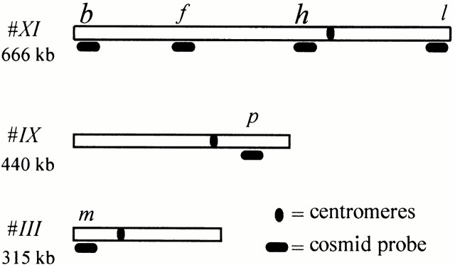
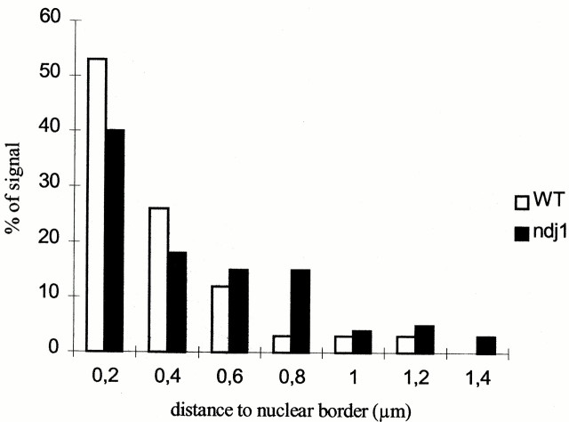
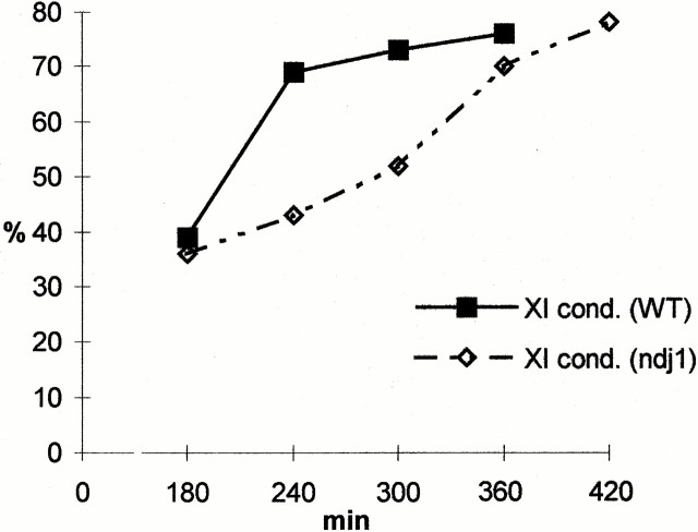
We have investigated the requirements for NDJ1 in meiotic telomere redistribution and clustering in synchronized cultures of Saccharomyces cerevisiae. On induction of wild-type meiosis, telomeres disperse from premeiotic aggregates over the nuclear periphery, and then cluster near the spindle pole body (bouquet arrangement) before dispersing again. In ndj1Delta meiocytes, telomeres are scattered throughout the nucleus and fail to form perinuclear meiosis-specific distribution patterns, suggesting that Ndj1p may function to tether meiotic telomeres to the nuclear periphery. Since ndj1Delta meiocytes fail to cluster their telomeres at any prophase stage, Ndj1p is the first protein shown to be required for bouquet formation in a synaptic organism. Analysis of homologue pairing by two-color fluorescence in situ hybridization with cosmid probes to regions on III, IX, and XI revealed that disruption of bouquet formation is associated with a significant delay (>2 h) of homologue pairing. An increased and persistent fraction of ndj1Delta meiocytes with Zip1p polycomplexes suggests that chromosome polarization is important for synapsis progression. Thus, our observations support the hypothesis that meiotic telomere clustering contributes to efficient homologue alignment and synaptic pairing. Under naturally occurring conditions, bouquet formation may allow for rapid sporulation and confer a selective advantage.


Figures

Figure 1
Figure 1
Physical map of chromosomes III, IX, and XI showing the location of the respective cosmid probes used for pairing analysis by FISH (see Materials and Methods for clone numbers).
Figure 2
Figure 2
FISH with a pan-centromere probe (fluorescein, green) and a pan-telomere probe (rhodamine, red) to undisrupted diploid nuclei (DAPI, blue) of wild-type and ndj1Δ SK1 cells. (a) Vegetative wild-type nucleus before induction of meiosis (t = 0 min) displays clustered centromeres that form a single green signal, while telomeres form four perinuclear clusters. (b) Premeiotic ndj1Δ nucleus with one centromere cluster and three large telomere clusters at the opposite pole of the nucleus, thereby resembling a Rabl orientation. (c) WT and (d) ndj1Δ meiocyte nuclei from two later time points (200 and 260 min, respectively) displaying centromere and telomere signals distributed throughout the nuclei. Few centromere signals are dissociated from the centromere cluster in nucleus (c), while in nucleus (d) exhibits dispersed centromere signals. Bar, 5 μm.
Figure 3
Figure 3
FISH analysis of the frequencies of vegetative/premeiotic centromere and telomere distribution patterns; i.e., one centromere cluster (cen cluster) and two to eight telomere clusters (two to eight telo signals) (Fig. 2, a and b), in spread ndj1Δ and wild-type SK1 nuclei at transfer to sporulation medium (0 min) and subsequent time points (minutes) in sporulation. The frequency (%) of nuclei with this hallmark of vegetative/premeiotic nuclear architecture drops similarly in ndj1Δ (ndj1) and wild-type (WT) meiotic time courses. Since meiotic divisions appeared at t = 300 min in the wild-type culture and complicated analysis, FISH analysis in the wild type was only conducted until 320 min.
Figure 4
Figure 4
(a–d) Double immunolabeling of SPB components (fluorescein, green) and of meiotic telomeres with antibod-ies against HA-tagged Ndj1p (rhodamine, red) reveals meiosis-specific telomere distribution patterns in mildly spread diploid wild-type SK1 nuclei. (a) Peripheral rim-like distribution of telomeres during early meiosis (140 min). (b and c) Meiocytes with telomeres accumulated at the SPB (bouquet arrangement). (d) Meiocyte nucleus from a later time point (240 min), which shows an SPB and dispersed telomere signals. (e–h) Mildly spread meiocyte nuclei from an independent FISH experiment with the XY′ repeat probe (see Materials and Methods) reveals telomere patterns similar to the ones obtained by Ndj1p IF. (e) Rim-like telomere distribution. (f and g) Bouquet nuclei with clustered telomere signals. (h) Advanced meiocyte from a later time point displays a scattered telomere distribution. Bar, 5 μm (applies to all details). The inset shows colocalization of Ndj1-HA IF signals (red) and XY′ telo-FISH signals (green) in a wild-type pachytene nucleus. Most of the Ndj1-HA and XY′ signals show significant overlap at chromosome ends (e.g., arrowheads). Ndj1 fluorescence is often seen beyond the telo-FISH signals and/or extends between telomere signals. Fewer IF signals in the Ndj1 channel may relate to loss of some epitopes during the FISH procedure.
Figure 5
Figure 5
Analysis of occurrence of meiosis-specific telomere distribution patterns during meiotic time courses (minutes) of wild-type (solid symbols) and ndj1Δ (open symbols, dotted lines) SK1 strains. Frequency of diploid nuclei (%) with telomeric FISH signals showing a rim-like distribution along the nuclear periphery (telos rim-like; Fig. 4 e) and of bouquet nuclei with a telomere cluster (bouquet; Fig. 4f and Fig. g). The frequency of nuclei with rim-like telomere distribution and with a bouquet arrangement is significantly reduced in ndj1Δ meiosis. Wild-type signal patterns are displayed until 360 min only, since meiotic divisions (anaphases) appeared after 300 min in the wild-type culture and complicated the signal analysis.
Figure 6
Figure 6
Confocal light optical sections at the equatorial planes of (a) a wild-type (taken at 240 min) and (b) an ndj1Δ nucleus (taken at 470 min) showing telomere-associated Rap1 signals (FITC, yellow) and DNA fluorescence (DAPI, blue). In the wild-type nucleus (a), telomeric Rap1p signals are confined to the nuclear periphery. The ndj1Δ meiocyte nuclear section (b) shows telomeric Rap1p signals at the periphery and within the nuclear interior. Bar, 2 μm.
Figure 7
Figure 7
Spatial analysis of Rap1p telomere signal distribution with respect to the nearest nuclear boundary segment. The frequency (%) of occurrence of Rap1/FITC signal centers situated at particular distances (given in 0.2-μm intervals) from the nearest nuclear periphery segment as determined at the equatorial plane from the corresponding confocal DAPI images. The values were derived from 146 signal spots for ndj1Δ (ndj1), and 86 for wild type (WT). In the wild type, most signal spot centers locate in the immediate vicinity of the nuclear periphery (open bars). An altered spatial distribution of telomeric Rap1 signals in ndj1Δ nuclei is reflected by a higher portion of telomeric signals being more distant from the nuclear border (solid bars).
Figure 8
Figure 8
Analysis of telomere FISH signal numbers in 20 randomly selected, mildly spread nuclei of diploid SK1 wild-type and ndj1Δ meiocytes. Nuclei were obtained 200 and 300 min after induction of meiosis in the wild type and ndj1Δ, respectively, to allow for a compensation of the delay in mutant prophase I. Ranking according to increasing numbers of telomere FISH signals/nucleoid reveals that ndj1Δ meiocytes display significantly larger telomere signal numbers/nucleus.
Figure 9
Figure 9
Costaining of telomeres by FISH (rhodamine, red) and Zip1p by IF (FITC, green) in mildly spread ndj1Δ meiocyte nuclei (DAPI, blue). (a and b) Spread nuclei (obtained at 210 min after induction of meiosis) display Zip1-signal stretches and a Zip1 polycomplex (bright green oblong, arrow), while telomere FISH signals are dispersed. (c) Nucleus with dispersed telomeres but without Zip1 polycomplex from a later time point (330 min). Bar, 5 μm.
Figure 10
Figure 10
Two-color FISH with cosmid probes m (III, green) and p (IX, red) to spread nuclei of SK1 meiocytes. Nuclei were taken at t = 180 min. (a) Spread nucleus with cosmid signals apart. (b) Meiocyte with both pairs of homologous cosmid signals paired. Therefore, the signals appear enlarged. Bar, 5 μm.
Figure 11
Figure 11
FISH analysis of homologue pairing during meiotic time courses of wild-type and ndj1Δ SK1 strains. Cosmid probe combinations m/p and f/l were hybridized to spread preparations obtained at the respective time points (minutes). Pairing values were obtained by determining the fraction of nuclei containing cosmid signals of the same color that touched each other or showed an enlarged coalesced signal. More than 200 FISH signal pairs were scored per time point, probe combination, and strain. Values were corrected for accidental heterologous contacts by subtracting 5% for cosmid combination m/p and 4% for probe combination l/f (see text). (A) Frequencies of nuclei with paired cos f signals (XI internal; Fig. 1) in wild-type (WT) and ndj1Δ (ndj1) time courses. (B) Frequencies (%) of paired cos l (XI, right telo) in wild-type and ndj1Δ time courses. (D) Frequencies of nuclei with paired cos p signals (IX, right arm) in wild-type and ndj1Δ time courses. (C) Frequencies of nuclei with paired cos m signals (III, HML) in wild-type and ndj1Δ time course. At all loci probed, the frequencies of nuclei with paired signals increase more gradually in the absence of Ndj1p, reaching nearly wild-type frequencies with a 2–3-h delay. In the wild type, signal patterns are displayed only until 320 min, since meiotic divisions (anaphases) appeared after 300 min and complicated the signal analysis.
Figure 12
Figure 12
Representative images of FISH signal patterns obtained with a painting probe for chromosomes XI (red) and the pan-telomere probe (green) in mildly spread nuclei from meiotic time courses of diploid wild type (wt) and ndj1Δ (ndj1) SK1 strains (a, 0 min; b and c, 260 min; d, 380 min). Images are aligned according to meiosis-specific changes in chromosome morphology during prophase I (see Trelles-Sticken et al. 1999). (a) Premeiotic wild-type and ndj1Δ nuclei (0 min) display separated variably shaped chromosomes XI territories and several telomere clusters. (b, wt) WT meiocyte nucleus with extended chromosome XI signal tracks and a rim-like distribution of telomere signals. (b, ndj1) Meiocyte with scattered telomere signals, while XI signal tracks extend across the nucleus and touch at one end. (c, wt) Nucleus with one large telomere signal cluster (bouquet arrangement). Chromosomes XI form one outstretched signal track along the telomere cluster. (c, ndj1) ndj1Δ meiocyte with scattered telomeres. Painted chromosomes XI are seen as a single extended signal track. (d) Wild-type and mutant pachytene nuclei both exhibit a condensed XI bivalent and scattered telomeres. Bar, 5 μm.
Figure 13
Figure 13
Frequency (%) of uninucleate meiocytes with condensed chromosome XI bivalent (Fig. 12 d) at given time points (minutes) in wild-type (WT) and ndj1Δ (ndj1) meiosis in the SK1 background. In ndj1Δ meiosis, the frequency of uninucleated meiocytes with a condensed XI bivalent increased more gradually than in the wild type.
Figure 14
Figure 14
Scheme showing the sequential steps of centromere and telomere redistribution that occur during earliest meiotic prophase of yeast. It is based on the observations made in this and earlier reports (see text). (a) In the premeiotic nucleus, telomeres (black) and centromeres (gray) form several telomere and one centromere cluster at the nuclear periphery. (b) In the wild type, induction of meiosis leads to dissolution of the centromere cluster and perinuclear telomere clusters. Centromeres become dispersed throughout the nuclear lumen, while telomeres disperse over the nuclear periphery. (c) Bouquet stage: telomeres tightly cluster at the spindle pole body, while centromeres are dispersed. (d) While premeiotic telomere topology is not affected, the early steps of meiotic telomere redistribution appear defective in the absence of Ndj1p, which leads to scattering of both telomeres and centromeres throughout the ndj1Δ meiocyte nucleus.

References

    1. Alani E., Padmore R., Kleckner N. Analysis of wild-type and rad50 mutants of yeast suggests an intimate relationship between meiotic chromosome synapsis and recombination. Cell. 1990;61:419–436. - PubMed
    1. Bass H.W., Marshall W.F., Sedat J.W., Agardand D.A., Cande W.Z. Telomeres cluster de novo before the initiation of synapsisa three-dimensional spatial analysis of telomere positions before and during meiotic prophase. J. Cell Biol. 1997;137:5–18. - PMC - PubMed
    1. Bélar K. Chromosomenreduktion. In: Baur E., Hartmann M., editors. Handbuch der Vererbungswissenschaft, die Cytologischen Grundlagen der Vererbung. Geb. Borntraeger; Berlin: 1928. pp. 168–201.
    1. Bascom-Slack C.A., Dawson D.S. The yeast motor protein, Kar3p, is essential for meiosis I. J. Cell Biol. 1997;139:459–467. - PMC - PubMed
    1. Blackburn E.H. Structure and function of telomeres. Nature. 1991;350:569–573. - PubMed

Publication types

Substances