. 2000 Oct 2;151(1):187-98.
doi: 10.1083/jcb.151.1.187.

Regulated interactions between dynamin and the actin-binding protein cortactin modulate cell shape

Affiliations

Regulated interactions between dynamin and the actin-binding protein cortactin modulate cell shape

M A McNiven et al. J Cell Biol. .

Abstract

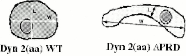
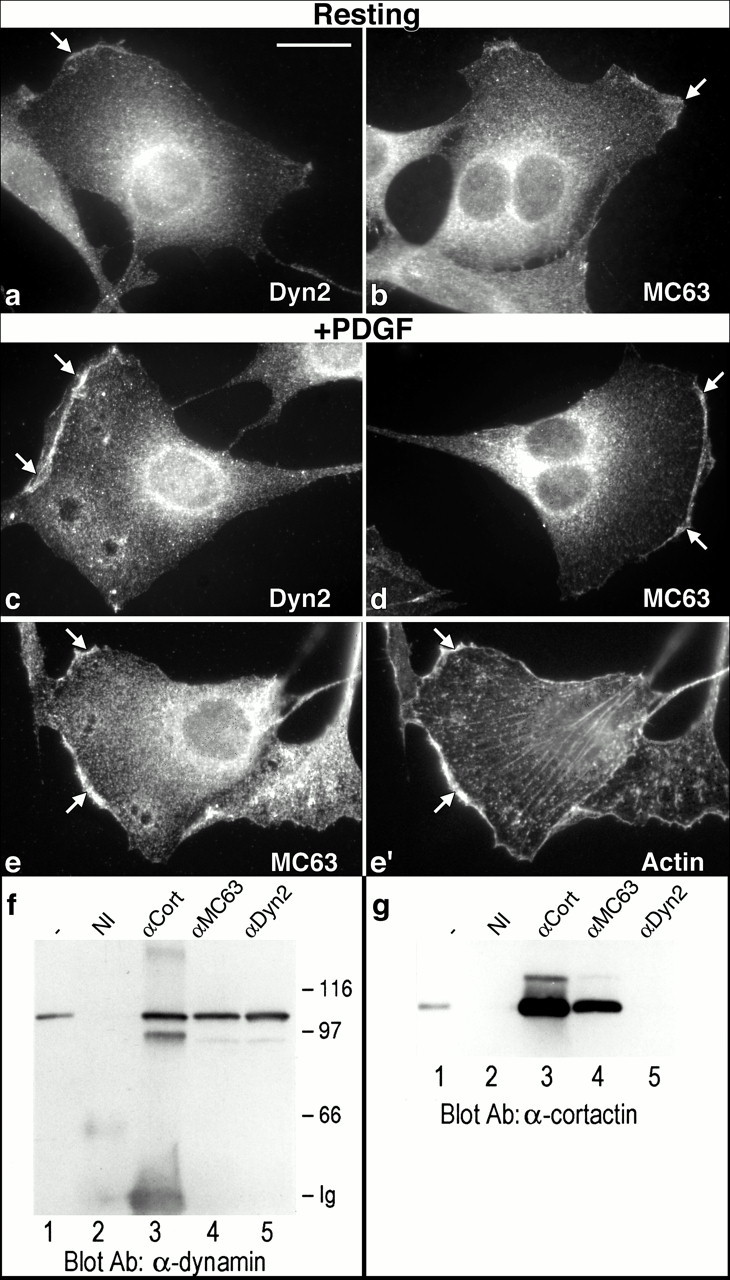
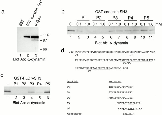
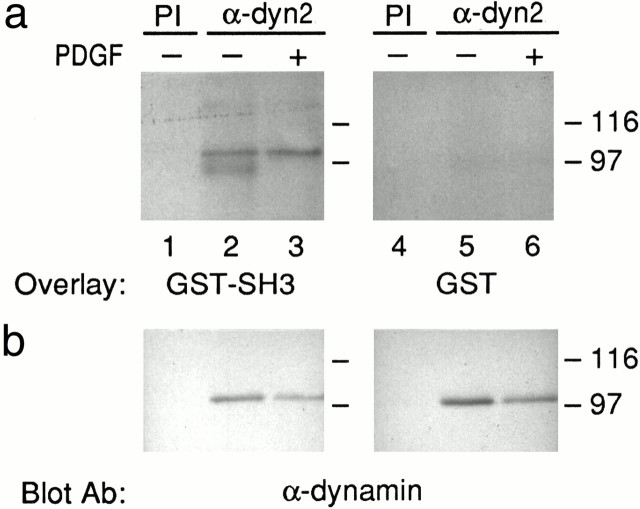
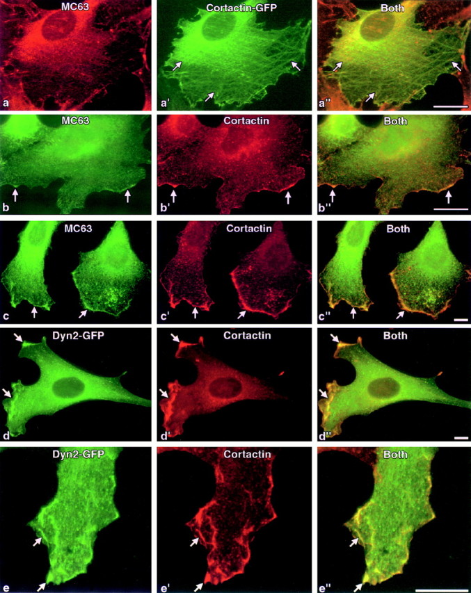
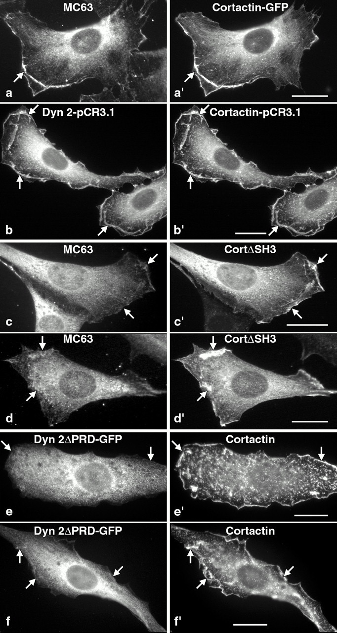
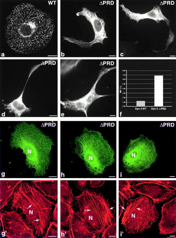
The dynamin family of large GTPases has been implicated in the formation of nascent vesicles in both the endocytic and secretory pathways. It is believed that dynamin interacts with a variety of cellular proteins to constrict membranes. The actin cytoskeleton has also been implicated in altering membrane shape and form during cell migration, endocytosis, and secretion and has been postulated to work synergistically with dynamin and coat proteins in several of these important processes. We have observed that the cytoplasmic distribution of dynamin changes dramatically in fibroblasts that have been stimulated to undergo migration with a motagen/hormone. In quiescent cells, dynamin 2 (Dyn 2) associates predominantly with clathrin-coated vesicles at the plasma membrane and the Golgi apparatus. Upon treatment with PDGF to induce cell migration, dynamin becomes markedly associated with membrane ruffles and lamellipodia. Biochemical and morphological studies using antibodies and GFP-tagged dynamin demonstrate an interaction with cortactin. Cortactin is an actin-binding protein that contains a well defined SH3 domain. Using a variety of biochemical methods we demonstrate that the cortactin-SH3 domain associates with the proline-rich domain (PRD) of dynamin. Functional studies that express wild-type and mutant forms of dynamin and/or cortactin in living cells support these in vitro observations and demonstrate that an increased expression of cortactin leads to a significant recruitment of endogenous or expressed dynamin into the cell ruffle. Further, expression of a cortactin protein lacking the interactive SH3 domain (CortDeltaSH3) significantly reduces dynamin localization to the ruffle. Accordingly, transfected cells expressing Dyn 2 lacking the PRD (Dyn 2(aa)DeltaPRD) sequester little of this protein to the cortactin-rich ruffle. Interestingly, these mutant cells are viable, but display dramatic alterations in morphology. This change in shape appears to be due, in part, to a striking increase in the number of actin stress fibers. These findings provide the first demonstration that dynamin can interact with the actin cytoskeleton to regulate actin reorganization and subsequently cell shape.


Figures

Scheme S1
Scheme S1
Figure 1
Figure 1
Dynamin associates with cortical membrane ruffles in growth factor-stimulated fibroblasts. a–e, Immunofluorescence staining of cultured NIH/3T3 fibroblasts with affinity-purified peptide antibodies made to a conserved region of the dynamin NH2 terminus (MC63) or to the proline rich COOH-terminal domain specific for Dyn 2. In resting cells (a and b), dynamin labeling is most prominent as bright spots on the plasma membrane and near the nucleus with a modest accumulation in the cell cortex (arrows). Upon stimulation with PDGF for 10–15 min (c and e), cells display a motile phenotype and dynamin staining becomes significantly enriched at the ruffled membrane at the leading edge of the cell (arrows). This cortical dynamin staining colocalizes markedly with actin in the leading of motile cells (arrows), as confirmed by double labeling with rhodamine phalloidin (e and e′). Immunoblot analysis of immunoprecipitates from cytosolic extract of NIH/3T3 cells using antibodies to cortactin or dynamin that were probed with mAbs to either dynamin (f) or cortactin (g). The lanes for each blot include: lane 1, starting cytosolic extract or IP with; lane 2, nonimmune serum (NI); lane 3, an anticortactin mAb; lane 4, the polyclonal Pan-dynamin antibody MC63; lane 5, the polyclonal antibody Dyn 2 specific for the COOH terminus of Dyn 2. Antibodies to cortactin immunoprecipitate a 105-kD immunoreactive dynamin band and a 95-kD band believed to be a proteolytic fragment of dynamin (f). Antibodies to dynamin immunoprecipitate the actin-binding protein cortactin from cell homogenates (g). The filter from f was stripped and reprobed with antibodies to cortactin (g). Interestingly, whereas the Pan-dynamin antibodies (MC63) immunoprecipitated cortactin (lane 4), the Dyn 2 tail-specific antibody (Dyn 2) did not (lane 5), suggesting that this antibody blocks dynamin–cortactin binding. Bar, 10 μm.
Figure 2
Figure 2
The SH3 domain of cortactin binds dynamin. a, A cytosolic extract of NIH/3T3 cells was incubated with GST or GST–cortactin SH3 domain that was immobilized on glutathione Sepharose beads (lanes 1 and 2) or immunoprecipitated with the Dyn 2-specific antibody (lane 3). The bound proteins were fractionated and analyzed by blotting with an antidynamin mAb. As shown by the prominent dynamin band in lane 2, a substantial amount of dynamin was retained on the GST–cortactin SH3 domain beads as compared with the GST beads alone (lane 1). b, The GST pull-down assay was carried out in the absence or presence of one of the five synthetic peptides (P1–P5) at 0.1- or 1-mM concentrations. Binding of dynamin to the immobilized GST–cortactin SH3 domain was measured by blotting with an antidynamin mAb. Note that the P2 and P1 peptides were the most effective in blocking dynamin binding to the GST–SH3 domain beads (c). As a control the GST pull-down assay was carried out using a GST fusion protein containing the SH3 domain of PLCγ-1, in the absence (lane 1) or presence (lanes 2–6) of 1 mM of each of the five peptides. Binding of dynamin by the PLCγ-1 SH3 domain was assayed by blotting with antidynamin antibodies. As for the cortactin–SH3 domain-associated beads, peptides P2 and P1 inhibited dynamin binding, and some inhibition was demonstrated by the P4 peptide. d, Peptide sequence of the Dyn 2 COOH terminus. The four proline rich sequences are underlined. The bottom shows the sequences of the five synthetic peptides used to test the inhibition of SH3–dynamin interactions. The consensus sequence PXXPSRP was found to inhibit Dyn 2 binding to the GST–cortactin SH3 domain.
Figure 3
Figure 3
Dynamin binds directly to the cortactin–SH3 domain. Cytosolic extracts of quiescent or PDGF-stimulated NIH/3T3 cells were immunoprecipitated with preimmune serum (PI; lane 1) or with the Dyn 2-specific tail antibody (lanes 2 and 3). The immunoprecipitated proteins were fractionated by SDS-PAGE and transferred to a filter (a). Duplicate filters were incubated with either GST or a fusion protein containing the SH3 domain of cortactin (GST-SH3). Binding of the GST or GST–SH3 to the filters was detected using anti-GST antibodies, followed by peroxidase-conjugated goat anti–rabbit IgG. The bottom panels show the same filters that were stripped and reprobed with antibodies to dynamin. Note that while all of the lanes containing immunoprecipitates using immune serum contained dynamin protein (b), the GST–cortactin SH3 domain probe, but not the GST probe alone, bound to the filter (a).
Figure 4
Figure 4
Dynamin colocalizes with cortactin in the lamellipodia of growth factor-stimulated fibroblasts via the PRD. Immunofluorescence localization of dynamin and cortactin in quiescent, and PDGF-treated NIH/3T3 cells. Labels indicate whether cells were transfected. Resting cells were transfected with a full-length cortactin–GFP construct and double-labeled for dynamin (a–a′′), or untransfected cells were fixed and double-labeled with antibodies to dynamin (MC63) and cortactin (b–b′′). Resting cells (a–a′′)transfected with the Cort–GFP construct displayed a nonpolarized phenotype with cortactin localized around the cell cortex and on actin stress fibers (arrows). The distribution of cortactin in untransfected cells was identical to that of cells transfected with cort-GFP, although there was a reduced localization to stress fibers (b–b′′). Upon stimulation with PDGF, there was a marked recruitment of both Dyn 2 (c) and cortactin (c′) into large cortical ruffles (arrows) situated at the leading edges of the actively migrating cells. An identical localization was observed in PDGF-stimulated NIH/3T3 cells expressing Dyn 2(aa)–GFP (d and e) that were fixed and stained for cortactin (d′ and e′). Similar to untransfected cells stained with antibodies, Dyn 2(aa)–GFP became concentrated in lamellipodia at the leading edge of PDGF stimulated, motile cells (d and e, arrows). Bars, 10 μm.
Figure 5
Figure 5
Alterations in cortactin change the distribution of Dyn 2 in living cells. NIH/3T3 cells were transfected with constructs encoding GFP-tagged (a), wt (b), or truncated (c–f) forms of cortactin and/or Dyn 2, allowed to recover for 24–48 h, then prepared for immunofluorescence microscopy to monitor changes in protein distribution. Labels denote if cells were transfected. a and b, Overexpression of cortactin–GFP induced a marked increase in ruffle formation and the recruitment of endogenous Dyn 2, even in unstimulated cells (a and a′). Lamellipodia formation and Dyn 2 recruitment was induced to high levels in stimulated cells that coexpressed both cortactin and Dyn 2 (b and b′). Stimulated cells expressing a mutant cortactin (CortΔSH3) protein lacking the interactive SH3 domain (c′ and d′) show high levels of cortactin in the lamellipodia and cortical ruffles, with low levels of endogenous Dyn 2 (c and d) recruited to these structures, particularly when compared with the cells expressing wt cortactin (a and b). Stimulated cells expressing a mutant Dyn 2 (Dyn 2ΔPRD–GFP) lacking the interactive PRD show cortactin is recruited to the lamellipodia and cortex of these cells (e′ and f′), whereas little of the expressed mutant protein (e and f) was found in these structures. Note the peculiar elongate shape of the Dyn 2ΔPRD–GFP expressing cell (f). A summary of these localization patterns is in Table . Bar, 10 μm.
Figure 6
Figure 6
Expression of a truncated dynamin (Dyn 2(aa)ΔPRD) induces profound changes in cell shape with a concomitant proliferation of actin stress fibers. Fluorescence micrographs of cultured clone 9 cells expressing either wt Dyn 2(aa)–GFP (a) or Dyn 2(aa)ΔPRD-GFP (b–e). Cells expressing the wt protein (a) display a normal discoidal shape and a punctate distribution of dynamin at both the plasma membrane and Golgi apparatus, identical to that of untransfected cells. In strong contrast, cells expressing the Dyn 2(aa)ΔPRD–GFP became elongated, sprouting long peculiar neurite-like appendages (b–e). Morphological measurements of >200 wt and mutant cells confirmed this shape change showing a six- to sevenfold increase in width versus length (f). To test if changes in actin organization might be responsible for these changes in shape, clone 9 cells expressing the Dyn 2(aa)ΔPRD–GFP (g–i) were fixed and stained for actin using rhodamine phalloidin (g′–i′). Whereas surrounding untransfected cells possessed a cortical ring of filamentous actin with few stress fibers, mutant cells displayed a reduction in cortical actin with a dramatic alteration in the actin cytoskeleton. Specifically, these cells possessed an extensive number of stress fibers that traversed the long axis and contributed to the shape malformation seen in isolated cells that were not grown in a monolayer (b–e). It should be noted that changes in the shape of these mutant cells are minimized in the confluent cultures shown here to provide a comparison of actin organization with the surrounding, untransfected cells (g–i). Shape changes are most prevalent in sparsely plated cells (b–e). Bars, 10 μm.

Similar articles

Cited by

References

    1. Bray D., White J.G. Cortical flow in animal cells. Science. 1988;239:883–888. - PubMed
    1. Cao H., Garcia F., McNiven M. Differential distribution of dynamin isoforms in mammalian cells. Mol. Biol. Cell. 1998;9:2595–2609. - PMC - PubMed
    1. Cook T.A., Urrutia R., McNiven M.A. Identification of dynamin 2, an isoform ubiquitiously expressed in rat tissues. Proc. Natl. Acad. Sci. USA. 1994;91:644–648. - PMC - PubMed
    1. Damke H., Baba T., Warnock D.E., Schmid S.L. Induction of mutant dynamin specifically blocks endocytic coated vesicle formation. J. Cell Biol. 1994;127:915–934. - PMC - PubMed
    1. Downing J.R., Reynolds A.B. PDGF, CSF-1, and EGF induce tyrosine phosphorylation of p120, a pp60src transformation-associated substrate. Oncogene. 1992;6:607–613. - PubMed

Publication types