. 2000 Oct;106(7):857-66.
doi: 10.1172/JCI8040.

Disruption of the myocardial extracellular matrix leads to cardiac dysfunction

Affiliations

Disruption of the myocardial extracellular matrix leads to cardiac dysfunction

H E Kim et al. J Clin Invest. 2000 Oct.

Abstract

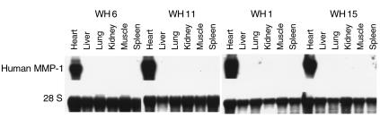
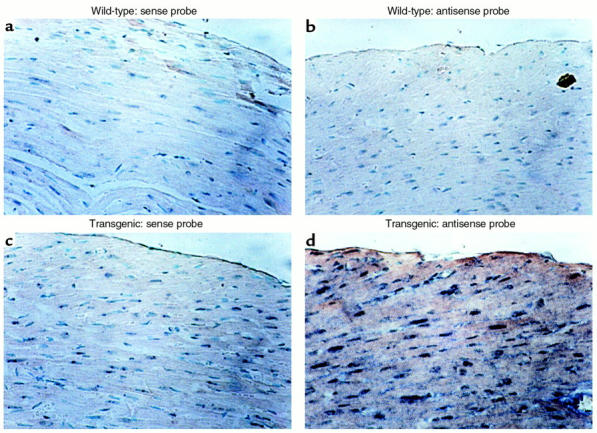
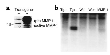
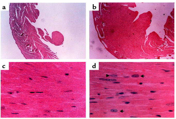
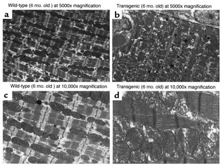
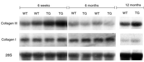
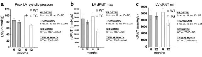
MMP activity with disruption of structural collagen has been implicated in the pathophysiology of dilated cardiomyopathy. To examine the role of this enzyme in cardiac function, a transgenic mouse was created that constitutively expressed human collagenase (MMP-1) in the heart. At 6 months of age, these animals demonstrated compensatory myocyte hypertrophy with an increase in the cardiac collagen concentration due to elevated transcription of type III collagen. Chronic myocardial expression of MMP-1 produced loss of cardiac interstitial collagen coincident with a marked deterioration of systolic and diastolic function at 12 months of age. This is the first animal model demonstrating that direct disruption of the extracellular matrix in the heart reproduces the changes observed in the progression of human heart failure.


Figures

Figure 1
Figure 1
Cardiac-specific expression of MMP-1 transgene. Northern blot analysis was performed using 10 μg total RNA isolated from the indicated tissues of transgenic lines WH 6, 11, 1, and 15 and probed with the transgene. The lower panel shows the same membrane probed with ribosomal 28S oligonucleotide to ensure that equal amounts of RNA were loaded in each lane.
Figure 2
Figure 2
Localization of the MMP-1 transgene expression to cardiac myocytes by in situ hybridization. Photomicrographs (± 200) of transverse sections through the anterior wall of the heart of a wild-type mouse (a and b) and a transgenic littermate (c and d) from line WH 1, probed with the sense MMP-1 probe (a and c) and probed with the antisense MMP-1 probe (b and d). Staining can be seen only in the myocytes of the transgenic heart (d).
Figure 3
Figure 3
(a) Presence of MMP-1 protein in transgenic mouse hearts. Western blot analysis was performed on ventricular extracts from 3-month-old transgenic mice (+) and control littermates (–) as described in Methods, and normalized for protein content. Equal amounts of protein were resolved by SDS-PAGE and subject to immunoblotting with anti–MMP-1 polyclonal antibodies. Arrows indicate the active and pro forms of MMP-1. (b) Collagenase activity in the homogenate from the heart of transgenic and control mice. Type I collagen was incubated with homogenate from the heart of a transgenic mouse treated with APMA (TG+) and without APMA (TG–) and the homogenate from the heart of a control littermate treated with APMA (WT+) and without APMA (WT–). As a control, 100 ng of active MMP-1 was incubated with type I collagen (MMP-1). The characteristic collagenase (one-quarter) degradation fragment can be seen only in the APMA-treated homogenate from the transgenic heart (TG+) and the positive control (MMP-1).
Figure 4
Figure 4
Histological analysis of mouse hearts from age-matched (3-month-old) littermates. Left ventricle from wild-type (a and c) and transgenic (b and d) hearts in cross-section. Note the increased thickness of the left ventricular free wall in the transgenic heart (b) compared with the wild-type (a). Enlarged hyperchromatic myocyte nuclei (arrows) with increased myofibrillar width consistent with cardiac hypertrophy is seen in the transgenic heart (d) compared with wild-type (c). Hematoxylin and eosin stains were used. a and b, ×40; c and d, ×400.
Figure 5
Figure 5
Representative transmission electron micrographs of wild-type and cardiac-collagenase transgenic mice. Ultrastructural analysis was performed on wild-type (a and c) and transgenic (b and d) hearts. The wild-type heart (a and c) shows an orderly row of myofibrils with neatly interposed mitochondria and sharp closely packed cristae. The transgenic heart (b and d) reveals changes consistent with pathological hypertrophy characterized by wider myofibrils, thicker Z-bands with breakdown of normal Z-band registration and myofibrillar disarray with disruption of the sarcomeric architecture. Note the numerous enlarged pleomorphic mitochondria distributed in a disorderly manner, all consistent with hypertrophy and reflective of the enormous metabolic demands of this tissue.
Figure 6
Figure 6
Upregulation of collagen type III but not collagen type I in cardiac-collagenase mice. Northern blot analysis was performed using 10 μg total RNA from heart tissue from transgenic (line WH 1) and wild-type littermate mice at 6-week, 6-month, and 12-month time points and probed with an xba I/Hind III fragment of the collagen type III cDNA (top). Increased collagen III message was detected in the transgenic mice compared with the wild-type littermates at the 6-week time point. No difference was noted by the 6- month or 12-month time point. The same membrane was probed with a Hind III fragment of the collagen type I cDNA (middle). No differences in type I collagen expression were observed between wild-type and transgenic mice at any time point. The lower panel shows the same membranes probed with ribosomal 28S oligonucleotide to ensure that equal amounts of RNA were loaded in each lane. WT, wild-type, TG, transgenic.
Figure 7
Figure 7
Hemodynamic measurements performed in wild-type and transgenic mice. Open chest measurements taken with a high-fidelity fluid-filled pressure transducer via a left ventricular apical puncture. (a) Increased peak left ventricular systolic pressure in transgenic mice from line WH 1 compared with controls at 6 months. (ac) Significant temporal reduction in left ventricular systolic and diastolic function in transgenic mice from line WH 1 comparing 6- and 12-month time points. Control and transgenic mice were compared using unpaired Student’s t test. Values are means ± SEM.

Comment in

References

    1. O’Connell JB, Bristow MR. Economic impact of heart failure in the United States: time for a different approach. J Heart Lung Transplant. 1994;13:S107–S112. - PubMed
    1. Katz, A.M. 1992. Heart failure. Raven Press. New York, New York, USA. 200–280.
    1. Weber KT. Cardiac interstitium in health and disease: the fibrillar collagen network. J Am Coll Cardiol. 1989;13:1637–1652. - PubMed
    1. Hasenfuss G. Animal models of human cardiovascular disease, heart failure and hypertrophy. Cardiovasc Res. 1998;39:60–76. - PubMed
    1. Franz WM, Mueller OJ, Hartong R, Frey N, Katus HA. Transgenic animal models: new avenues in cardiovascular physiology. J Mol Med. 1997;75:115–129. - PubMed

Publication types