. 2000 Oct;106(7):873-8.
doi: 10.1172/JCI10489.

Complete antithrombin deficiency in mice results in embryonic lethality

Affiliations

Complete antithrombin deficiency in mice results in embryonic lethality

K Ishiguro et al. J Clin Invest. 2000 Oct.

Abstract

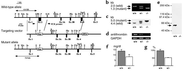
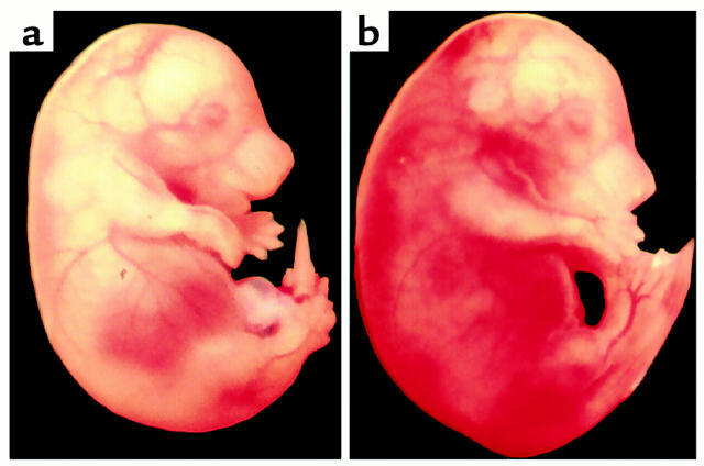
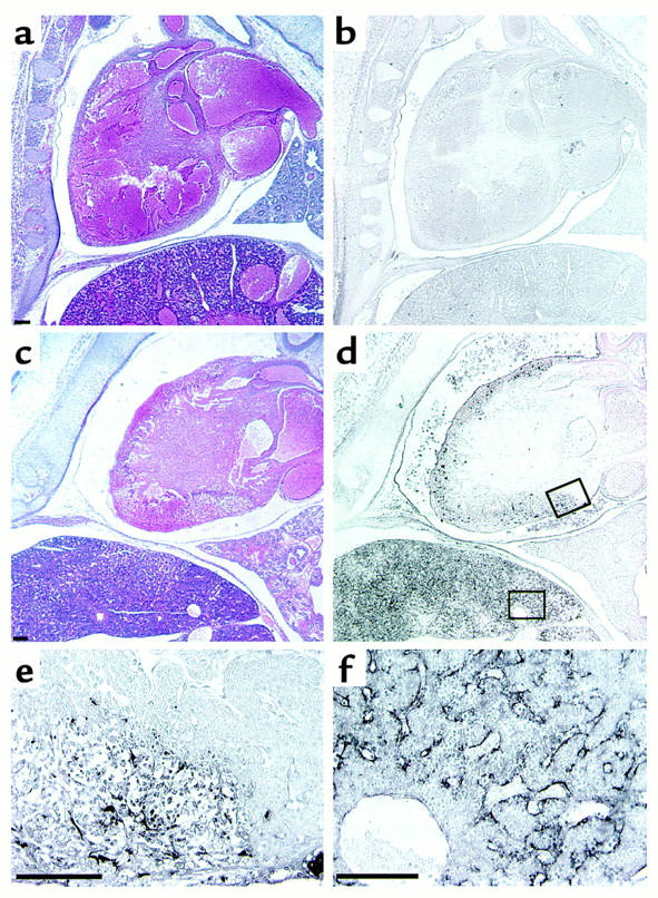
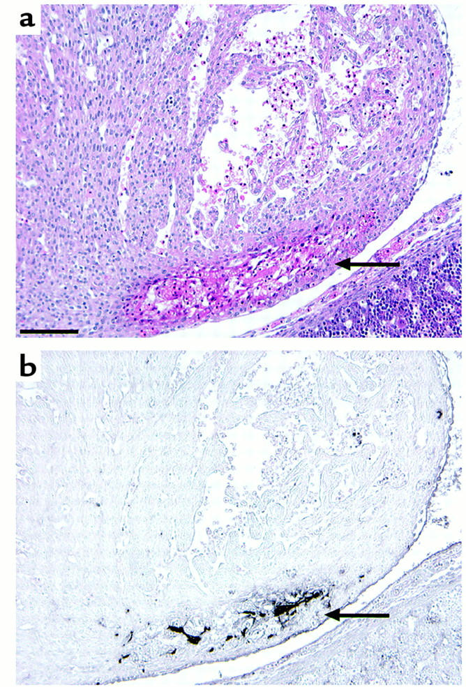
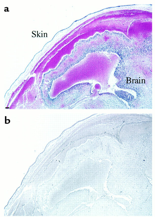
Antithrombin is a plasma protease inhibitor that inhibits thrombin and contributes to the maintenance of blood fluidity. Using targeted gene disruption, we investigated the role of antithrombin in embryogenesis. Mating mice heterozygous for antithrombin gene (ATIII) disruption, ATIII(+/-), yielded the expected Mendelian distribution of genotypes until 14.5 gestational days (gd). However, approximately 70% of the ATIII(-/-) embryos at 15.5 gd and 100% at 16.5 gd had died and showed extensive subcutaneous hemorrhage. Histological examination of those embryos revealed extensive fibrin(ogen) deposition in the myocardium and liver, but not in the brain or lung. Furthermore, no apparent fibrin(ogen) deposition was detected in the extensive hemorrhagic region, suggesting that fibrinogen might be decreased due to consumptive coagulopathy and/or liver dysfunction. These findings suggest that antithrombin is essential for embryonic survival and that it plays an important role in regulation of blood coagulation in the myocardium and liver.


Figures

Figure 1
Figure 1
Antithrombin gene-disruption in mice. (a) Antithrombin replacement construct and partial restriction map. P, PvuII; H, HindIII; X, XbaI; Ss, SspI; E, EcoRI; Sm, SmaI; Ex, exon; Neo, neomycin resistance gene. (b) PCR. External primers (PCR primers in a) were used. The mutant allele gave a 1.3-kb band, whereas the wild-type allele gave a 2.0-kb band. +/+, ATIII+/+; +/–, ATIII+/–; –/–, ATIII–/– embryos. (c) Southern blot analysis after PvuII digestion. An external probe (probe in a) was used. The mutant allele gave a 5.0-kb band, whereas the wild-type allele gave a 4.4-kb band. (d) RT-PCR demonstrating that mRNA expression of antithrombin was detected in total RNA derived from the whole of an ATIII+/+ or ATIII+/– embryo, whereas it was not detected in total RNA derived from the whole ATIII–/– embryo at 14.5 gd. (e) Western blot analysis demonstrating that protein expression of antithrombin was detected in homogenate derived from the whole liver of an ATIII+/+ embryo, whereas it was not detected in homogenate derived from that of an ATIII–/– embryo at 14.5 gd. The arrow indicates the position where antithrombin migrates. (f) The amount of antithrombin antigen in plasma was significantly reduced in ATIII+/– mice compared with ATIII+/+ mice (n = 8, P < 0.001). The values are means ± SD. The P value was calculated with Student’s t test. (g) The functional activity of antithrombin in plasma was significantly reduced in ATIII+/– mice compared with ATIII+/+ mice (n = 8, P < 0.001). The values are means ± SD. The P value was calculated with Student’s t test.
Figure 2
Figure 2
Macroscopic observation of ATIII+/+ (a) and dead ATIII–/– (b) embryos at 15.5 gd. A dead ATIII–/– embryo at 15.5 gd showed extensive subcutaneous hemorrhage.
Figure 3
Figure 3
Microscopic observation of ATIII+/+ (a and b) and dead ATIII–/– (cf) embryos at 15.5 gd. (a and c) Hematoxylin-eosin and (b and d) immunochemical staining with antifibrin(ogen) Ab. (e and f) Magnification of the areas indicated in d. (e) Myocardium. (f) Liver. A dead ATIII–/– embryo at 15.5 gd showed degeneration of the myocardium and liver (c). Fibrin(ogen) deposition was observed in the myocardium and sinusoids of the liver (d, e, and f). Size bars, 100 μm.
Figure 4
Figure 4
Hematoxylin-eosin (a) and immunohistochemical staining with anti-fibrin(ogen) Ab (b) of the myocardium of a living ATIII–/– embryo at 14.5 gd. Arrows indicate partial degeneration (a) and fibrin(ogen) deposition (b) in the myocardium.
Figure 5
Figure 5
Hematoxylin-eosin (a) and immunohistochemical staining with anti-fibrin(ogen) Ab (b) of the head of a dead ATIII–/– embryo at 16.5 gd. Extensive hemorrhage was observed subcutaneously and intracranially (a), but there was no apparent fibrin(ogen) deposition (b). Size bar, 100 μm.

References

    1. Lane DA, Caso R. Antithrombin: structure, genomic organization, function and inherited deficiency. Baillieres Clin Haematol. 1989;2:961–998. - PubMed
    1. O’Reilly MS, Pirie-Shepherd S, Lane WS, Folkman J. Antiangiogenic activity of the cleaved conformation of the serpin antithrombin. Science. 1999;285:1926–1928. - PubMed
    1. Rosenberg RD, Damus PS. The purification and mechanism of action of human antithrombin-heparin cofactor. J Biol Chem. 1973;248:6490–6505. - PubMed
    1. Marcum JA, Fritze L, Galli SJ, Karp G, Rosenberg RD. Microvascular heparin-like species with anticoagulant activity. Am J Physiol. 1983;245:H725–H733. - PubMed
    1. Marcum JA, Rosenberg RD. Anticoagulantly active heparin-like species molecules from vascular tissue. Biochemistry. 1984;23:1730–1737. - PubMed

Publication types

Substances