. 2000 Oct;157(4):1239-46.
doi: 10.1016/S0002-9440(10)64639-1.

Antibody-mediated resolution of light chain-associated amyloid deposits

Affiliations

Antibody-mediated resolution of light chain-associated amyloid deposits

R Hrncic et al. Am J Pathol. 2000 Oct.

Abstract

Primary light-chain-associated (AL) amyloidosis is characterized by the deposition in tissue of monoclonal light chains as fibrils. With rare exception, this process is seemingly irreversible and results in progressive organ dysfunction and eventually death. To determine whether immune factors can effect amyloid removal, we developed an experimental model in which mice were injected with amyloid proteins extracted from the spleens or livers of patients with AL amyloidosis. Notably, the resultant amyloidomas were rapidly resolved, as compared to controls, when animals received injections of an anti-light-chain monoclonal antibody having specificity for an amyloid-related epitope. The reactivity of this monoclonal antibody was not dependent on the V(L) or C(L) isotype of the fibril, but rather seemed to be directed toward a beta-pleated sheet conformational epitope expressed by AL and other amyloid proteins. The amyloidolytic response was associated with a pronounced infiltration of the amyloidoma with neutrophils and putatively involved opsonization of fibrils by the antibody, leading to cellular activation and release of proteolytic factors. The demonstration that AL amyloid resolution can be induced by passive administration of an amyloid-reactive antibody has potential clinical benefit in the treatment of patients with primary amyloidosis and other acquired or inherited amyloid-associated disorders.


Figures
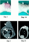
Figure 1.
Figure 1.
Human AL amyloidoma model. Top: Appearance of a mouse injected subcutaneously between the scapulae with 200 mg of a human ALκ amyloid extract (left) and resolution of the amyloidoma after 14 days (right). Bottom: Radiographic (CT scan) images of a mouse after injection of a human ALλ amyloid extract (left) and 21 days later (right).
Figure 2.
Figure 2.
Infiltration of regressing amyloidoma by polymorphonuclear leukocytes. Amyloid tumor excised on day 10 and formalin-fixed, paraffin-embedded sections stained with Congo red (left); hematoxylin-eosin (middle); and napthol AS-D chloroacetate (right). Original magnifications, ×400, ×400, ×1,000, respectively).
Figure 3.
Figure 3.
Humoral immune response to AL amyloidoma. Detection by immunoblotting of murine anti-human AL fibril antibodies. Top: ALκ HIG amyloid extract stained with Coomassie blue (lane 2) and blotted with an anti-κ light chain mAb (lane 3), nonimmune serum obtained from a normal mouse (lane 4), serum obtained from a mouse 20 days after HIG amyloidoma induction (lane 5), and serum obtained from a mouse 20 days after BAL amyloidoma induction (lane 6). Bottom: ALλ BAL extract stained with Coomassie blue (lane 2) and blotted with an anti-λ light chain mAb (lane 3), nonimmune serum obtained from a normal mouse (lane 4), serum obtained from a mouse 20 days after BAL amyloidoma induction (lane 5), and serum obtained from a mouse 20 days after HIG amyloidoma induction (lane 6). The Mrs of the molecular mass markers located in lane 1 are indicated in kd.
Figure 4.
Figure 4.
Reactivity of mAb 11-1F4 with ALκ and ALλ amyloid tissue deposits. Histochemical and immunohistochemical analyses of liver and spleen tissue obtained from ALκ1 patient HIG (top) and ALλ1 patient SHE (bottom), respectively. Left: Polarizing microscopy of Congo red-stained sections. Right: Immunoperoxidase staining with the mAb 11-1F4. Original magnifications, ×200).
Figure 5.
Figure 5.
Localization of fluorescein-labeled mAb 11-1F4 within an ALκ amyloidoma (fluorescent microscopy, original magnification, ×400).
Figure 6.
Figure 6.
Monoclonal anti-amyloid antibody-mediated resolution of human AL amyloidomas. Top: Appearance of residual ALκ amyloid tumor on day 4 in a mouse given a single 100-μg injection of mAb 11-1F4 at the time of amyloidoma induction (left) and in an untreated animal (right). Bottom: Appearance of residual ALλ amyloid tumor on day 7 in a mouse given 100-μg injections of mAb 11-1F4 at the time of amyloidoma induction (day 0) and then again on days 2, 4, and 6 (left) and in an untreated animal (right).

References

    1. Solomon A, Weiss DT: Protein and host factors implicated in the pathogenesis of light chain amyloidosis (AL amyloidosis). Amyloid. Int J Exp Clin Invest 1995, 2:269-279
    1. Kyle RA, Gertz MA: Primary systemic amyloidosis: clinical and laboratory features in 474 cases. Semin Hematol 1995, 32:45-59 - PubMed
    1. Dhodapkar MV, Merlini G, Solomon A: Biology and therapy of immunoglobulin deposition diseases. Hematol Oncol Clin North Am 1997, 11:89-110 - PubMed
    1. Falk RH, Comenzo RL, Skinner M: The systemic amyloidoses. N Engl J Med 1997, 337:898-909 - PubMed
    1. Gertz MA, Kyle RA: Amyloidosis: prognosis and treatment. Semin Arthritis Rheum 1994, 24:124-138 - PubMed

Publication types