. 2000 Oct;157(4):1247-58.
doi: 10.1016/S0002-9440(10)64640-8.

p75(NTR) mediates neurotrophin-induced apoptosis of vascular smooth muscle cells

Affiliations

p75(NTR) mediates neurotrophin-induced apoptosis of vascular smooth muscle cells

S Wang et al. Am J Pathol. 2000 Oct.

Abstract

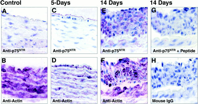
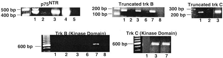
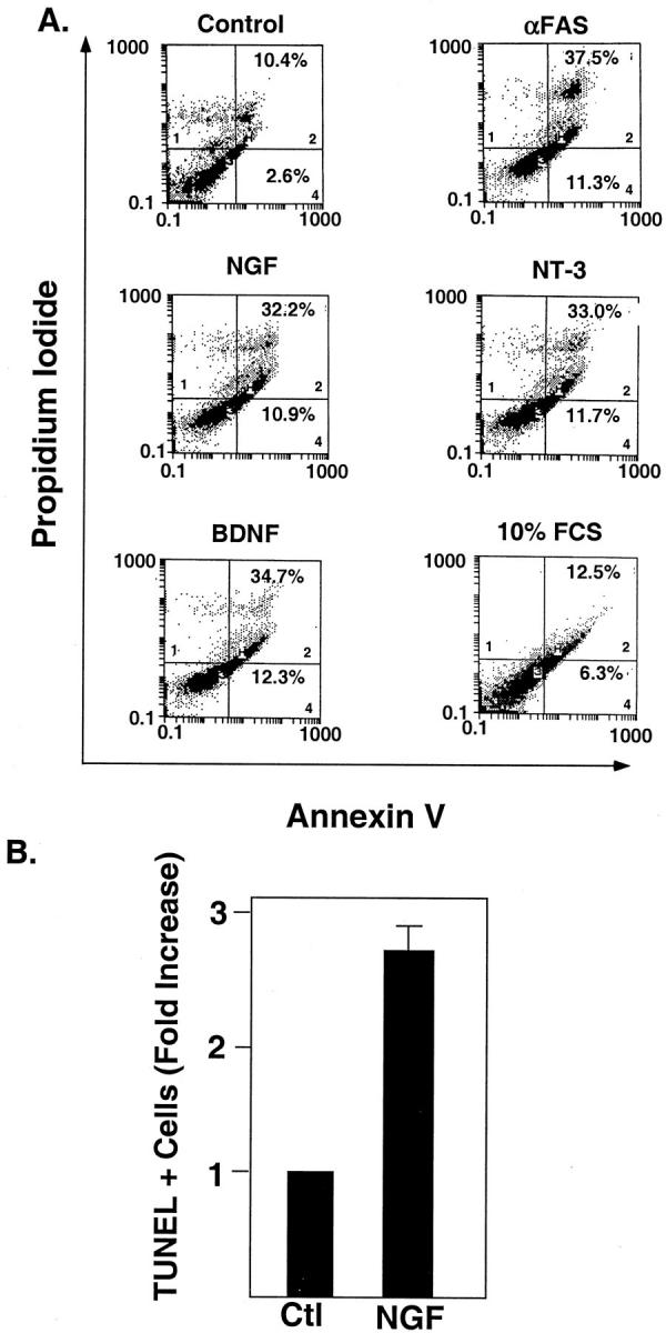
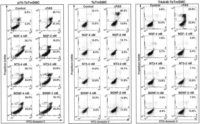
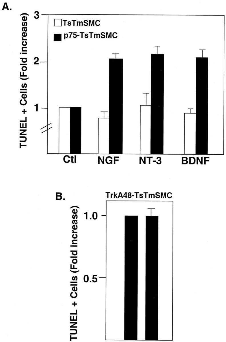
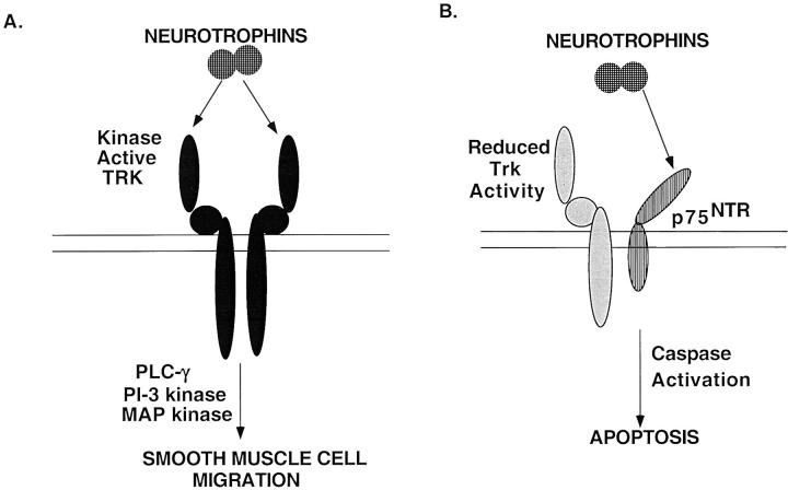
The development of atherosclerotic lesions results from aberrant cell migration, proliferation, and extracellular matrix production. In advanced lesions, however, cellular apoptosis, leading to lesion remodeling, predominates. During lesion formation, the neurotrophins and the neurotrophin receptor tyrosine kinases, trks B and C, are induced and mediate smooth muscle cell migration. Here we demonstrate that a second neurotrophin receptor, p75(NTR), is expressed by established human atherosclerotic lesions and late lesions that develop after balloon injury of the rat thoracic aorta. The p75(NTR), a member of the tumor necrosis factor/FAS receptor family, can modulate trk receptor function as well as initiate cell death when expressed in cells of the nervous system that lack kinase-active trk receptors. p75(NTR) expression colocalizes to neointimal cells, which express smooth muscle cell alpha-actin and are expressed by cultured human endarterectomy-derived cells (HEDC). Areas of the plaque expressing p75(NTR) demonstrate increased TUNEL positivity, and HEDC undergo apoptosis in response to the neurotrophins. Finally, neurotrophins also induced apoptosis of a smooth muscle cell line genetically manipulated to express p75(NTR), but lacking trk receptor expression. These studies identify the regulated expression of neurotrophins and p75(NTR) as an inducer of smooth muscle cell apoptosis in atherosclerotic lesions.


Figures

Figure 1.
Figure 1.
Immunohistochemical analysis of p75NTR in vascular lesions of the rat thoracic aorta 2 weeks after balloon de-endothelialization. Sections of rat thoracic aorta from either control animals (A and B), or 5 (C and D) and 14 days (E and F) after balloon de-endothelialization were incubated with the indicated antibodies. Immunoreactive proteins were visualized using avidin-biotin-based horseradish peroxidase kit using Vector VIP as a chromogenic substrate; the sections were counterstained with hematoxylin. Original magnification, ×90.
Figure 2.
Figure 2.
Immunohistochemical analysis of p75NTR expression and in situ TUNEL analysis in vascular lesions of human atherosclerotic lesions. A: Hematoxylin and eosin stain of atherectomy specimen derived from a carotid artery. Panels represented by the insets are indicated. The outer black line represents where the medial smooth muscle layer would extend to form the entire cross-section of the vessel. The inner black line represents the potential boundary of the lumen of the vessel. Panels B-M were treated as indicated. Immunoreactive proteins were detected as in Figure 1 ▶ . Areas marked off in E, H, and K are shown at higher magnification in F, I, and L, respectively. Open arrowheads in C are calcifications stained with hematoxylin. Arrows in F, cells expressing high levels p75NTR, shown at higher magnification in G; arrows in I, nuclei of TUNEL-positive cells, shown at higher maginification in J; arrowhead in I, nuclei of TUNEL-positive cells sectioned obliquely, shown at higher magnification in J. M, magnified area of L represented in G and J. Insets: F, control section incubated with anti-p75NTR preabsorbed with blocking peptide; J, control section incubated with fluorescenated-dUTP in the absence of TdT; L, control section incubated with mouse IgG.
Figure 3.
Figure 3.
Expression of p75NTR, truncated trk B, and truncated trk C mRNA in human atherectomy-derived smooth muscle cells. Total RNA was subjected to reverse transcription polymerase chain reaction and amplified products detected following cycling with p75NTR-, truncated trk B-, or truncated trk C-specific primer pairs and Taq polymerase. Sizes of amplified product: p75NTR, 463 bp; truncated trk B, 161 bp; truncated trk C, 158 bp; kinase-active trk B, 571 bp; kinase-active trk C, 571 bp. Lanes 1, 3–5, and 7, RNA subjected to reverse transcription. Lanes 2, 6, and 8, RNA not subjected to reverse transcription. Lanes 1 and 2, HEDC-12. Lanes 3 and 6, HEDC-201. Lane 4, p75-TsTmSMC (positive control, see below). Lane 5, TsTmSMC. Lanes 7 and 8, bone marrow.
Figure 4.
Figure 4.
Neurotrophin-induced apoptosis of HEDC. A: Flow cytometric analysis of annexin V binding to HEDC. HEDC were treated with either NGF (2 nmol/L), NT-3 (2 nmol/L), BDNF (2 nmol/L), FAS-activating antibody (CH11; 100 ng/ml), or 10% FCS. After 24 hours, a cell suspension was prepared and the cells were stained with FITC-conjugated annexin V and propidium iodide as described in Methods, and flow cytometric analysis was performed. x axis, FITC-conjugated annexin V binding; y axis, propidium iodide incorporation. As has been previously demonstrated, cells in quadrant 3 (lower left corner) represent live cells that do not bind annexin V or incorporate propidium iodide. Cells in quadrant 4 (lower right corner) represent cells in the early phases of apoptosis, in which annexin V binds to phosphatidyl serine exposed on the outer surface of apoptotic cells, but there is no incorporation of propidium iodide because membrane integrity is maintained. Cells in quadrant 2 (upper right corner) represent cells in the later stages of apoptosis, which bind annexin V and incorporate propidium iodide due to a loss of membrane integrity. Cells in quadrant 1 represent cells that have completed the apoptotic pathway. Histogram tracings from 1 of 3 duplicate experiments using 2 different HEDC culture lines is shown. Quadrants 1, 2, and 4 are labeled in the lower left (quadrant 1) and lower right (quadrants 2 and 4) corners. The percentage of the total cell population in either quadrant 2 (late stage, secondary necrotic) or quadrant 4 (early apoptotic phase) are given in the appropriate quadrant. B: Quantitative analysis of TUNEL for HEDC. In each experiment there were two replicates per treatment group. Data are the mean ± SE of 3 separate experiments using 3 different cell lines.
Figure 5.
Figure 5.
Western blot analysis of p75NTR, trk, and FAS in native TsTmSMC and p75-TsTmSMC. For FAS Western blot analysis African green monkey kidney cells and the murine lymphocyte cell line A20 are used as a negative and positive control, respectively. The pheochromocytoma cell line PC12, genetically manipulated to overexpress trk receptors, was used as a positive control for trk and p75NTR Western blot analysis. This cell line expresses approximately 40,000 trk receptors and 40,000 p75NTR cell. Lane 1, TsTmSMC. Lane 2, A20 cells. Lane 3, COS cells. Lane 4, PC12. Lane 5, p75-TsTmSMC. Lane 6, p75-TsTmSMC-2.
Figure 6.
Figure 6.
Flow cytometric analysis of annexin V binding to p75-TsTmSMC, native TsTmSMC, and trk48A-TsTmSMC. TsTmSMC cell lines were grown for 3 days at 39.5°C followed by treatment with the neurotrophins at the indicated doses. After 15 hours, a cell suspension was prepared and the cells were stained with FITC-conjugated annexin V and propidium iodide, as described in Methods, and flow cytometric analysis was performed. Histograms from one of three duplicate experiments for each cell line is shown. Quadrants 1, 2, and 4 are labeled in the lower left (quadrant 1) and lower right (quadrants 2 and 4) corners. The percentage of cells in the early and late stages of apoptosis are indicated in quadrants 4 and 2, respectively.
Figure 7.
Figure 7.
A: Quantitative analysis of the TUNEL assay for TsTmSMC and p75NTR-TsTmSMC after treatment with the neurotrophins (each 4 nmol/L). Data are presented as -fold increase of TUNEL-positive cells over control, nontreated cells and are the means ± SE of one of three duplicate experiments for both native TsTmSMC and p75-TsTmSMC. In each experiment, there were two replicates per treatment group. Percentage of TUNEL-positive cells in the absence of exogenous ligand: TsTmSMC = 2.1 ± 0.4%; p75TsTmSMC = 4.6 ± 0.9%. B: Quantitative analysis of TUNEL assay for trkA48-TsTmSMC. Data are presented as in A and are from one of two duplicate experiments, two replicates per experimental group. Percentage of TUNEL-positive cells in the absence of ligand = 1.9 ± 0.4%.
Figure 8.
Figure 8.
Immunofluorescent analysis for expression of activated caspases in p75-TsTmSMC. Cells were counterstained with DAPI for detection of condensed chromatin as an index of apoptosis.
Figure 9.
Figure 9.
Model to assess the role of the neurotrophins in vascular lesion development. A: Migratory response of the neurotrophins on smooth muscle cells expressing full length, kinase-active trk receptors. B: Ligand-induced activation of p75NTR, in the presence of reduced trk activity, results in apoptosis of smooth muscle cells.

References

    1. Chao MV, Hempstead BL: p75 and trk: a two-receptor system. Trends Neurosci 1995, 18:321-326 - PubMed
    1. Donovan MJ, Miranda RC, Kraemer R, Mccaffrey TA, Tessarollo L, Mahadeo D, Sharif S, Kaplan DR, Tsoulfas P, Parada L, Toran-Allerand D, Hajjar DP, Hempstead BL: Neurotrophin and neurotrophin receptors in vascular smooth muscle cells: regulation of expression in response to injury. Am J Pathol 1995, 147:309-324 - PMC - PubMed
    1. Klein R, Lamballe F, Bryant S, Barbacid M: The trkB tyrosine protein kinase is a receptor for neurotrophin 4. Neuron 1992, 8:947-956 - PubMed
    1. Cordon-Cardo C, Tapley P, Jing S, Nanduri V, O’Rourke E, Lamballe F, Kovary K, Klein K, Jones KR, Reichardt LF, Barbacid M: The trk tyrosine protein kinase mediates the mitogenic properties of nerve growth factor and neurotrophin-3. Cell 1991, 66:173-183 - PMC - PubMed
    1. Kraemer R, Ngyuen H, March KL, Hempstead BL: NGF activates similar intracellular signaling pathways in vascular smooth muscle cells as PDGF-BB but elicits different biological respones. Arterioscler Thromb Vasc Biol 1999, 19:1041-1050 - PubMed

Publication types

MeSH terms