Review
. 2000 Oct;13(4):571-87.
doi: 10.1128/CMR.13.4.571.

Rubella virus replication and links to teratogenicity

Affiliations
Review

Rubella virus replication and links to teratogenicity

J Y Lee et al. Clin Microbiol Rev. 2000 Oct.

Abstract

Rubella virus (RV) is the causative agent of the disease known more popularly as German measles. Rubella is predominantly a childhood disease and is endemic throughout the world. Natural infections of rubella occur only in humans and are generally mild. Complications of rubella infection, most commonly polyarthralgia in adult women, do exist; occasionally more serious sequelae occur. However, the primary public health concern of RV infection is its teratogenicity. RV infection of women during the first trimester of pregnancy can induce a spectrum of congenital defects in the newborn, known as congenital rubella syndrome (CRS). The development of vaccines and implementation of vaccination strategies have substantially reduced the incidence of disease and in turn of CRS in developed countries. The pathway whereby RV infection leads to teratogenesis has not been elucidated, but the cytopathology in infected fetal tissues suggests necrosis and/or apoptosis as well as inhibition of cell division of critical precursor cells involved in organogenesis. In cell culture, a number of unusual features of RV replication have been observed, including mitochondrial abnormalities, and disruption of the cytoskeleton; these manifestations are most probably linked and play some role in RV teratogenesis. Further understanding of the mechanism of RV teratogenesis will be brought about by the investigation of RV replication and virus-host interactions.


Figures

FIG. 1
FIG. 1
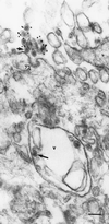
Attachment and entry of RV into Vero cells. Vero cells were inoculated with RV at a multiplicity of infection of 50 and incubated at 37°C. At 3, 5, 15, 30, and 45 min after the addition of virus, the cell monolayer was harvested and processed for TSEM as described by Lee (81). (A) An RV virion (solid arrow), comprising an electron-lucent core surrounded by a host-derived lipid envelope, can be seen attached to a cell surface projection (SP) adjacent to a coated pit (open arrow). (B) A virion (solid arrow) can be seen located within a coated vesicle (open arrow). Bars, 100 nm. PM, plasma membrane.
FIG. 2
FIG. 2
Detection of an RV virion within an endosome-like vacuole. RV-infected Vero cells were harvested at 4 h p.i. and processed for immunogold-labeling EM using polyclonal antibodies to RV as described previously (84). Gold particles (diameter, 10 nm) were found associated with virions located extracellularly. Note the virion (short solid arrow) attached near a coated pit (open arrow). An unlabeled RV virion (long solid arrow) is seen in an endosome-like vacuole (v). Since the cells were harvested during the viral latent period, it is unlikely that the virion within the endosome-like vacuole represented a newly assembled particle. Bar, 100 nm.
FIG. 3
FIG. 3
Schematic representation of the translation and processing strategy of the RV ns and structural proteins. The RV genome comprises two long nonoverlapping ORFs, with the 5′ ORF coding for the ns proteins and the 3′ ORF coding for the structural proteins. A polyprotein precursor, p200, is translated from the 5′ ORF of the RV genomic RNA and undergoes cis cleavage to produce two ns proteins, p150 and p90. The locations of the putative amino acid motifs for methyltransferase (M), X motif, papain-like cysteine protease (P), helicase (H), and replicase (R) are indicated on the 5′ ORF. The RV structural proteins are synthesized from a 24S subgenomic RNA transcribed from the 3′ ORF. A polyprotein precursor, p100, is translated from the subgenomic RNA and undergoes several posttranslational modifications to ultimately produce the mature capsid (C), E2, and E1.
FIG. 4
FIG. 4
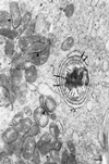
Cellular changes in RV-infected cells. A typical replication complex is observed with the characteristic vesicles (double-headed arrows) and the close association of the RER (open arrow). RV core particles (long solid arrows) can be seen at the cytoplasmic side of the vesicles of the replication complex. Core particles (small solid arrows) can also be detected in association with the outer membrane of mitochondria. Electron-dense zones (arrowheads) are frequently observed between the outer membranes of adjacent mitochondria. Note the clustering of mitochondria near the replication complex. Bar, 200 nm.
FIG. 5
FIG. 5
Schematic representation of the role of vesicles within the RV replication complex as precise sites of viral RNA synthesis. The vesicles of the replication complex are postulated to provide a protective environment for the synthesis of nascent viral genomic RNA. Newly synthesized viral RNA is then rapidly encapsidated by RV capsid proteins, which are synthesized from the adjacent RER. The mechanisms involved in the translocation of the resulting nucleocapsids for interaction with RV E2-E1 heterodimers have not been defined.
FIG. 6
FIG. 6
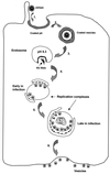
Schematic representation of the biogenesis of RV replication complexes. Step 1, The RV virion attaches to the cell surface and is translocated to the coated pit. Step 2, The coated pit then pinches off to form a coated vesicle that contains the virion. Step 3, The virion passes through a series of endosomes with progressively acidic pH until it arrives at an endosome where the environment is sufficiently acidic to trigger the uncoating process. The E1 and capsid proteins undergo conformational changes that result in the release of the viral genomic RNA into the cytoplasm. Step 4, Release of the viral RNA triggers the transformation of the endosome, and vesicles are induced to form within the endosome. This leads to the formation of the replication complex. Concomitantly, the RER migrates to the vicinity of the virus-modified endosome. At this early stage of the infection, the RER is associated with the side of the vacuole where the vesicles are located. Step 5, As infection progresses, the RER surrounds the entire vacuole, which is lined internally with vesicles. While these events are occurring, the virus-modified endosome fuses to a lysosome as part of its life cycle. Step 6, The replication complex continues in its life cycle as a virus-modified lysosome and eventually expels its lysosomal contents, including the vesicles, after fusion of the lysosomal vacuole membrane to the plasma membrane.
FIG. 7
FIG. 7
Mitochondrial changes in RV-infected cells. Electron-dense zones (short arrows) are frequently observed between the outer membrane of an adjacent mitochondrion (m). The composition of the electron-dense zone is not known. Note an RV virion (long arrow) within a lumen of an ER. Bar, 100 nm.

References

    1. Abe T, Nakada T, Hatanka H, Tajima M, Hiraiwa M, Ushijima H. Myoclonus in a case of suspected progressive rubella panencephalitis. Arch Neurol. 1983;40:98–100. - PubMed
    1. Acheson N H, Tamm I. Replication of Semliki Forest virus: an electron microscope study. Virology. 1967;32:128–143. - PubMed
    1. Atreya C D, Lee N S, Forng R-Y, Höfmann J, Washington G, Marti G, Nakhasi H L. The rubella virus putative replicase interacts with the retinoblastoma tumor suppressor protein. Virus Genes. 1998;16:177–183. - PubMed
    1. Atreya C D, Singh N K, Nakhasi H L. The rubella virus binding activity of human calreticulin is localized to the N-terminal domain. J Virol. 1995;69:3848–3851. - PMC - PubMed
    1. Balachandran S, Kim C N, Yeh W C, Mak T W, Bhalla K, Barber G N. Activation of the dsRNA-dependent protein kinase, PKR, induces apoptosis through FADD-mediated death signaling. EMBO J. 1998;17:6888–6902. - PMC - PubMed

Publication types