Review
. 2000 Oct;13(4):662-85.
doi: 10.1128/CMR.13.4.662.

Fungal and parasitic infections of the eye

Affiliations
Review

Fungal and parasitic infections of the eye

S A Klotz et al. Clin Microbiol Rev. 2000 Oct.

Abstract

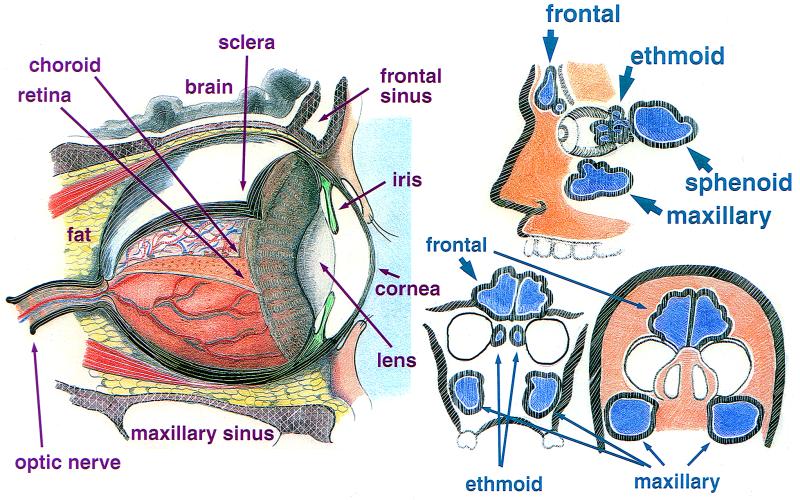
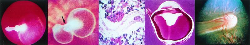
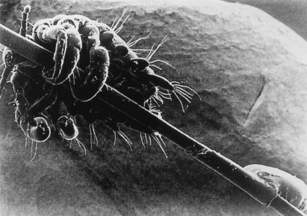
The unique structure of the human eye as well as exposure of the eye directly to the environment renders it vulnerable to a number of uncommon infectious diseases caused by fungi and parasites. Host defenses directed against these microorganisms, once anatomical barriers are breached, are often insufficient to prevent loss of vision. Therefore, the timely identification and treatment of the involved microorganisms are paramount. The anatomy of the eye and its surrounding structures is presented with an emphasis upon the association of the anatomy with specific infection of fungi and parasites. For example, filamentous fungal infections of the eye are usually due to penetrating trauma by objects contaminated by vegetable matter of the cornea or globe or, by extension, of infection from adjacent paranasal sinuses. Fungal endophthalmitis and chorioretinitis, on the other hand, are usually the result of antecedent fungemia seeding the ocular tissue. Candida spp. are the most common cause of endogenous endophthalmitis, although initial infection with the dimorphic fungi may lead to infection and scarring of the chorioretina. Contact lens wear is associated with keratitis caused by yeasts, filamentous fungi, and Acanthamoebae spp. Most parasitic infections of the eye, however, arise following bloodborne carriage of the microorganism to the eye or adjacent structures.
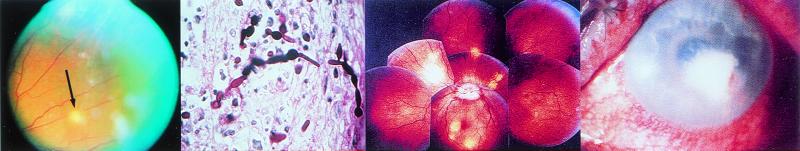
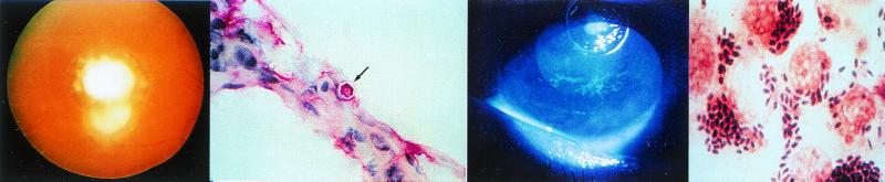
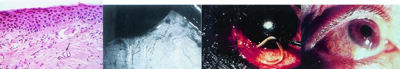

Figures

FIG. 1
FIG. 1
(Left) The human eye in situ with the tunics peeled back, exposing a portion of the vasculature of the retina, lens, and anterior chamber as seen from the side. (Right) The relationship of the paranasal sinuses to the eye. The top figure shows a side view. (Bottom left) Relationship of the sinuses with the orbit as seen by coronal section just posterior to the bridge of the nose. (Bottom right) Relationship of the sinuses with the orbits with the head tilted backwards.
FIG. 2
FIG. 2
(Left) View of the fundus of a patient with C. parapsilosis fungemia complicating total parenteral nutrition for esophageal cancer. A chorioretinal lesion is shown (arrow), and there is accompanying vitreous haze. (Middle left) C. albicans in retinal tissue with both blastoconidia and pseudohyphae present. (Photo courtesy of F. G. LaPiana.) (Middle right) Fundoscopic montage of the eye of a patient with P. carinii. There are edema of the optic nerve head (the circular whitish area in the center from which the blood vessels originate) and scattered multifocal choroidal mass lesions (whitish to yellowish circular lesions). (Reprinted from reference with permission from the publisher.) (Right) C. albicans keratitis in an immunocompromised patient. The dense yellow-white stromal infiltrate resembles bacterial keratitis. It partially covers the pupil. There is a small hypopyon present. (Reprinted from reference with permission from the publisher.)
FIG. 3
FIG. 3
(Left) Recurrent toxoplasmosis around the periphery of an old scar, which is the white central disc. (photo courtesy of F. G. LaPiana.) (Middle left) Corneal scraping stained with Masson trichrome stain demonstrating a polygonal cyst of Acanthamoeba (arrow). (Middle right) Dendritiform epithelial lesions of the lower part of the cornea demonstrated by fluorescein staining in a patient with Acanthamoeba keratitis. (Right) Gram stain of a conjunctival scraping from a patient with microsporidial keratoconjunctivitis demonstrating large gram-positive ovoid microorganisms within conjunctival epithelial cells. (The last three panels reprinted from reference with permission from the publisher.)
FIG. 4
FIG. 4
(Left) Microfilaria of O. volvulus (arrow) in the superficial corneal stroma (hematoxylin and eosin stain). (Middle left) L. loa beneath the conjunctiva. (Left and middle left panels reprinted from reference with permission from the publisher.) (Middle right) Adult L. loa being extracted from the subconjunctival space. (Right) A serpentine Dirofilaria migrating beneath the lateral aspect of the bulbar conjunctiva. (Middle right and right panels reprinted from reference with permission from the publisher.)
FIG. 5
FIG. 5
(Left) Cysticercosis of the eye, fundoscopic view. While being observed by the ophthalmologist, the protoscolex inverted and everted rhythmically. Here it is everted and pointed to the left. (Reprinted from reference with permission from the publisher.) (Middle left) Two cystercerci in the vitreous. One shows an evaginated protoscolex (arrow) (Reprinted from reference with permission from the publisher.) (Middle) Toxocara endophthalmitis. Serial section of the eye shows an intact Toxocara larva. (Middle right) Enucleated eye from a patient with toxocaral endophthalmitis. Note the large retrolental mass (white) associated with a funnel-shaped retinal detachment. (Middle and middle right panels reprinted from reference with permission from the publisher.) (Right) The optic disk of the eye “dragged” by a T. canis larva. (Photo courtesy of F. G. LaPiana).
FIG. 6
FIG. 6
Scanning electron micrograph of P. pubis adult and nit attached to an eyelash. (Reprinted from reference with permission from the publisher.)

References

    1. Adams A D. The morphology of human conjunctival mucus. Arch Ophthalmol. 1979;97:730–734. - PubMed
    1. Agger W A. Sporotrichosis. In: Fraunfelder F T, Roy F H, editors. Current ocular therapy. W. B. Philadelphia, Pa: Saunders Company; 1995. pp. 79–80.
    1. Ainbinder D, Parmley V C, Mader T H, Nelson M L. Infectious crystalline keratopathy caused by Candida guilliermondii. Am J Ophthalmol. 1998;125:723–725. - PubMed
    1. Akham O, Bilgi S, Akata D, Kiratli H, Ozmen M. Percutaneous treatment of an orbital hydatic cyst: a new therapeutic approach. Am J Ophthalmol. 1998;125:827–829. - PubMed
    1. Akler M, Vellend H, McNeely D M, Walmsley S L, Gold W L. Use of fluconazole in the treatment of candidal endophthalmitis. Clin Infect Dis. 1995;20:657–664. - PubMed

MeSH terms

LinkOut - more resources