. 2000 Nov;74(21):9916-27.
doi: 10.1128/jvi.74.21.9916-9927.2000.

Global analysis of herpes simplex virus type 1 transcription using an oligonucleotide-based DNA microarray

Affiliations

Global analysis of herpes simplex virus type 1 transcription using an oligonucleotide-based DNA microarray

S W Stingley et al. J Virol. 2000 Nov.

Abstract

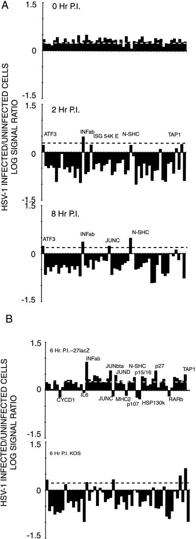
More than 100 transcripts of various abundances and kinetic classes are expressed during phases of productive and latent infections by herpes simplex virus (HSV) type 1. To carry out rapid global analysis of variations in such patterns as a function of perturbation of viral regulatory genes and cell differentiation, we have made DNA microchips containing sets of 75-mer oligonucleotides specific for individual viral transcripts. About half of these are unique for single transcripts, while others function for overlapping ones. We have also included probes for 57 human genes known to be involved in some aspect of stress response. The chips efficiently detect all viral transcripts, and analysis of those abundant under various conditions of infection demonstrates excellent correlation with known kinetics of mRNA accumulation. Further, quantitative sensitivity is high. We have further applied global analysis of transcription to an investigation of mRNA populations in cells infected with a mutant virus in which the essential immediate-early alpha27 (U(L)54) gene has been functionally deleted. Transcripts expressed at 6 h following infection with this mutant can be classified into three groups: those whose abundance is augmented (mainly immediate-early transcripts) or unaltered, those whose abundance is somewhat reduced, and those where there is a significant reduction in transcript levels. These do not conform to any particular kinetic class. Interestingly, levels of many cellular transcripts surveyed are increased. The high proportion of such transcripts suggests that the alpha27 gene plays a major role in the early decline in cellular gene expression so characteristic of HSV infection.


Figures

FIG. 1
FIG. 1
Demonstration of microarray specificity. The maximum hybridization signal in arbitrary (Arb) units attained with nick-translated HSV-1 DNA fluorescently labeled with Cy3dCTP and hybridized with the probes listed in Table 1 at 68°C is plotted against the G+C content of each probe.
FIG. 2
FIG. 2
Hybridization of oligo(dT)-primed cDNA synthesized from HeLa cells under various conditions of infection to an HSV-1 specific microarray. Each panel is based on a single experiment; a summary showing median values is shown in Table 2. (A) Hybridization of cDNA synthesized to RNA abundant in HeLa cells 3 h following infection in the presence of cycloheximide (50 μg/ml). (B) Hybridization of cDNA synthesized to RNA abundant in HeLa cells 2 h following infection. (C) Hybridization of cDNA synthesized to RNA abundant in HeLa cells 8 h following infection. Arb, arbitrary.
FIG. 2
FIG. 2
Hybridization of oligo(dT)-primed cDNA synthesized from HeLa cells under various conditions of infection to an HSV-1 specific microarray. Each panel is based on a single experiment; a summary showing median values is shown in Table 2. (A) Hybridization of cDNA synthesized to RNA abundant in HeLa cells 3 h following infection in the presence of cycloheximide (50 μg/ml). (B) Hybridization of cDNA synthesized to RNA abundant in HeLa cells 2 h following infection. (C) Hybridization of cDNA synthesized to RNA abundant in HeLa cells 8 h following infection. Arb, arbitrary.
FIG. 3
FIG. 3
The log10 of the ratio of late to early infected HeLa and rabbit skin cell viral RNA is plotted to emphasize time-specific differences in abundance. The late and early times for HeLa cells are 8 and 2 h p.i., respectively; those for rabbit skin cells are 7 and 3 h p.i.
FIG. 4
FIG. 4
Abundance of selected cellular transcripts under various conditions of HSV-1 infection of HeLa cells. The relative abundance of transcripts hybridizing to the cellular probes used in this study is shown in Table 5. The log10 of the ratio of the signal seen in mock-infected to infected cells is plotted here. (A) Relative levels of cellular transcripts present under various conditions of infection with the 17syn+ strain of HSV-1. The dashed line is the average ratio of mock-infected to infected cell signal for RNA isolated immediately following a 30-min virus adsorption period (top). (B) Effect of the α27 (UL54) immediate-early protein on cellular RNA abundance. The dashed line shows the average values of the 0-h control in panel A. The top panel shows the ratio of abundance of cellular RNAs in HeLa cells 6 h following infection and mock infection with the 27lacZ mutant of the KOS strain of HSV-1; the bottom panel shows the same ratio for cells infected and mock infected with the wt parental KOS strain.

References

    1. Anderson K P, Frink R J, Devi-Rao G B, Gaylord B H, Costa R H, Wagner E K. Detailed characterization of the mRNA mapping in the HindIII fragment K region of the herpes simplex virus type 1 genome. J Virol. 1981;37:1011–1027. - PMC - PubMed
    1. Arnosti D N, Preston C M, Hagmann M, Schaffner W, Hope R G, Laughlan G, Luisi B F. Specific transcriptional activation in vitroby the herpes simplex virus protein VP16. Nucleic Acids Res. 1993;21:5570–5576. - PMC - PubMed
    1. Ben-Hur T, Moyal M, Rosen-Wolff A, Darai G, Becker Y. Characterization of RNA transcripts from herpes simplex virus-1 DNA fragment BamHI-B. Virology. 1989;169:1–8. - PubMed
    1. Bruni R, Roizman B. Open reading frame P—a herpes simplex virus gene repressed during productive infection encodes a protein that binds a splicing factor and reduces synthesis of viral proteins made from spliced mRNA. Proc Natl Acad Sci USA. 1996;93:10423–10427. - PMC - PubMed
    1. Carter K L, Ward P L, Roizman B. Characterization of the products of the UL43 gene of herpes simplex virus 1: potential implications for regulation of gene expression by antisense transcription. J Virol. 1996;70:7663–7668. - PMC - PubMed

Publication types

LinkOut - more resources