. 2000 Nov;74(21):10006-17.
doi: 10.1128/jvi.74.21.10006-10017.2000.

Alphaherpesvirus proteins related to herpes simplex virus type 1 ICP0 affect cellular structures and proteins

Affiliations

Alphaherpesvirus proteins related to herpes simplex virus type 1 ICP0 affect cellular structures and proteins

J Parkinson et al. J Virol. 2000 Nov.

Abstract

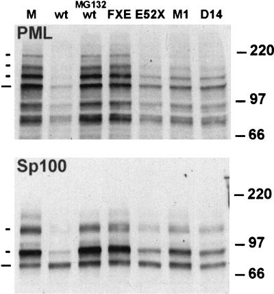
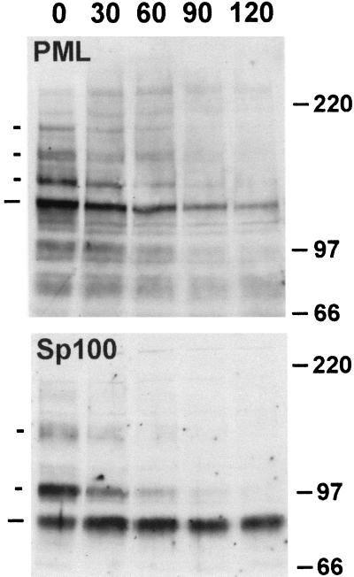
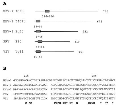
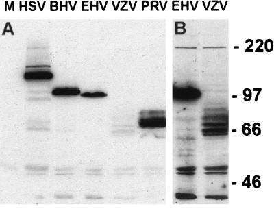
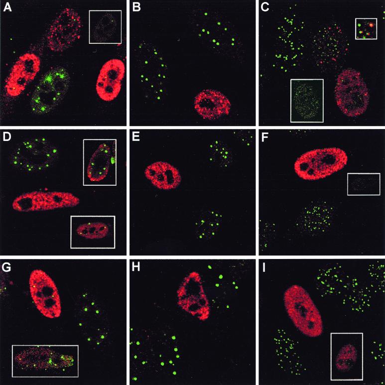
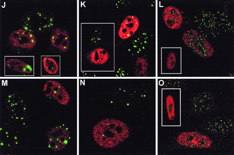
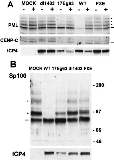
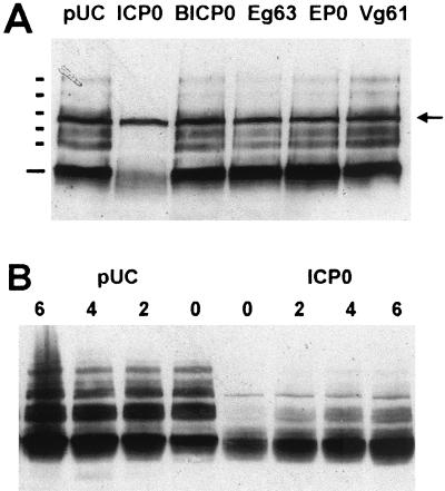
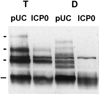
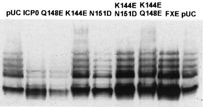
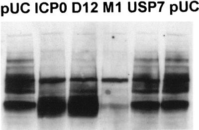
The herpes simplex virus type 1 (HSV-1) immediate-early protein ICP0 interacts with several cellular proteins and induces the proteasome-dependent degradation of others during infection. In this study we show that ICP0 is required for the proteasome-dependent degradation of the ND10 protein Sp100 and, as with the other target proteins, the ICP0 RING finger domain is essential. Further, comparison of the kinetics and ICP0 domain requirements for the degradation of PMI and Sp100 suggests that a common mechanism is involved. Homologues of ICP0 are encoded by other members of the alphaherpesvirus family. These proteins show strong sequence homology to ICP0 within the RING finger domain but limited similarity elsewhere. Using transfection assays, we have shown that all the ICP0 homologues that we tested have significant effects on the immunofluorescence staining character of at least one of the proteins destabilized by ICP0, and by using a recombinant virus, we found that the equine herpesvirus ICP0 homologue induced the proteasome-dependent degradation of endogenous CENP-C and modified forms of PML and Sp100. However, in contrast to ICP0, the homologue proteins had no effect on the distribution of the ubiquitin-specific protease USP7 within the cell, consistent with their lack of a USP7 binding domain. We also found that ICP0 by itself could induce the abrogation of SUMO-1 conjugation and then the proteasome-dependent degradation of unmodified exogenous PML in transfected cells, thus demonstrating that other HSV-1 proteins are not required. Surprisingly, the ICP0 homologues were unable to cause these effects. Overall, these data suggest that the members of the ICP0 family of proteins may act via a similar mechanism or pathway involving their RING finger domain but that their intrinsic activities and effects on endogenous and exogenous proteins differ in detail.


Figures

FIG. 1
FIG. 1
Comparative demodification and degradation of PML and Sp100 induced by ICP0 during HSV-1 infection. HFL cells were mock infected (lane M) or infected at 10 PFU per cell with HSV-1 strain 17syn+ in the absence (lane wt) or presence (lane MG132 wt) of proteasome inhibitor MG132 (5 μM final concentration) and the ICP0 mutant viruses FXE, E52X, M1, and D14, as indicated. The cells were harvested for Western blotting 4 h postabsorption. Western blots were probed for PML using MAb 5E10 at a dilution of 1/5 (upper panel), and the filter was then stripped and reprobed for Sp100 using rabbit serum SpGH at a dilution of 1/1,000 (lower panel). To the left of the panels, the short dashes indicate the major SUMO-1-modified isoforms of PML and Sp100 and the longer ones indicate the major, presumed unmodified, forms of the proteins. The positions of the 220-, 97-, and 66-kDa molecular mass markers are indicated to the right.
FIG. 2
FIG. 2
Time course of demodification and degradation of PML and Sp100 induced by HSV-1 infection. HFL cells were infected with HSV-1 strain 17syn+ at 10 PFU per cell and then harvested 30, 60, 90, and 120 min postabsorption, as indicated. The lane marked 0 contains proteins from a mock-infected sample. Total cell proteins were analyzed for PML and Sp100, and the results were annotated as described for Fig. 1.
FIG. 3
FIG. 3
Comparisons of ICP0 and the alphaherpesvirus homologues. (A) Diagram to show the position of the RING finger domain in ICP0 and the homologues. (B) Alignment of the RING finger domains. The first and last cysteine residues of the ICP0 RING finger are numbered, and the conserved residues and positions of similar hydrophobic residues (∗) are shown underneath.
FIG. 4
FIG. 4
Expression of tagged ICP0 homologues. (A) HEp-2 cells were transfected with pUC9 (lane M) or plasmids expressing pp65-ICP0 or the pp65 homologues, as indicated. Samples were harvested into SDS-gel loading buffer at 24 h posttransfection and analyzed by Western blotting using MAb anti-pp65 at a dilution of 1/750. The positions of the molecular weight markers are indicated. (B) Longer exposure of the EHV and VZV tracts of the blot.
FIG. 5
FIG. 5
Effect of tagged ICP0 and the tagged homologues on cellular proteins seen by confocal microscopy. HEp-2 cells were transfected with pp65-ICP0 (A to C), pp65-BICP0 (D to F), pp65-Eg63 (G to I), pp65-EP0 (J to L), or pp65-Vg61 (M to O). At 24 h posttransfection, cells were processed for confocal microscopy and costained with MAb anti-pp65 at a dilution of 1/1,000 and either polyclonal anti-PML r8 at a dilution of 1/1,000 (A, D, G, J, and M), polyclonal anti-Sp100 SpGh at a dilution of 1/1,000 (B, E, H, K, and N), or polyclonal anti-CENP-C r554 at a dilution of 1/500 (C, F, I, L, and O). Secondary antibodies used were FITC-conjugated goat anti-rabbit IgG (Sigma) at 1/100 and Cy3-conjugated goat anti-mouse (Amersham) at 1/1,000.
FIG. 5
FIG. 5
Effect of tagged ICP0 and the tagged homologues on cellular proteins seen by confocal microscopy. HEp-2 cells were transfected with pp65-ICP0 (A to C), pp65-BICP0 (D to F), pp65-Eg63 (G to I), pp65-EP0 (J to L), or pp65-Vg61 (M to O). At 24 h posttransfection, cells were processed for confocal microscopy and costained with MAb anti-pp65 at a dilution of 1/1,000 and either polyclonal anti-PML r8 at a dilution of 1/1,000 (A, D, G, J, and M), polyclonal anti-Sp100 SpGh at a dilution of 1/1,000 (B, E, H, K, and N), or polyclonal anti-CENP-C r554 at a dilution of 1/500 (C, F, I, L, and O). Secondary antibodies used were FITC-conjugated goat anti-rabbit IgG (Sigma) at 1/100 and Cy3-conjugated goat anti-mouse (Amersham) at 1/1,000.
FIG. 6
FIG. 6
Effect of ICP0 on USP7. HEp-2 cells were transfected with pp65-ICP0 (A and B) or pCIM1 (C and D), processed for confocal microscopy after 24 h, and costained with MAb anti-ICP0 11060 at 1/5,000 (A and C) and polyclonal anti-USP7 r201 at 1/200 (B and D). Secondary antibodies used were FITC-conjugated goat anti-rabbit IgG (Sigma) at 1/100 and Cy3-conjugated goat anti-mouse (Amersham) at 1/1,000 or Cy5-conjugated goat anti-mouse (Amersham) at 1/500.
FIG. 7
FIG. 7
Effect of 17Eg63 on cellular proteins. (A) HFL cells were mock infected or infected with wild-type 17syn+ virus (lane WT) or ICP0 mutants and recombinant as shown at 10 PFU per cell in the presence (+) or absence (−) of 5 μM MG132. Cells were harvested into SDS-gel loading buffer at 4 h postadsorption and analyzed by Western blotting. The blot was probed with anti-PML antibody E510 at 1/5 and reprobed with anti-CENP-C antibody r554 at 1/1,000 and anti-ICP4 MAb 10176 at 1/5,000. (B) A further blot was probed with polyclonal anti-Sp100 SpGH at 1/1,000 and reprobed with MAb anti-ICP4 10176 at 1/5,000. The positions of the PML and Sp100 isoforms and CENP-C are indicated, as are molecular weight markers. Long and short dashes indicate unmodified and modified proteins, respectively.
FIG. 8
FIG. 8
Effect of ICP0 and its homologues on the SUMO-1 conjugation of PML. Using Lipofectamine PLUS, HEp-2 cells were (A) cotransfected with pPML(F) and either pUC9 (lane pUC), pp65-ICP0 (lane ICP0), or the pp65-homologue plasmids (remaining lanes) shown, and harvested into SDS-gel loading buffer at 24 h posttransfection and analyzed by Western blotting using MAb anti-F at a dilution of 1/5,000 or (B) cotransfected with pPML(F) and either pUC9 or pp65-ICP0 and treated with 5 μM MG132 at 24 h posttransfection and harvested over the hourly time course as indicated. The positions of unmodified and SUMO-1-conjugated PML(F) bands are indicated by the long and short bars, respectively, in panel A, and the arrow (A) indicates a background antibody-detected band present in untransfected cells.
FIG. 9
FIG. 9
Effect of ICP0 on the SUMO-1 conjugation of PML in the presence of exogenous SUMO-1. Using Lipofectamine PLUS, HEp-2 cells were cotransfected with pPML(F) and either pUC9 or pp65-ICP0 (lanes D) or triple transfected with pPML(F), pCIPIC1, and either pUC9 or pp65-ICP0 (lanes T). Samples were harvested into SDS-gel loading buffer at 24 h posttransfection and analyzed by Western blotting using MAb anti-F at a dilution of 1/5,000. The positions of unmodified PML(F) and PML(F) modified by exogenous SUMO-1 in the triple transfections are indicated by long and short bars, respectively.
FIG. 10
FIG. 10
The RING finger of ICP0 is required for abrogation of SUMO-1 conjugation of PML. HEp-2 cells were cotransfected with plasmids as shown and pPML(F), using Lipofectamine PLUS. Samples were harvested into SDS-gel loading buffer at 24 h posttransfection and analyzed by Western blotting using MAb anti-F at a dilution of 1/5,000. p110N151D caused a significant level of transfected-cell mortality in this experiment, which explains why the level of PML(F) is reduced in this lane (lane N151D); however, staining for ICP0 indicated that the remaining cotransfected cells expressed ICP0.
FIG. 11
FIG. 11
Effect of ICP0 USP7 binding mutants on the SUMO-1 conjugation of PML. HEp-2 cells were cotransfected with pPML(F) and the plasmids as shown, using Lipofectamine PLUS. Samples harvested 24 h posttransfection were analyzed by Western blotting using MAb anti-F at a dilution of 1/5,000.

References

    1. Ahn J-H, Hayward G S. The major immediate-early proteins IE1 and IE2 of human cytomegalovirus colocalize with and disrupt PML-associated nuclear bodies at very early times in infected permissive cells. J Virol. 1997;71:4599–4613. - PMC - PubMed
    1. Ahn J-H, Brignole III E J, Hayward G S. Disruption of PML subnuclear domains by the acidic IE1 protein of human cytomegalovirus is mediated through interaction with PML and may modulate a RING finger-dependent cryptic transactivator function of PML. Mol Cell Biol. 1998;18:4899–4913. - PMC - PubMed
    1. Ali S, Lutz Y, Bellocq J P, Chenard-Neu M P, Rouyer N, Metzger D. Production and characterization of monoclonal antibodies recognising defined regions of the human oestrogen receptor. Hybridoma. 1993;12:391–405. - PubMed
    1. Ascoli C A, Maul G G. Identification of a novel nuclear domain. J Cell Biol. 1991;112:785–795. - PMC - PubMed
    1. Boddy M N, Howe K, Etkin L D, Solomon E, Freemont P S. PIC1, a novel ubiquitin-like protein which interacts with the PML component of a multiprotein complex that is disrupted in acute promyelocytic leukaemia. Oncogene. 1996;13:971–982. - PubMed

Publication types

MeSH terms

Substances