. 2000 Nov;74(21):10018-24.
doi: 10.1128/jvi.74.21.10018-10024.2000.

Incoming human cytomegalovirus pp65 (UL83) contained in apoptotic infected fibroblasts is cross-presented to CD8(+) T cells by dendritic cells

Affiliations

Incoming human cytomegalovirus pp65 (UL83) contained in apoptotic infected fibroblasts is cross-presented to CD8(+) T cells by dendritic cells

G Arrode et al. J Virol. 2000 Nov.

Abstract

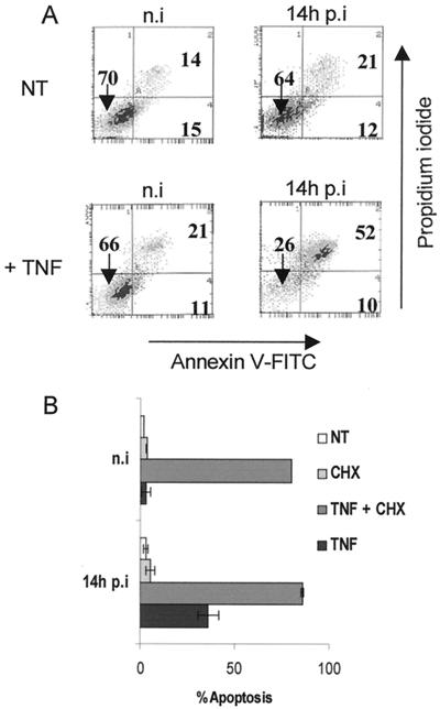
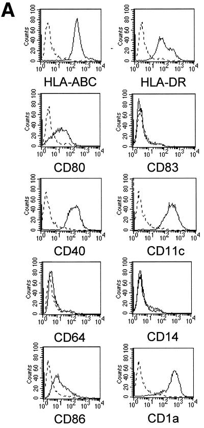
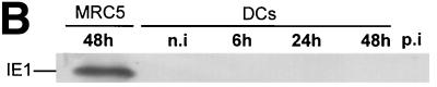
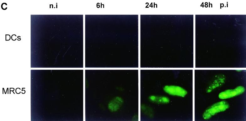
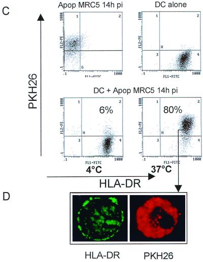
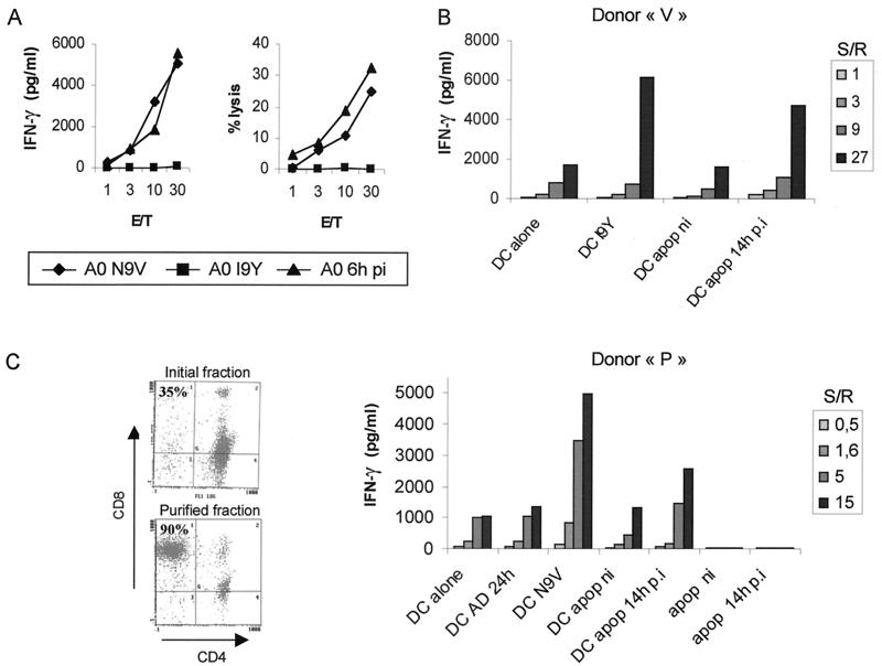
Human cytomegalovirus (HCMV) infection is well controlled mainly by cytotoxic CD8(+) T lymphocytes (CTL) directed against the matrix protein pp65 despite the numerous immune escape mechanisms developed by the virus. Dendritic cells (DCs) are key antigen-presenting cells for the generation of an immune response which have the capacity to acquire antigens via endocytosis of apoptotic cells and thus present peptides to major histocompatibility complex class I-restricted T cells. We examined whether this mechanism could contribute to the activation of anti-pp65 CTL. In this study, we show that infection by HCMV AD169 induced sensitization of MRC5 fibroblasts to tumor necrosis factor alpha-mediated apoptosis very early after virus inoculation and that pp65 contained in apoptotic cells came from the delivery of the matrix protein into the cell. We observed that immature DCs derived from peripheral monocytes were not permissive to HCMV AD169 infection but were able to internalize pp65-positive apoptotic infected MRC5 cells. We then demonstrated that following exposure to these apoptotic bodies, DCs could activate HLA-A2- or HLA-B35-restricted anti-pp65 CTL, suggesting that they acquired and processed properly fibroblast-derived pp65. Together, our data suggest that cross-presentation of incoming pp65 contained in apoptotic cells may provide a quick and efficient way to prime anti-HCMV CD8(+) T cells.


Figures

FIG. 1
FIG. 1
HCMV sensitizes MRC5 fibroblasts to TNF-α-mediated apoptosis very early after infection. MRC5 cells were either mock infected (n.i [not infected]) or infected for 6 h with HCMV AD169 at an MOI of 3. Then cells were not treated (NT) or treated with CHX, with TNF-α alone (TNF), or with TNF-α supplemented with CHX (TNF + CHX) for an additional 8 h. The indicated times correspond to the total time p.i. including TNF-α and/or CHX treatment. The presence of apoptotic cells was detected by flow cytometry analysis using PI and FITC-conjugated AV (A) or by fluorescence microscopy using Hoechst 33342 staining to reveal condensed nuclei (B). Histograms show mean values (±standard deviation) of the percentages of condensed nuclei from four independent experiments.
FIG. 2
FIG. 2
Immature DCs are not permissive to HCMV AD169. DCs were derived from peripheral blood monocytes after 7 days of differentiation in AIMV medium supplemented with GM-CSF and IL-13. Filled histograms represent labeling with specific antibody against the indicated surface marker; dotted lines represent staining with the control isotype antibody (A). MRC5 cells and immature DCs were not infected (n.i) or infected with HCMV AD169 (MOI of 3) for the indicated times p.i. Viral IE protein expression was detected by Western blotting using a mouse anti-IE MAb (B), and the matrix protein pp65 was visualized by intracellular staining with anti-pp65 antibody under a Leitz fluorescence microscope (C). Results are representative of two experiments.
FIG. 2
FIG. 2
Immature DCs are not permissive to HCMV AD169. DCs were derived from peripheral blood monocytes after 7 days of differentiation in AIMV medium supplemented with GM-CSF and IL-13. Filled histograms represent labeling with specific antibody against the indicated surface marker; dotted lines represent staining with the control isotype antibody (A). MRC5 cells and immature DCs were not infected (n.i) or infected with HCMV AD169 (MOI of 3) for the indicated times p.i. Viral IE protein expression was detected by Western blotting using a mouse anti-IE MAb (B), and the matrix protein pp65 was visualized by intracellular staining with anti-pp65 antibody under a Leitz fluorescence microscope (C). Results are representative of two experiments.
FIG. 3
FIG. 3
Apoptotic cells containing incoming HCMV pp65 protein are internalized by immature DCs. MRC5 cells either infected (14h pi) or not infected (n.i) were induced to undergo apoptosis (Apop) by treatment with TNF-α plus CHX (T+C). The expression of pp65 in purified apoptotic cells was detected by intracellular staining with mouse anti-pp65 MAb and flow cytometry analysis (A). Lysates from [35S]methionine-[35S]cysteine-labeled MRC5 cells that had been infected with HCMV as indicated were immunoprecipitated with anti-pp65 antibody, submitted to SDS-PAGE, and exposed to autoradiography (B). PKH26-dyed red apoptotic infected MRC5 cells (Apop MRC5 14h pi) were cocultured with immature DCs for 10 h at 4 or 37°C at a DC/apoptotic cell ratio of 1:5. Uptake of apoptotic MRC5 by HLA-DR+ DCs was analyzed by flow cytometry where dot plots were gated on FL1 high positive cells (DCs), thus excluding the uncleared apoptotic material (C). Internalization of PKH26-labeled apoptotic cells was visually confirmed by fluorescence microscopy using flow cytometry samples as starting material (D). Similar results were obtained in three independent experiments.
FIG. 3
FIG. 3
Apoptotic cells containing incoming HCMV pp65 protein are internalized by immature DCs. MRC5 cells either infected (14h pi) or not infected (n.i) were induced to undergo apoptosis (Apop) by treatment with TNF-α plus CHX (T+C). The expression of pp65 in purified apoptotic cells was detected by intracellular staining with mouse anti-pp65 MAb and flow cytometry analysis (A). Lysates from [35S]methionine-[35S]cysteine-labeled MRC5 cells that had been infected with HCMV as indicated were immunoprecipitated with anti-pp65 antibody, submitted to SDS-PAGE, and exposed to autoradiography (B). PKH26-dyed red apoptotic infected MRC5 cells (Apop MRC5 14h pi) were cocultured with immature DCs for 10 h at 4 or 37°C at a DC/apoptotic cell ratio of 1:5. Uptake of apoptotic MRC5 by HLA-DR+ DCs was analyzed by flow cytometry where dot plots were gated on FL1 high positive cells (DCs), thus excluding the uncleared apoptotic material (C). Internalization of PKH26-labeled apoptotic cells was visually confirmed by fluorescence microscopy using flow cytometry samples as starting material (D). Similar results were obtained in three independent experiments.
FIG. 4
FIG. 4
Immature DCs cross-present incoming pp65 contained in apoptotic cells. Anti-pp65 CTL lines were raised as described in Materials and Methods. Specific stimulation was assayed through either 51Cr release or IFN-γ secretion in the presence of HLA-A2+ U373MG (A0) cells either pulsed with HLA-A2 binding peptide (N9V) or irrelevant peptide (I9Y) or infected with HCMV at an MOI of 1 for 6 h (A0 6h pi) (A). E/T, effector-to-target ratio. MRC5 (HLA-A2) cells infected (14h p.i) or not infected (ni) with HCMV were induced to undergo apoptosis by TNF-α–CHX treatment. Apoptotic (apop) purified cells were fed to immature DCs at a ratio of 1:1. After 24 h of incubation, DCs were collected and cocultured with an HLA-B35-restricted anti-pp65 CTL line from donor V (B) and HLA-A2-restricted purified CD8+ CTL from donor P (C) at different stimulator-to-responder ratios (S/R). Alternatively, HCMV-pulsed DCs (DC AD 24h, MOI of 3), and purified apoptotic MRC5 cells either uninfected (apop ni) or infected (apop 14h p.i) were used as stimulator cells. Additional controls consisted of immature DCs which either had or had not been pulsed with the HLA-A2 (N9V) and HLA-B35 (I9Y) peptides at 1 μM. Stimulation of CTL was assayed by secretion of IFN-γ using ELISA. Results shown are representative of two to four independent experiments.

References

    1. Albert M L, Sauter B, Bhardwaj N. Dendritic cells acquire antigen from apoptotic cells and induce class I-restricted CTLs. Nature. 1998;392:86–89. - PubMed
    1. Bell D, Young J, Banchereau J. Dendritic cells. Adv Immunol. 1999;72:255–324. - PubMed
    1. Britt W J, Alford C. Cytomegalovirus. In: Fields B N, Knipe D M, Howley P M, et al., editors. Fields virology. Philadelphia, Pa: Lippincott-Raven Publishers; 1996. pp. 2493–2523.
    1. Chaudhuri A R, St. Jeor S, Maciejewski J P. Apoptosis induced by human cytomegalovirus infection can be enhanced by cytokines to limit the spread of virus. Exp Hematol. 1999;27:1194–1203. - PubMed
    1. Chu Z L, McKinsey T A, Liu L, Gentry J J, Malim M H, Ballard D W. Suppression of tumor necrosis factor-induced cell death by inhibitor of apoptosis c-IAP2 is under NF-kappaB control. Proc Natl Acad Sci USA. 1997;94:10057–10062. - PMC - PubMed

Publication types

Substances