. 2000 Nov;74(21):10055-62.
doi: 10.1128/jvi.74.21.10055-10062.2000.

Characterization of hepatitis C virus (HCV) and HCV E2 interactions with CD81 and the low-density lipoprotein receptor

Affiliations

Characterization of hepatitis C virus (HCV) and HCV E2 interactions with CD81 and the low-density lipoprotein receptor

S Wünschmann et al. J Virol. 2000 Nov.

Abstract

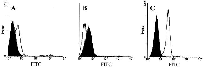
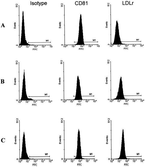
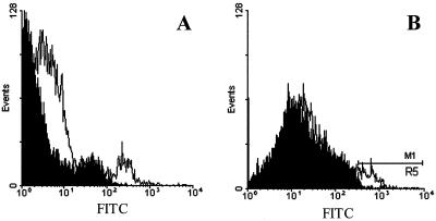
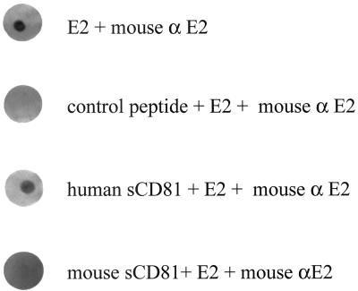
Hepatitis C virus (HCV) or HCV-low-density lipoprotein (LDL) complexes interact with the LDL receptor (LDLr) and the HCV envelope glycoprotein E2 interacts with CD81 in vitro. However, E2 interactions with LDLr and HCV interactions with CD81 have not been clearly described. Using sucrose gradient-purified low-density particles (1.03 to 1.07 g/cm(3)), intermediate-density particles (1. 12 to 1.18 g/cm(3)), recombinant E2 protein, or control proteins, we assessed binding to MOLT-4 cells, foreskin fibroblasts, or LDLr-deficient foreskin fibroblasts at 4 degrees C by flow cytometry and confocal microscopy. Viral entry was determined by measuring the coentry of alpha-sarcin, a protein synthesis inhibitor. We found that low-density HCV particles, but not intermediate-density HCV or controls bound to MOLT-4 cells and fibroblasts expressing the LDLr. Binding correlated with the extent of cellular LDLr expression and was inhibited by LDL but not by soluble CD81. In contrast, E2 binding was independent of LDLr expression and was inhibited by human soluble CD81 but not mouse soluble CD81 or LDL. Based on confocal microscopy, we found that low-density HCV particles and LDL colocalized on the cell surface. The addition of low-density HCV but not intermediate-density HCV particles to MOLT-4 cells allowed coentry of alpha-sarcin, indicating viral entry. The amount of viral entry also correlated with LDLr expression and was independent of the CD81 expression. Using a solid-phase immunoassay, recombinant E2 protein did not interact with LDL. Our data indicate that E2 binds CD81; however, virus particles utilize LDLr for binding and entry. The specific mechanism by which HCV particles interact with LDL or the LDLr remains unclear.


Figures

FIG. 1
FIG. 1
HCV and HCV-E2 binding to MOLT-4 cells. HCV low-density particles (A), HCV intermediate-density particles (B), and HCV-E2 (C) were evaluated for binding to MOLT-4 cells (open graphs). Mock sucrose gradient preparations (shaded graphs [A and B]) or the HCV nonstructural protein NS3/NS4 (shaded graph [C]) served as negative controls. Cell-bound virus or HCV-E2 protein was visualized with HCV-specific antiserum and anti-human IgG Oregon green (FITC).
FIG. 2
FIG. 2
Flow cytometric characterization of CD81 and LDLr expression on MOLT-4 cells. MOLT-4 cells were grown for 48 h in lipoprotein-rich medium (A) or for 24 h (B) or 48 h (C) in lipoprotein-deficient medium. Background fluorescence was measured with an irrelevant isotype-matched control MAb. CD81 expression was measured with JS64 MAb, and LDLr expression was measured with C7 MAb. Cell-bound antibodies were detected with anti-mouse IgG Oregon green (FITC).
FIG. 3
FIG. 3
HCV binding to FSF. HCV low-density particles were evaluated for binding to FSF (A) and LDLr-deficient FSF (B). Mock sucrose gradient preparations (shaded graphs) served as negative controls. Cell-bound virus was visualized with HCV specific antiserum and anti-human IgG Oregon green (FITC).
FIG. 4
FIG. 4
Specificity of HCV-E2 interactions with human CD81. Soluble human or mouse CD81 (10 μg/ml) applied to nitrocellulose was incubated with HCV-E2 (10 μg/ml). E2 applied to nitrocellulose served as the positive control, and an irrelevant control peptide served as the negative control. Interactions of E2 with each protein were detected with anti HCV-E2 MAb, anti-mouse IgG-AP, and 5-bromo-4-chloro-3-indolyl phosphate–nitroblue tetrazolium substrate.
FIG. 5
FIG. 5
Immunofluorescence detection of HCV, LDL, and HCV-E2 on MOLT-4 cells. MOLT-4 cells were incubated with low-density Mock fractions (A), low-density HCV (B and D to F), or HCV-E2 (10 μg/ml) (C) at 4°C. HCV and HCV-E2 were localized with human HCV-specific antiserum followed by anti-human IgG-Oregon green (B, C, E, and G), whereas LDL was localized with mouse anti-LDL MAb and anti-mouse IgG-Texas red (A, D, and F). Incubation of cells with HCV low-density particles followed by dual staining showed colocalization of LDL (D) and HCV (E) as yellow fluorescence in the overlay (F). Dual staining of MOLT-4 cells incubated with Mock sucrose gradient fractions is shown in panel A.
FIG. 6
FIG. 6
HCV-E2 does not interact with LDL. HCV-E2, HCV NS3/4, human soluble CD81, or mouse soluble CD81 spotted on nitrocellulose were incubated with LDL (10 μg/ml). The positive control consisted of goat-anti LDL polyclonal antibody spotted on nitrocellulose. Interaction of spotted proteins with LDL was detected with mouse anti-LDL MAb and anti-mouse IgG AP followed by 5-bromo-4-chloro-3-indolyl phosphate–nitroblue tetrazolium substrate. Binding was quantitated by densitometry (AlphaImager 2000; Alpha Innotech Corp., San Leandro, Calif.), and results represent density units × 100.

References

    1. Afonao A M R, Jiang J, Penin F, Tareau C, Samuel D, Petit M-A, Bismuth H, Dussaix E, Feray C. Nonrandom distribution of hepatitis C virus quasispecies in plasma and peripheral blood mononuclear cells subsets. J Virol. 1999;73:9213–9221. - PMC - PubMed
    1. Agnello V, Abel G, Elfahal M, Knight G B, Zhang X. Hepatitis C virus and other flaviviridae viruses enter cells via low density lipoprotein receptor. Proc Natl Acad Sci USA. 1999;96:12766–12771. - PMC - PubMed
    1. Agnello V, Chung R T, Kaplan L M. A role for hepatitis C virus infection in type II cryoglobulinemia. N Engl J Med. 1992;327:1490–1495. - PubMed
    1. Bouffard P, Hayashi P, Acevedo R, Levy N, Zeldis J. Hepatitis C virus is detected in a monocyte/macrophage subpopulation of peripheral blood mononuclear cells of infected patients. J Infect Dis. 1992;166:1276–1280. - PubMed
    1. Bradley D W, McCaustland K, Cook E H. Posttransfusion non-A, non-B hepatitis in chimpanzees: physicochemical evidence that the tubule forming agent is a small, enveloped virus. Gastroenterology. 1985;88:773–779. - PubMed

Publication types

LinkOut - more resources