

doi: 10.1128/jvi.74.21.10202-10206.2000.
Restricting expression prolongs expression of foreign genes introduced into animals by retroviruses
Affiliations
- PMID: 11024149
- PMCID: PMC102059
- DOI: 10.1128/jvi.74.21.10202-10206.2000
Item in Clipboard
Restricting expression prolongs expression of foreign genes introduced into animals by retroviruses
J Virol.
2000 Nov.
Abstract
If foreign genes are ubiquitously expressed in mice using a viral vector, expression is abrogated by CD8(+) cells in 2 to 4 weeks. However, if the expression of the genes is confined to skeletal muscle cells, the CD8(+) T-cell response is much weaker and expression is maintained for more than 6 weeks. These data show that restricting the expression of foreign genes to skeletal muscle cells and presumably to other cells that are inefficient at antigen presentation can prolong the expression of a foreign gene product.
Figures
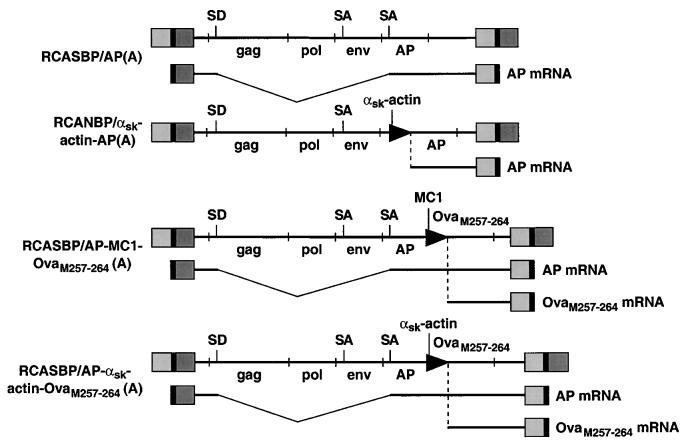
Schematic representation of the ASLV proviral DNAs and mRNAs coding for the reporter genes, AP and OVAM257–264. The viral genes gag, pol, and env are shown (not to scale), as are the genes AP and OVAM257–264. Internal promoters are shown as arrowheads. The positions of splice donors (SD) and splice acceptors (SA) are also shown. The retroviral vectors RCASBP(A) and RCANBP(A) and the Cla12 adapter plasmid have been described elsewhere (18, 27), as has RCASBP/AP(A) (10). The αsk-actin promoter was cloned into the ClaI-EcoRI site of the Cla12 adapter plasmid, and the AP cDNA was cloned into the EcoRI-SalI site of the same adapter plasmid. The ClaI fragment containing the αsk-actin AP fragment was excised from the Cla12 adapter and cloned into the ClaI site of RCANBP(A) to produce RCANBP/αsk-actin AP(A). The MC1 promoter, kindly provided by Mario Capecchi, contains a polyomavirus enhancer and a minimal TK promoter. The two oligonucleotides 5′-CCCGCCTCTAGACTCGAGCAGTGTGGTTTTCAAGAGG-3′ and 5′-CCCGCCGTCGACTCAGAGCTTCTCGAAGTTGATGATCGACATGGTTGCAGGGTCGCTCGG-3′ were used to produce the MC1 OvaM257–264 PCR product. This product was cloned into the XbaI-SalI site of Cla12 that contained AP in the EcoRI site. The ClaI fragment that contained the AP-MC1-OvaM257–264 was excised from the Cla12 adapter and cloned into the ClaI site of RCASBP(A) to produce RCASBP/AP-MC1-OvaM257–264(A). The αsk-actin promoter was cloned into the SmaI-EcoRI site of pBluescript SK(+). The two oligonucleotides 5′-AATTCACCATGTCGATCATCAACTTCGAGAAGCTCTGAG-3′ and 5′-TCGACTCAGAGCTTCTCGAAGTTGATGATCGACATGGTG-3′ that code for the peptide were cloned into the EcoRI-SalI site of pBluescript SK(+) that contained the αsk-actin promoter. The XbaI-SalI fragment that contained the αsk-actin promoter linked to OvaM257–264 was cloned into the XbaI-Sa1I site of Cla12 that contained AP in the EcoRI site. The ClaI fragment that contained the AP–αsk-actin–OvaM257–264 segment was excised from the Cla12 adapter and cloned into the ClaI site of RCASBP(A) to produce RCASBP/AP–αsk-actin–OvaM257–264(A).
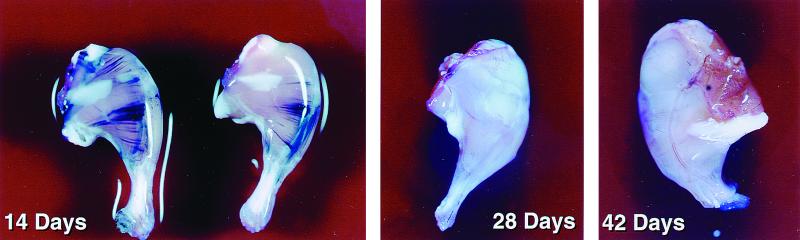
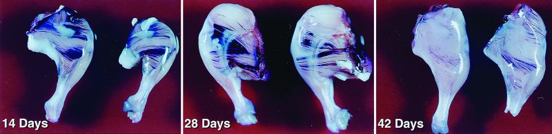
βAKE mice infected with RCASBP/AP(A). The mice were sacrificed on days 14, 28, and 42, and their legs were stained for AP (see the text). Muscle fibers that express AP can be seen as purple streaks.
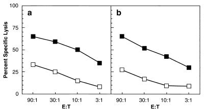
Splenocytes from adult C57BL/6 mice infected with OVAM257–264 VV (a) or 2-week-old βAKE mice injected on day 1 with RCASBP/AP-MC1 OVAM257–264(A) (b) virus-producing DF-1 cells were restimulated in vitro with 0.1 μM SIINFEKL and subsequently assayed for cytolytic activity against control (open boxes) or peptide-pulsed (closed boxes) targets at various E:T ratios (see the text).
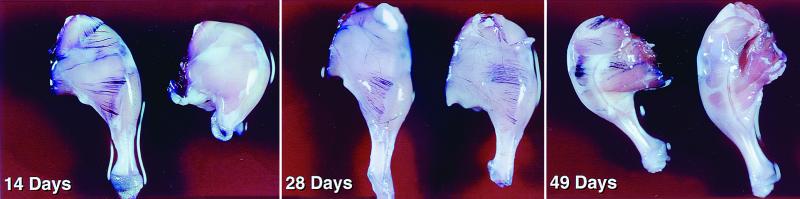
TAP1−/− mice carrying the Tva receptor infected with RCASBP/AP(A). The mice were sacrificed on days 14, 28, and 49, and their legs were stained for AP (see the text). Two of the legs (14 and 49 days) are negative for AP staining. These legs are from mice that do not have the subgroup A receptor.
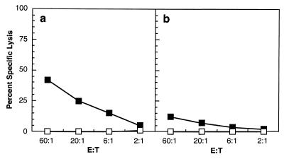
Splenocytes from 2-week-old βAKE mice injected on day 1 with RCASBP/AP-MC1 OVAM257–264(A) virus-producing DF-1 cells (a) or 2-week-old βAKE mice injected on day 1 with RCASBP/AP-αsk OVAM257–264(A) virus-producing DF-1 cells (b) were restimulated in vitro with 0.1 μM SIINFEKL and subsequently assayed for cytolytic activity against control (open boxes) or peptide-pulsed (closed boxes) targets at various E:T ratios (see the text).
βAKE mice infected with RCANBP/αskAP(A). The mice were sacrificed on days 14, 28, and 42, and their legs were stained for AP.
Similar articles
-
Immediate-early expression of a recombinant antigen by modified vaccinia virus ankara breaks the immunodominance of strong vector-specific B8R antigen in acute and memory CD8 T-cell responses.J Virol. 2010 Sep;84(17):8743-52. doi: 10.1128/JVI.00604-10. Epub 2010 Jun 10. J Virol. 2010. PMID: 20538860 Free PMC article.
-
Intrinsic transgene immunogenicity gears CD8(+) T-cell priming after rAAV-mediated muscle gene transfer.Mol Ther. 2015 Apr;23(4):697-706. doi: 10.1038/mt.2014.235. Epub 2014 Dec 10. Mol Ther. 2015. PMID: 25492560 Free PMC article.
-
Silencing of T lymphocytes by antigen-driven programmed death in recombinant adeno-associated virus vector-mediated gene therapy.Blood. 2009 Jan 15;113(3):538-45. doi: 10.1182/blood-2008-01-131375. Epub 2008 Jun 19. Blood. 2009. PMID: 18566327 Free PMC article.
-
Modified Vaccinia Virus Ankara Can Induce Optimal CD8+ T Cell Responses to Directly Primed Antigens Depending on Vaccine Design.J Virol. 2019 Oct 15;93(21):e01154-19. doi: 10.1128/JVI.01154-19. Print 2019 Nov 1. J Virol. 2019. PMID: 31375596 Free PMC article.
-
The production of foreign proteins in mammalian cells.Genet Eng. 1988;(7):91-127. Genet Eng. 1988. PMID: 3078408 Review.
Cited by
-
Use of avian retroviral vectors to introduce transcriptional regulators into mammalian cells for analyses of tumor maintenance.Proc Natl Acad Sci U S A. 2003 Jul 22;100(15):8764-9. doi: 10.1073/pnas.1133333100. Epub 2003 Jul 11. Proc Natl Acad Sci U S A. 2003. PMID: 12857957 Free PMC article.
-
A Cre-loxP-based mouse model for conditional somatic gene expression and knockdown in vivo by using avian retroviral vectors.Proc Natl Acad Sci U S A. 2008 Jul 22;105(29):10137-42. doi: 10.1073/pnas.0800487105. Epub 2008 Jul 11. Proc Natl Acad Sci U S A. 2008. PMID: 18621715 Free PMC article.
-
Osterix overexpression in mesenchymal stem cells stimulates healing of critical-sized defects in murine calvarial bone.Tissue Eng. 2007 Oct;13(10):2431-40. doi: 10.1089/ten.2006.0406. Tissue Eng. 2007. PMID: 17630878 Free PMC article.
References
-
- Bacik I, Cox J H, Anderson R, Yewdell J W, Bennink J R. Tap-independent presentation of endogenously synthesized peptides is enhanced by endocytoplasmic reticulum insertion sequences located at the amino but not carboxy terminus of the peptide. J Immunol. 1994;152:381–338. - PubMed
-
- Ehrenforth S, Kreuz W, Scharrer I, Linde R, Funk M, Gungor T, Krackhardt B, Kornhuber B. Incidence of development of factor VIII and factor IX inhibitors in haemophiliacs. Lancet. 1992;339:594–598. - PubMed
Publication types
MeSH terms
Substances
LinkOut - more resources
Full Text Sources
Other Literature Sources
Research Materials
