. 2000 Nov;74(21):10212-6.
doi: 10.1128/jvi.74.21.10212-10216.2000.

Interaction of the rabies virus P protein with the LC8 dynein light chain

Affiliations

Interaction of the rabies virus P protein with the LC8 dynein light chain

H Raux et al. J Virol. 2000 Nov.

Abstract

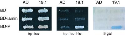
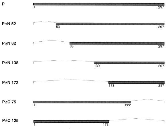
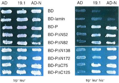
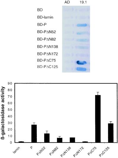
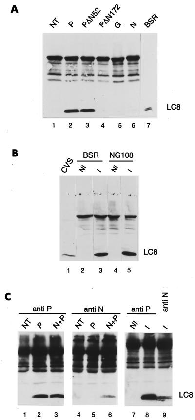
The rabies virus P protein is involved in viral transcription and replication but its precise function is not clear. We investigated the role of P (CVS strain) by searching for cellular partners by using a two-hybrid screening of a PC12 cDNA library. We isolated a cDNA encoding a 10-kDa dynein light chain (LC8). LC8 is a component of cytoplasmic dynein involved in the minus end-directed movement of organelles along microtubules. We confirmed that this molecule interacts with P by coimmunoprecipitation in infected cells and in cells transfected with a plasmid encoding P protein. LC8 was also detected in virus particles. Series of deletions from the N- and C-terminal ends of P protein were used to map the LC8-binding domain to the central part of P (residues 138 to 172). These results are relevant to speculate that dynein may be involved in the axonal transport of rabies virus along microtubules through neuron cells.


Figures

FIG. 1
FIG. 1
Interaction of P with LC8. L40 yeast cells expressing the indicated bait and prey pairs were streaked onto plates containing minimal medium lacking tryptophan and leucine (trp leu) for double transformants or lacking tryptophan, leucine, and histidine (trp leu his) to assay the activation of the HIS3 reporter gene. The induction of the lacZ reporter gene was assayed by the appearance of blue colonies as follows: an X-Gal mixture containing 0.5% agar, 0.1% SDS, 6% dimethylformamide, and 0.04% X-Gal was overlaid on fresh transformants grown on Trp Leu dishes, and blue clones were detected after 60 min to 18 h at 30°C. Note that 19.1 corresponds to the AD-LC8 clone.
FIG. 2
FIG. 2
Schematic diagram of the truncated P proteins fused to the LexA BD. Dark bars represent the protein product of each deleted P gene, with amino acid positions indicated. Each deleted region is indicated by a thin line. The constructs pLex-PΔN52, pLex-PΔN82, pLex-PΔN138, and pLex-PΔN172 differed from pLex-P by deletion at the 5′ terminus of the P gene of 186, 276, 444, and 546 bp, respectively. The constructs pLex-PΔC75 and pLex-PΔC125 differed from the wild-type P gene by deletion of 225 and 375 nucleotides from the 3′ terminus of the P gene, respectively. These deletions were created by PCR amplification of the wild-type P protein using specific oligonucleotides.
FIG. 3
FIG. 3
Analysis of P-N and P-LC8 interactions by induction of the HIS3 gene. L40 yeast cells expressing the indicated bait and prey pairs were streaked onto Trp Leu plates for double transformants and Trp Leu His plates for assaying the activation of the HIS3 reporter gene. The interaction between P and N was used as a positive control.
FIG. 4
FIG. 4
Qualitative and quantitative analyses of P-LC8 interactions by induction of the lacZ reporter gene. L40 yeast cells were cotransformed with the indicated combinations of plasmids. The interaction was assessed by the appearance of blue colonies in the presence of X-Gal (top) and by assaying the β-galactosidase activity of yeast grown in liquid medium (bottom). Quantitative results were obtained from three independent yeast cotransformants assayed with o-nitrophenyl-β-d-galactopyranoside (ONPG) as substrate (20). β-galactosidase activity was expressed in units and calculated using the following formula: (A420 · 1,000)/A600 · T · V), where A420 is the absorbance of the reaction mixture, A600 is the cell density of the culture, T is the reaction time (in minutes), and V is the volume (in milliliters) used for the assay. Error bars indicate the standard deviation.
FIG. 5
FIG. 5
Detection of P-LC8 complex in transfected cells and infected cells. (A) BSR cells were infected with vTF7-3 (lanes 1 to 6) and transfected with plasmids encoding P (lane 2), PΔN52 (lane 3), PΔN172 (lane 4), G (lane 5), or N (lane 6). These plasmids have been described elsewhere (10). Twenty hours after infection, samples of cytoplasmic cell extracts were immunoprecipitated with the murine polyclonal anti-P antibody (lanes 1 to 4), a monoclonal anti-G antibody (lane 5), or a monoclonal anti-N antibody (lane 6). Immunoprecipitated proteins were analyzed by Western blotting (SDS–15% PAGE). The blot was immunostained with a rabbit polyclonal anti-LC8 antibody (26) and with an anti-rabbit peroxidase-labeled antibody. An aliquot of uninfected BSR cell extract was used to indicate the position of LC8 in the gel (lane 7). (B) BSR (lanes 2 and 3) and NG108 (lanes 4 and 5) cells were uninfected (lanes 2 and 4) or infected with CVS rabies virus (lanes 2 and 4). Twenty hours after infection, cell extracts were immunoprecipitated with the murine polyclonal anti-P antibody and then treated as in panel A. Lane 1, proteins from purified virus. (C) BSR cells were infected with vTF7-3 (lanes 1 to 6) and transfected with plasmids encoding P (lanes 2 and 5) or cotransfected with plasmids encoding P and N (lanes 3 and 6). Cells were also infected with rabies virus. At 20 h after infection, identical samples of cytoplasmic cell extracts were immunoprecipitated with the anti-P antibody (lanes 1 to 3, 7, and 8) or anti-N antibody (lanes 4 to 6 and 9) and then treated as in panel A.

References

    1. Altschul S F, Madden T L, Schaffer A A, Zhang J, Zhang W, Miller W, Lipman D J. A new generation of protein database search programs. Nucleic Acids Res. 1997;25:3389–3402. - PMC - PubMed
    1. Banerjee A K. Transcription and replication of rhabdoviruses. Microbiol Rev. 1987;51:66–87. - PMC - PubMed
    1. Barik S, Banerjee A K. Phosphorylation by cellular casein kinase II is essential for the transcriptional activity of vesicular stomatitis virus phosphoprotein P. Proc Natl Acad Sci USA. 1992;89:6570–6574. - PMC - PubMed
    1. Barik S, Banerjee A K. Sequential phosphorylation of the phosphoprotein of vesicular stomatitis virus by cellular and viral protein kinases is essential for transcription activation. J Virol. 1992;66:1109–1118. - PMC - PubMed
    1. Beckwith S M, Roghi C H, Liu B, Morris N R. The “8kD” cytoplasmic dynein light chain is required for nuclear migration and for dynein heavy chain localization. J Cell Biol. 1998;143:1239–1247. - PMC - PubMed

Publication types

MeSH terms

LinkOut - more resources