. 2000 Oct 15;28(20):3880-6.
doi: 10.1093/nar/28.20.3880.

Mitochondrial DNA ligase III function is independent of Xrcc1

Affiliations

Mitochondrial DNA ligase III function is independent of Xrcc1

U Lakshmipathy et al. Nucleic Acids Res. .

Abstract

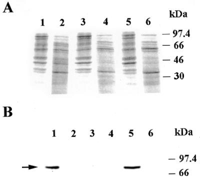
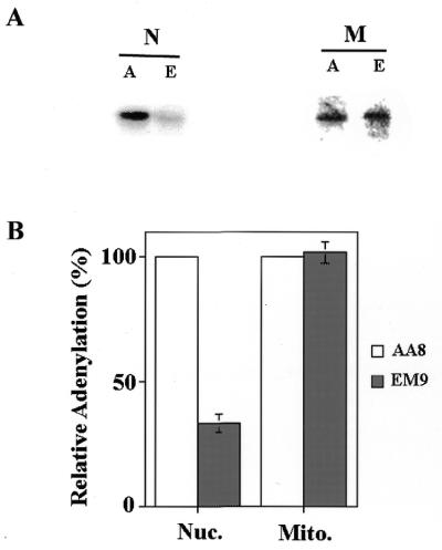
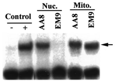
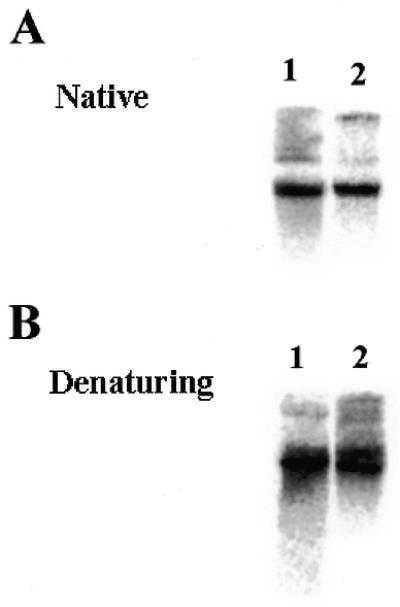
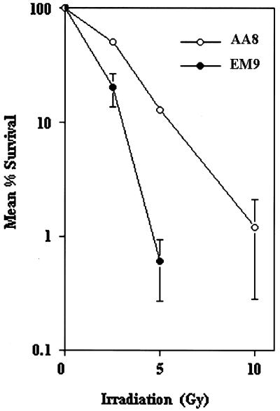
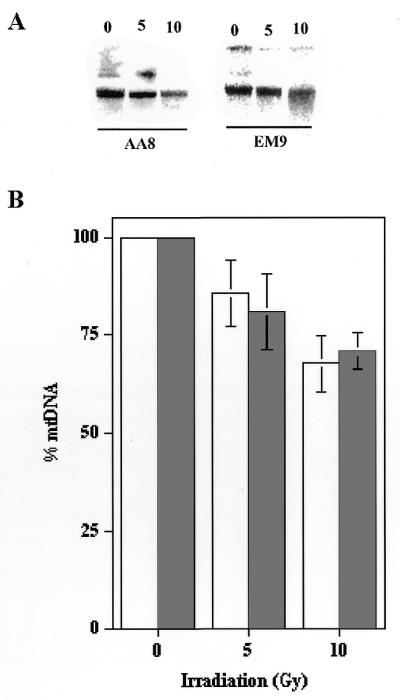
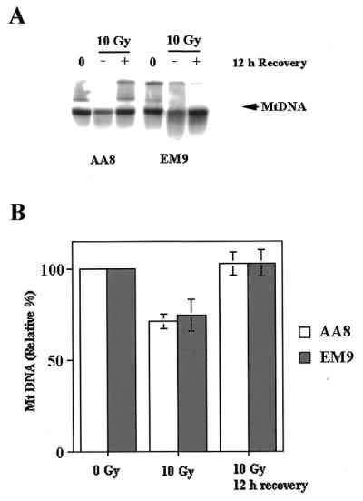
Hamster EM9 cells, which lack Xrcc1 protein, have reduced levels of DNA ligase III and are defective in nuclear base excision repair. The Xrcc1 protein stabilizes DNA ligase III and may even play a direct role in catalyzing base excision repair. Since DNA ligase III is also thought to function in mitochondrial base excision repair, it seemed likely that mitochondrial DNA ligase III function would also be dependent upon Xrcc1. However, several lines of evidence indicate that this is not the case. First, western blot analysis failed to detect Xrcc1 protein in mitochondrial extracts. Second, DNA ligase III levels present in mitochondrial protein extracts from EM9 cells were indistinguishable from those seen in similar extracts from wild-type (AA8) cells. Third, the mitochondrial DNA content of both cell lines was identical. Fourth, EM9 cells displayed no defect in their ability to repair spontaneous mitochondrial DNA damage. Fifth, while EM9 cells were far more sensitive to the cytotoxic effects of ionizing radiation due to a defect in nuclear DNA repair, there was no apparent difference in the ability of EM9 and AA8 cells to restore their mitochondrial DNA to pre-irradiation levels. Thus, mitochondrial DNA ligase III function is independent of the Xrcc1 protein.


Figures

Figure 1
Figure 1
The Xrcc1 protein in not present in mitochondrial protein extracts. (A) Coomassie blue stained gel of 10 µg of nuclear (lanes 1, 3 and 5) or mitochondrial extracts (lanes 2, 4 and 6) from AA8 (lanes 1 and 2), EM9 (lanes 3 and 4) and V79 (lanes 5 and 6) cells. (B) Western blot analysis using an Xrcc1-specific monoclonal antibody on the same samples described in (A). The position of Xrcc1 is indicated with an arrow.
Figure 2
Figure 2
Mitochondrial protein extracts from EM9 cells have DNA ligase activity but nuclear protein extracts do not. (A) Adenylation experiments were performed on 5 µg of nuclear (N) or mitochondrial (M) extracts from AA8 (A) and EM9 (E) cells. (B) Adenylation experiments were performed on multiple nuclear (Nuc.) and mitochondrial (Mito.) extracts from AA8 and EM9 cells. The amount of adenylation activity in extracts from EM9 cells (shaded bars) was normalized relative to that present in extracts from AA8 cells (open bars). Error bars represent the standard error of the mean (SEM, n = 3).
Figure 3
Figure 3
Nick-sealing activity of nuclear and mitochondrial extracts from EM9 and AA8 cells. A nick-seal assay was carried out on 5 µg samples of nuclear (Nuc.) and mitochondrial (Mito.) protein extracts as described in Materials and Methods. As a positive control, the substrate was incubated with 1 U of T4 DNA ligase. – Control, substrate alone; + control, substrate plus T4 DNA ligase; Nuc. AA8, substrate plus nuclear extract from AA8 cells; Nuc. EM9, substrate plus nuclear extract from EM9 cells; Mito. AA8, substrate plus mitochondrial extract from AA8 cells; Mito. EM9, substrate plus mitochondrial extract from EM9 cells. The arrow indicates the position of mobility of the sealed product.
Figure 4
Figure 4
Mitochondrial DNA copy number and integrity is similar in AA8 and EM9 cells. (A) Total cellular DNA (1 µg) from AA8 (lane 1) or EM9 (lane 2) cells was digested with the restriction enzyme PvuII to linearize the mitochondrial DNA and resolved on a 0.5% agarose gel. The gel was transferred onto a nylon membrane and hybridized with a mitochondrial DNA probe. (B) Cellular DNA was isolated from AA8 and EM9 cells and treated as above. The DNA was treated with 0.1 N NaOH for 30 min at 37°C prior to electrophoresis. The gel was then transferred to nylon and hybridized with a mitochondrial DNA probe.
Figure 5
Figure 5
EM9 cells are hypersensitive to ionizing radiation. EM9 (closed circles) and AA8 (open circles) cells were exposed to ionizing radiation, and the percent survival determined as described in Materials and Methods. Error bars indicate the SEM (n = 3).
Figure 6
Figure 6
Sensitivity of mitochondrial DNA from EM9 and AA8 cells to ionizing radiation. EM9 and AA8 cells were exposed to 0, 5 or 10 Gy of ionizing radiation. Total genomic DNA was immediately purified, cut with PvuII, resolved by agarose gel electrophoresis and hybridized to a mitochondrial DNA specific probe. (A) The results from a representative experiment. (B) Four experiments similar to that presented in (A) were performed and the relative amounts of mitochondrial DNA present were quantitated. Open bars, AA8 cells; shaded bars, EM9 cells. The error bars represent the SEM (n = 4).
Figure 7
Figure 7
Post-irradiation mitochondrial DNA recovery kinetics in AA8 and EM9 cells is similar. AA8 and EM9 cells were treated with ionizing radiation and their mitochondrial DNA was analyzed immediately, or following a 12 h recovery period, as described in the text. (A) Representative experiment. 0, pre-irradiation; –, immediately post-irradiation; +, 12 h post-irradiation. (B) Three experiments similar to that depicted in (A) were performed and mitochondrial DNA content measured by Southern blot hybridization. Opens bars, AA8 cells; shaded bars, EM9 cells. The error bars represent the SEM (n = 3).

References

    1. Caldecott K.W., McKeown,C.K., Tucker,J.D., Ljunquist,S. and Thompson,L.H. (1994) Mol. Cell. Biol., 14, 68–76. - PMC - PubMed
    1. Caldecott K.W., McKeown,C.K., Tucker,J.D., Stanker,L. and Thompson,L.H. (1995) Nucleic Acids Res., 23, 4836–4843. - PMC - PubMed
    1. Ljungquist S., Kenne,K., Olsson,L. and Sandstrom,M. (1994) Mutat. Res., 314, 117–186. - PubMed
    1. Thompson L.H., Brookman,K.W., Jones,N.J., Allen,S.A. and Carrano,A.V. (1990) Mol. Cell. Biol., 10, 6160–6171. - PMC - PubMed
    1. Caldecott K.W., Aoufouchi,S., Johnson,P. and Shall,S. (1996) Nucleic Acids Res., 24, 4387–4394. - PMC - PubMed

Publication types

MeSH terms