. 2000 Oct 15;28(20):E87.
doi: 10.1093/nar/28.20.e87.

Solid phase DNA amplification: characterisation of primer attachment and amplification mechanisms

Affiliations

Solid phase DNA amplification: characterisation of primer attachment and amplification mechanisms

C Adessi et al. Nucleic Acids Res. .

Abstract

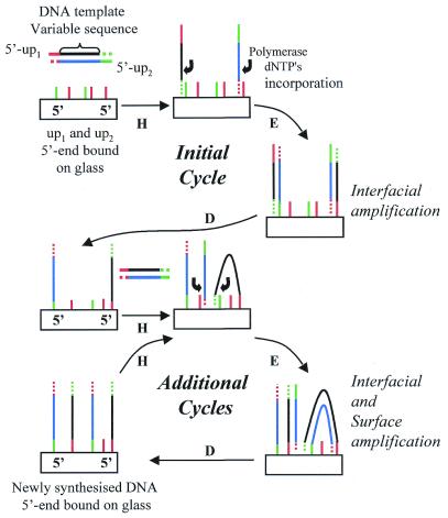
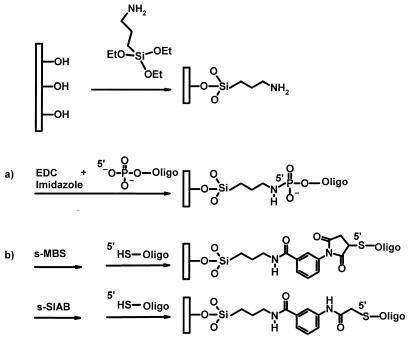
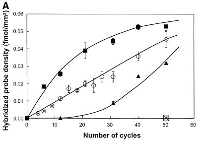
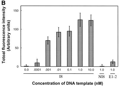
Different chemical methods used to attach oligonucleotides by their 5'-end on a glass surface were tested in the framework of solid phase PCR where surface-bound instead of freely-diffusing primers are used to amplify DNA. Each method was first evaluated for its capacity to provide a high surface coverage of oligonucleotides essentially attached via a 5'-specific linkage that satisfyingly withstands PCR conditions and leaves the 3'-ends available for DNA polymerase activity. The best results were obtained with 5'-thiol-modified oligonucleotides attached to amino-silanised glass slides using a heterobifunctional cross-linker reagent. It was then demonstrated that the primers bound to the glass surface using the optimal chemistry can be involved in attaching and amplifying DNA molecules present in the reaction mix in the absence of freely-diffusing primers. Two distinct amplification processes called interfacial and surface amplification have been observed and characterised. The newly synthesised DNA can be detected and quantified by radioactive and fluorescent hybridisation assays. These new surface amplification processes are seen as an interesting approach for attachment of DNA molecules by their 5'-end on a solid support and can be used as an alternative route for producing DNA chips for genomic studies.


Figures

Figure 1
Figure 1
Amplification of DNA by solid phase PCR. DNA primers (up1 or up2) are 5′-end covalently attached to the solid support. Thermocycling the surface (H, hybridisation; E, elongation; D, denaturation) in the presence of a solution of DNA target template, nucleotides and a thermostable polymerase leads to the amplification of DNA molecules on the solid surface. During the initial cycle only interfacial amplification occurs, as DNA target template is present only in solution. During subsequent cycles, in addition to interfacial amplification, surface amplification also occurs between the copied DNA template (5′-end attached to the surface) and attached primers in the vicinity.
Figure 2
Figure 2
Process to covalently attach nucleic acid molecules to a glass support. Hydroxylated glass surfaces were silanised using amino-silane reagents exemplified here by ATS. EDC and imidazole reagents lead to the formation of a phophoramidate linkage between amino-derivatised glass surfaces and 5′-phosphate modified oligonucleotides in a one-step reaction (a). For cross-linker chemistry (b), an amide bond was formed between amino groups of the surface and the succinimidyl ester moiety of the cross-linker compounds exemplified with s-MBS or s-SIAB reagents. Oligonucleotides, 5′-thiol modified, reacted with either the maleimide portion of s-MBS-like compounds or the iodoacetamide portion of s-SIAB.
Figure 3
Figure 3
Effect of the concentration of oligonucleotides. A mix of two primers, 5′-SH up1 and 5′-SH up2, was applied to a glass surface derivatised with ATS and functionalised with cross-linker s-MBS. Concentration of the mixed primer solution ranged from 1 to 100 µM. Glass slides were submitted to solid phase PCR with 1 nM template E1-2 (square) or I8 (circle) in solution or with no template in solution (triangle). Radioactively-labelled nucleotides were incorporated during PCR to the solid phase amplified DNA. Glass slides were exposed for phosphor imaging (A, top left) and density of the radioactive signal analysed (A). The loading density of up1, after PCR, at different concentrations was analysed by hybridisation using radioactively-labelled Rup1 complementary oligonucleotide (B).
Figure 3
Figure 3
Effect of the concentration of oligonucleotides. A mix of two primers, 5′-SH up1 and 5′-SH up2, was applied to a glass surface derivatised with ATS and functionalised with cross-linker s-MBS. Concentration of the mixed primer solution ranged from 1 to 100 µM. Glass slides were submitted to solid phase PCR with 1 nM template E1-2 (square) or I8 (circle) in solution or with no template in solution (triangle). Radioactively-labelled nucleotides were incorporated during PCR to the solid phase amplified DNA. Glass slides were exposed for phosphor imaging (A, top left) and density of the radioactive signal analysed (A). The loading density of up1, after PCR, at different concentrations was analysed by hybridisation using radioactively-labelled Rup1 complementary oligonucleotide (B).
Figure 4
Figure 4
Effect of the concentration of DNA template in solution. A 50 µM primer solution (5′-SH up1 and 5′-SH up2) was applied on glass slides derivatised with ATS and functionalised with the bi-functional cross-linker s-MBS. (A) Glass slides were submitted to solid phase PCR with increasing concentrations of template I8: 0.1 nM (triangle), 1 nM (circle), 10 nM (square). The loading density of DNA molecules (fmol/mm2) amplified on glass surface was determined by hybridisation with radioactively-labelled probe. The negative control, 1 nM of NI8 template (open square), has the same sequence as I8 but does not contain up1 or up2 primer sequences. To check the specificity of the hybridisation assay a non-specific radioactive probe was used (cross). (B) Fifty PCR cycles were run with I8 template (0, 0.0001, 0.001, 0.01, 0.1, 1 and 10 nM) or non-specific templates (1 nM of NI8 or E1-2). Glass slides were submitted to hybridisation using specific I8 digoxygenin-labelled long probe and the fluorescence signal was measured using the epi-fluorescence microscope.
Figure 4
Figure 4
Effect of the concentration of DNA template in solution. A 50 µM primer solution (5′-SH up1 and 5′-SH up2) was applied on glass slides derivatised with ATS and functionalised with the bi-functional cross-linker s-MBS. (A) Glass slides were submitted to solid phase PCR with increasing concentrations of template I8: 0.1 nM (triangle), 1 nM (circle), 10 nM (square). The loading density of DNA molecules (fmol/mm2) amplified on glass surface was determined by hybridisation with radioactively-labelled probe. The negative control, 1 nM of NI8 template (open square), has the same sequence as I8 but does not contain up1 or up2 primer sequences. To check the specificity of the hybridisation assay a non-specific radioactive probe was used (cross). (B) Fifty PCR cycles were run with I8 template (0, 0.0001, 0.001, 0.01, 0.1, 1 and 10 nM) or non-specific templates (1 nM of NI8 or E1-2). Glass slides were submitted to hybridisation using specific I8 digoxygenin-labelled long probe and the fluorescence signal was measured using the epi-fluorescence microscope.

References

    1. Ramsay G. (1998) Nature Biotechnol., 16, 40–44. - PubMed
    1. Pease A.C., Solas,D., Sullivan,E.J., Cronin,M.T., Holmes,C.P. and Fodor,S.P.A. (1994) Proc. Natl Acad. Sci. USA, 91, 5022–5026. - PMC - PubMed
    1. Shchepinov M.S., Case-Green,S.C. and Southern,E.M. (1997) Nucleic Acids Res., 25, 1155–1161. - PMC - PubMed
    1. Mir K.U. and Southern,E.M. (1999) Nature Biotechnol., 17, 788–792. - PubMed
    1. Kwiatkowski M., Fredriksson,S., Isaksson,A., Nilsson,M. and Landegren,U. (1999) Nucleic Acids Res., 27, 4710–4714. - PMC - PubMed

MeSH terms