. 2000 Oct 15;20(20):7505-9.
doi: 10.1523/JNEUROSCI.20-20-07505.2000.

Deficiency of complement defense protein CD59 may contribute to neurodegeneration in Alzheimer's disease

Affiliations

Deficiency of complement defense protein CD59 may contribute to neurodegeneration in Alzheimer's disease

L B Yang et al. J Neurosci. .

Abstract

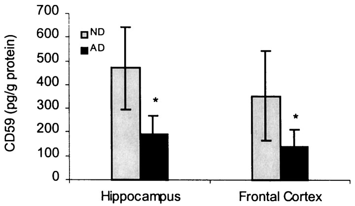
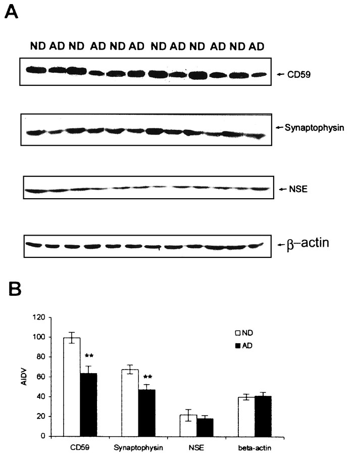
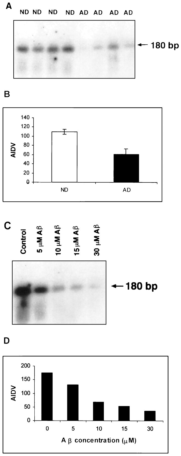
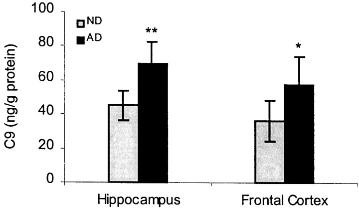
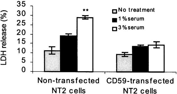
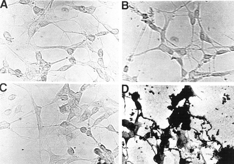
Complement defense 59 (CD59) is a cell surface glycophosphoinositol (GPI)-anchored protein that prevents complement membrane attack complex (MAC) assembly. Here, we present evidence from ELISA assays that CD59 protein levels are significantly decreased in the frontal cortex and hippocampus of Alzheimer's disease (AD) compared with nondemented elderly (ND) patients, whereas complement component 9, a final component to form MAC, is significantly increased. To further confirm the CD59 deficit, PI-specific phospholipase C (PIPLC) was used to cleave the CD59 GPI anchor at the cell surface in intact slices from AD and ND cortex. CD59 released by PIPLC cleavage was significantly reduced in AD compared with ND samples. By the use of a ribonuclease protection technique, amyloid beta-peptide was found to downregulate CD59 expression at the mRNA level, suggesting a partial explanation of CD59 deficits in the AD brain. To evaluate the pathophysiological significance of CD59 alterations in neurons, we exposed cultured NT2 cells, which normally underexpress CD59, and NT2 cells transfected to overexpress CD59 to homologous human serum. Lactic acid dehydrogenase assays revealed significant complement-induced cell lysis in CD59-underexpressing NT2 cells and significant protection from such lysis in CD59-overexpressing NT2 cells. Moreover, cells expressing normal levels of CD59 showed no evidence of MAC assembly or damage after exposure to homologous serum, whereas pretreatment of these cells with a CD59-neutralizing antibody resulted in MAC assembly at the cell surface and morphological damage. Taken together, these data suggest that CD59 deficits may play a role in the neuritic losses characteristic of AD.


Figures

Fig. 1.
Fig. 1.
Reduction of CD59 by ELISA in the AD and ND brains. The data represent the mean (± SE) quantification analysis of 20–22 cases using a specific ELISA assay showing that CD59 immunoreactivity was decreased by 52% in AD compared with ND (nondemented aged-matched controls). Results from three independent measurements displayed a significant difference between AD and ND samples (*p < 0.05).
Fig. 2.
Fig. 2.
Relationship of CD59 deficiency to neuronal or synaptic loss in AD brains. A, Western blot studies on CD59, synaptophysin, and neuron-specific enolase (NSE) in AD and ND brains are shown. B, Data represent the mean (± SD) quantification analysis using densitometry imaging (Chemimager 4000) showing that CD59 immunoreactivity was decreased by 39% in AD compared with ND, whereas synaptophysin was decreased ∼26%. There is no significant change inNSE expression levels. Results from three independent measurements displayed a significant difference between AD and ND samples (**p < 0.01). AIDV, Average integrated density value.
Fig. 3.
Fig. 3.
CD59 mRNA expression in the AD brain and Aβ-treated neurons by the use of the RNase protection assay. Total RNA was recovered from 0.5 gm of tissue or 2 × 10⁶ cells after stimulation with Aβ(1–42) for 16 hr. The RNAs were hybridized with the CD59 antisense probe. After treatment with RNase A and T1, protected bands were run through an 8% polyacrylamide gel, and the dried gel was exposed to x-ray film.A, CD59 mRNA from AD and ND brains is shown.C, CD59 mRNA expression is downregulated in a dose-dependent manner in Aβ(1–42)-treated cells. Data from both experiments show that CD59 at the mRNA level is decreased in AD brains and Aβ-treated neurotypic cells. B, D,Data represent quantitative analysis results from A andC, respectively, using Chemimager 4000.
Fig. 4.
Fig. 4.
C9 production in AD brains. Data represent the mean (± SE) quantification analysis of 20–22 cases using a specific ELISA assay showing that C9 immunoreactivity was increased by 35% in AD compared with ND. Results from repeated measurements displayed a significant difference between AD and ND samples (*p < 0.05; **p < 0.01).
Fig. 5.
Fig. 5.
Overexpression of CD59 protects neurons against cell lysis by complement. Human neuronal NT2 cells were transfected with human CD59 cDNA and then exposed to 1 and 3% human complement for 24 hr. Values represent the mean (± SE) of four separate determinations (**p < 0.01 when compared with transfected and nontransfected cells by Student's pairedt test).
Fig. 6.
Fig. 6.
Detection of MAC from human neuronal cells by silver-staining enhancement for peroxidase–DAB. A, Cells after supplementation with human C9-deficient serum yielded minimal MAC detection in cells expressing CD59. B–D,Anti-CD59 antibody treatment alone for 18 hr (B), Aβ(1–42) treatment alone for 18 hr (C), and C9 transfection plus C9-deficient serum treatment for 18 hr after CD59-anchored protein was blocked by anti-CD59 antibody (D) are shown.B, C, Thus, treatment with the antibody of CD59 or Aβ alone produced little MAC detection in cells expressing CD59.D, Conversely, when CD59-anchored protein was blocked by anti-CD59 antibody, cells overexpressing C9 with C9-deficient serum treatment show that MAC detection was prevalent.

References

    1. Eikelenboom P, Stam FC. Immunoglobulins and complement factors in senile plaques. Acta Neuropathol (Berl) 1982;57:239–242. - PubMed
    1. Gouras GK, Tsai J, Naslund J, Vincent B, Edgar M, Checler F, Greenfield JP, Haroutunian V, Buxbaum JD, Xu H, Greengard P, Relkin NR. Intraneuronal Abeta42 accumulation in human brain. Am J Pathol. 2000;156:15–20. - PMC - PubMed
    1. Itagaki S, Akiyama H, Saito H, McGeer PL. Ultrastructural localization of complement membrane attack complex (MAC)-like immunoreactivity in brains of patients with Alzheimer's disease. Brain Res. 1994;645:78–84. - PubMed
    1. Kim SH, Darney DF, Hammer CH, Shin ML. Nucleated cell killing by complement: effects of C5b-9 channel size and extracellular Ca2+ on the lytic process. J Immunol. 1987;138:1530–1536. - PubMed
    1. Masters CL, Simms G, Weiman NA, Multhaup G, McDonald BL, Beyreuther K. Amyloid plaque core protein in Alzheimer disease and Down syndrome. Proc Natl Acad Sci USA. 1985;82:4245–4249. - PMC - PubMed

Publication types

MeSH terms