. 2000 Oct 15;20(20):7622-30.
doi: 10.1523/JNEUROSCI.20-20-07622.2000.

Akt-mediated survival of oligodendrocytes induced by neuregulins

Affiliations

Akt-mediated survival of oligodendrocytes induced by neuregulins

A I Flores et al. J Neurosci. .

Abstract

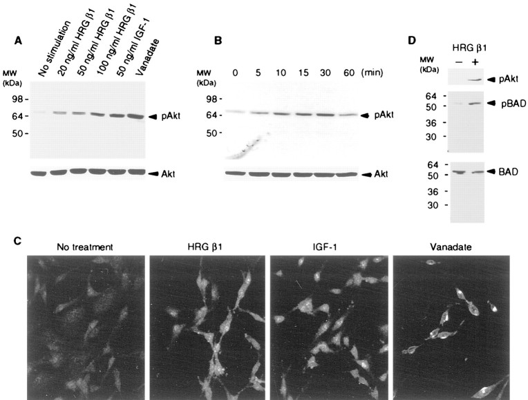
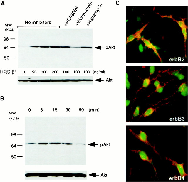
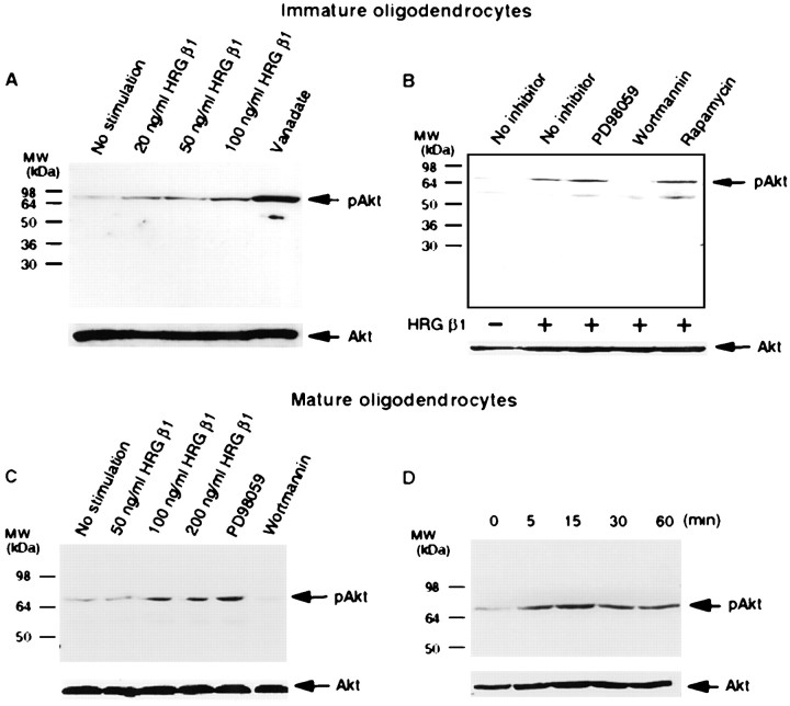
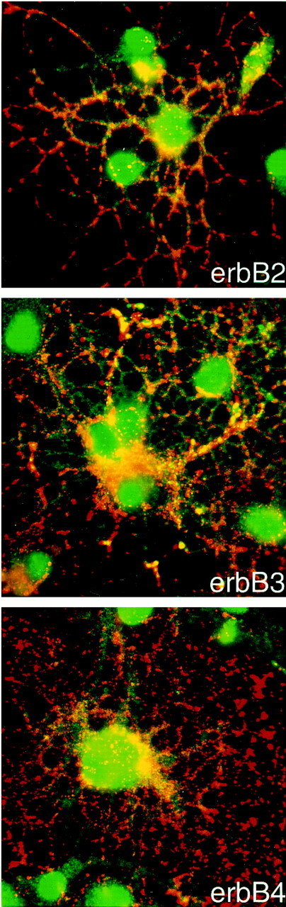
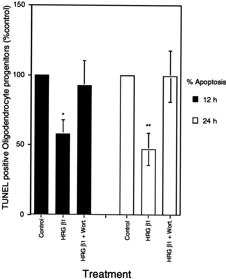
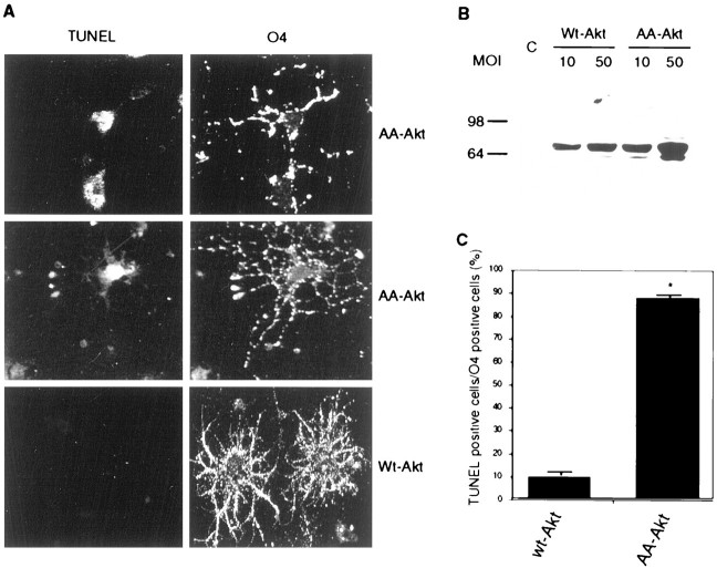
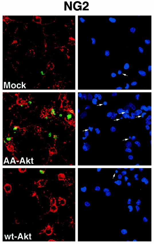
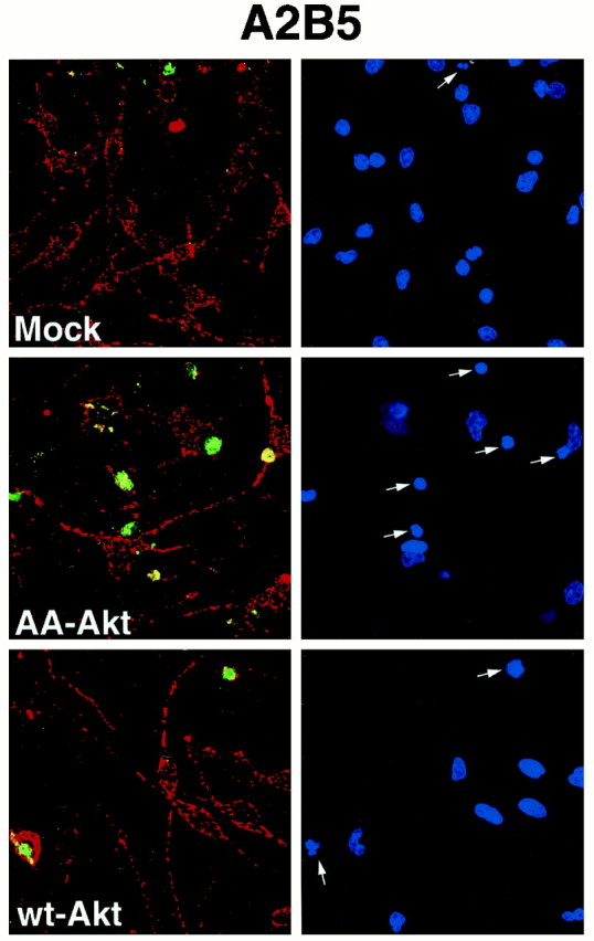
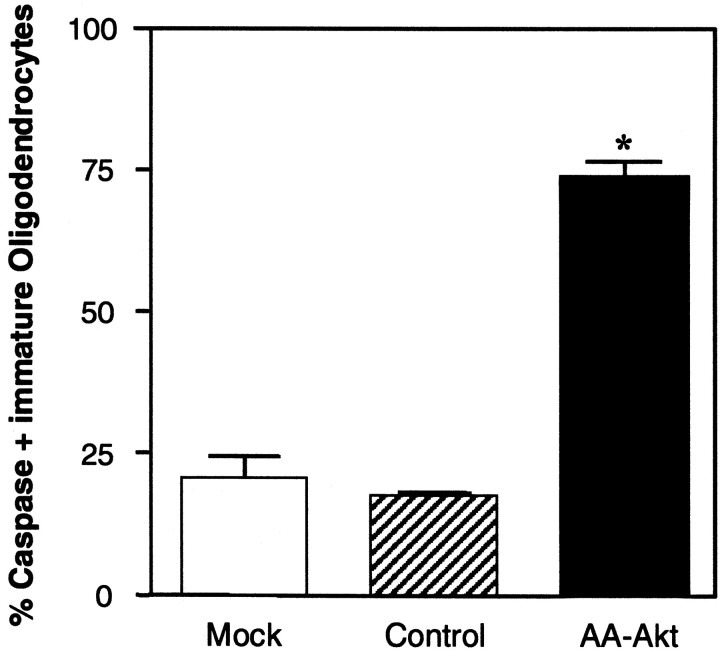
Neuregulins have been implicated in a number of events in cells in the oligodendrocyte lineage, including enhanced survival, mitosis, migration, and differentiation. At least two signaling pathways have been shown to be involved in neuregulin signaling: the phosphatidylinositol (PI)-3 kinase and the mitogen-activated protein kinase pathways. In the present studies, we examined the signaling pathway involved in the survival function of heregulin, focusing on heregulin-induced changes in Akt activity in cultured glial cells, and the consequences of Akt activation in cells in the oligodendrocyte lineage. Heregulin binds erbB receptors, and in our studies, primary cultures of both oligodendrocyte progenitor cells and differentiating oligodendrocytes expressed erbB2, erbB3, and erbB4 receptors. In C6 glioma cells and primary cultures of oligodendrocytes, heregulin induced time- and dose-dependent Akt phosphorylation at Ser(473) in a wortmannin-sensitive manner. To investigate further the signaling pathway for heregulin in glial cells, BAD was overexpressed in C6 glioma cells. In these cells, heregulin induced phosphorylation of BAD at Ser(136). Apoptosis of oligodendrocyte progenitor cells induced by growth factor deprivation was effectively blocked by heregulin in a wortmannin-sensitive manner. Overexpression of dominant negative Akt but not of wild-type Akt by adenoviral gene transfer in primary cultures of both oligodendrocytes and their progenitors induced significant apoptosis through activation of the caspase cascade. The present data suggest that the survival function of heregulin is mediated through the PI-3 kinase/Akt pathway in cells in the oligodendrocyte lineage and that the Akt pathway may be quite important for survival of cells in this lineage.


Figures

Fig. 1.
Fig. 1.
Effect of HRG β1 on Akt in C6 glioma cells. A, C6 glioma cells were treated with HRG β1, IGF-1, and pervanadate for 30 min. Cell lysates were subjected to immunoblot with anti-phospho Akt (Ser473) (top panel) or anti-total Akt (bottom panel). HRG β1 dose-dependently promoted phosphorylation of Akt.B, C6 glioma cells were treated with 100 ng/ml HRG β1 at different times, and Akt phosphorylation was assessed by immunoblot with anti-phospho Akt (Ser473) antibody (top panel) or anti-total Akt (bottom panel). HRG β1 promoted Akt phosphorylation in a time-dependent manner.C, C6 glioma cells were treated with HRG β1, IGF-1, or pervanadate for 30 min and subjected to immunocytochemistry with anti-phospho Akt (Ser473) antibody. HRG β1 induced phosphorylation of Akt in a similar distribution and intensity in cells to that of IGF-1. Pervanadate induced more intense phosphorylation, which was localized in a rather discrete region inside the cells.D, GST-BAD cDNA was transfected into C6 glioma cells, and 48 hr later the cells were treated with 100 ng/ml HRG β1 for 30 min. Cells were subjected to immunoblot with anti-phospho-Ser473 specific Akt (top panel), anti-phospho-Ser136 specific BAD (middle panel), anti-BAD antibodies (bottom panel). HRG β1 promoted BAD phosphorylation at Ser136.
Fig. 2.
Fig. 2.
Effect of HRG β1 on Akt in primary cultures of oligodendrocyte progenitors. A, Primary cultures of oligodendrocyte progenitor cells were treated with different concentrations of HRG β1 for 30 min or they were pretreated with different kinase inhibitors (20 μm PD098059, 200 nm wortmannin, and 20 ng/ml rapamycin), and then stimulated for an additional 30 min with 100 ng/ml HRG β1. Cell lysates were subjected to immunoblot with anti-phospho Akt (Ser473) (top panel) and anti-total Akt (bottom panel). HRG β1 dose-dependently promoted phosphorylation of Akt in primary cultures of oligodendrocytes progenitors, which was selectively blocked by wortmannin. B, Oligodendrocytes progenitors were treated with 100 ng/ml HRG β1 at different times, and Akt phosphorylation was assessed by immunoblot with anti-phospho Akt (Ser473) antibody (top panel) or anti-total Akt (bottom panel). HRG β1 promoted Akt phosphorylation in a time-dependent manner. C, Immunofluorescence microscopy of oligodendrocyte progenitor cultures double-stained with anti-A2B5 (red) and anti-erbB2, anti-erbB3, or anti-erbB4 antibodies (green) is shown. Oligodendrocytes progenitors express erbB2, erbB3, and erbB4 receptors.
Fig. 3.
Fig. 3.
Effect of HRG β1 on Akt in primary cultures of immature and mature oligodendrocytes. A, Primary cultures of immature oligodendrocytes were treated with different concentrations of HRG β1 or vanadate for 30 min. Cell lysates were subjected to immunoblot with anti-phospho Akt (Ser473) (top panel) or anti-total Akt (bottom panel). HRG β1 dose-dependently promoted phosphorylation of Akt.B, Primary cultures of immature oligodendrocytes were pretreated with various protein kinase inhibitors (20 μmPD098059, 200 nm wortmannin, 20 ng/ml rapamycin) for 30 min and then stimulated with 50 ng/ml HRG β1 for an additional 30 min. Cells lysates were subjected to immunoblot with anti-phospho Akt (Ser473) (top panel) or anti-total Akt (bottom panel). HRG β1-induced Akt phosphorylation was selectively blocked by wortmannin. C, Mature oligodendrocytes were treated with different concentrations of HRG β1 for 30 min or pretreated with different kinase inhibitors (20 μm PD098059, 200 nm wortmannin) and then stimulated with 100 ng/ml HRG β1 for an additional 30 min.D, Mature oligodendrocytes were treated with 100 ng/ml HRG β1 at different times, and Akt phosphorylation was assessed by immunoblot with anti-phospho Akt (Ser473) antibody (top panel) or anti-total Akt (bottom panel). HRG β1 promoted Akt phosphorylation in a time-dependent manner in mature oligodendrocytes.
Fig. 4.
Fig. 4.
Mature oligodendrocytes express erbB2, erbB3, and erbB4 receptors. Mature oligodendrocytes were obtained after incubation in CNTF/T3 as described in Materials and Methods. Immunofluorescence microscopy of mature oligodendrocyte cultures double-stained with anti-O4 (red) and anti-erbB2, anti-erbB3, or anti-erbB4 antibodies (green) is shown. Note stain for erbB receptors on processes of oligodendrocytes, in addition to cell body stain.
Fig. 5.
Fig. 5.
HRG β1 maintains cell survival of oligodendrocyte progenitors. Immunopanned oligodendrocyte progenitors were plated in PDGF/bFGF for 2 d and switched to DMEM without any growth factors for 12 or 24 hr (control), with 150 ng/ml HRG β1 with or without a 30 min pretreatment with wortmannin. Apoptotic cells were detected by TUNEL staining and FACS analysis. In each experiment, the number of TUNEL-positive cells was determined for each condition. The absolute number of TUNEL-positive cells varied in different experiments, reaching as much as 50% in some experiments. To combine data from multiple experiments, the number of TUNEL-positive cells in control cultures was therefore defined as 100%. The number of TUNEL-positive oligodendrocyte progenitors in each experimental condition was then expressed as a percentage of control values. Each bar represents the mean ± SEM of five different experiments (*p < 0.0267; **p < 0.0198).
Fig. 6.
Fig. 6.
Dominant-negative Akt induced apoptosis in primary cultures of immature oligodendrocytes. Adenoviruses encoding wild-type Akt or a dominant-negative Akt were used to express recombinant proteins in primary cultures of immature oligodendrocytes. Adenovirus infection was performed on oligodendrocyte precursors 24 hr after incubation in PDGF/T3. A, Cells were subjected to O4 staining and TUNEL analysis 72 hr after infection. The wild-type Akt infection did not induce apoptosis (bottom panel), but dominant-negative Akt infection did induce apoptosis (top and middle panels). B, Expression of wild-type Akt and dominant-negative Akt was assessed by immunoblot with anti-HA antibody; 50 MOI for wild-type Akt and 10 MOI for dominant-negative Akt showed similar levels of Akt expression. Thus, 50 MOI of wild-type Akt and 10 MOI of dominant-negative Akt were used for TUNEL experiments in A. Lysate of cells infected with adenovirus encoding green fluorescent protein was used for control (C). C, O4-positive cells were examined randomly in 20 different fields. In total, 241 cells were examined for dominant-negative Akt virus infection and 171 cells for wild-type Akt virus infection. TUNEL positivity is shown as a percentage of the total number of O4-positive cells. Dominant-negative Akt infection caused apoptosis in O4-positive oligodendrocytes at a significantly higher rate compared to wild-type Akt infection (*p < 0.001).
Fig. 7.
Fig. 7.
Dominant-negative Akt induced caspase activation in primary cultures of NG2-positive oligodendrocyte progenitors. Oligodendrocyte progenitors were mock-infected or infected with adenovirus expressing wt-Akt (50 MOI) or AA-Akt (10 MOI) for 24 hr as described in Materials and Methods. After infection, cells were subjected to caspase activation analysis (green) and NG2 staining (red). Condensed nuclei were detected by DAPI staining (indicated by arrows). Representative data from one of three independent experiments is shown.
Fig. 8.
Fig. 8.
Dominant-negative Akt induced caspase activation in primary cultures of A2B5-positive oligodendrocyte progenitors. Oligodendrocyte progenitors were mock-infected or infected with adenovirus expressing wt-Akt (50 MOI) or AA-Akt (10 MOI) for 24 hr as described in Materials and Methods. After infection, cells were subjected to caspase activation analysis (green) and A2B5 staining (red). Condensed nuclei were detected by DAPI staining (indicated by arrows). Representative data from one of three independent experiments is shown.
Fig. 9.
Fig. 9.
Dominant-negative Akt induced caspase activation in primary cultures of immature oligodendrocytes in the presence of HRG β1. Oligodendrocyte progenitor cells were mock-infected or infected with 10 MOI of a control adenovirus (Ad.RSV.alphos) or an adenovirus expressing AA-Akt the presence of 150 ng/ml HRG β1. Oligodendrocyte precursors were grown in the presence of PDGF/bFGF for 24 hr; growth factors were then removed, HRG β1 was added to the media, and adenovirus infection was performed. After 48 hr infection in the presence of HRG β1, the virus was removed, and cells were cultured in DMEM plus HRG β1 for an additional 20–24 hr before caspase inhibitor staining. A total of 1000 cells were counted, and the caspase-positive cells were expressed as a percentage of total number of cells visualized by DAPI staining. Data are the mean of a representative experiment made in duplicate. Data are shown as mean ± SEM (n = 2); *p < 0.05.

References

    1. Andjelkovic M, Alessi DR, Meier R, Fernandez A, Lamb NJC, Frech M, Cron P, Cohen P, Lucocq JM, Hemmings BA. Role of translocation in the activation and function of protein kinase B. J Biol Chem. 1997;272:31515–31524. - PubMed
    1. Baek SY, Kim SU. Proliferation of human Schwann cells induced by neu differentiation factor isoforms. Dev Neurosci. 1998;20:512–517. - PubMed
    1. Barres BA, Hart IK, Coles HSR, Burne JF, Voyvodic JT, Richardson WD, Raff MC. Cell death and control of cell survival in the oligodendrocyte lineage. Cell. 1992;70:31–46. - PubMed
    1. Basu S, Bayoumy S, Zhang Y, Lozano J, Kolesnick R. BAD enables ceramide to signal apoptosis via Ras and Raf-1. J Biol Chem. 1998;273:30419–30426. - PubMed
    1. Burden S, Yarden Y. Neuregulins and their receptors: a versatile signaling module in organogenesis and oncogenesis. Neuron. 1997;18:847–855. - PubMed

Publication types

MeSH terms