. 2000 Oct 15;20(20):7664-71.
doi: 10.1523/JNEUROSCI.20-20-07664.2000.

Facial visceral motor neurons display specific rhombomere origin and axon pathfinding behavior in the chick

Affiliations

Facial visceral motor neurons display specific rhombomere origin and axon pathfinding behavior in the chick

J Jacob et al. J Neurosci. .

Abstract

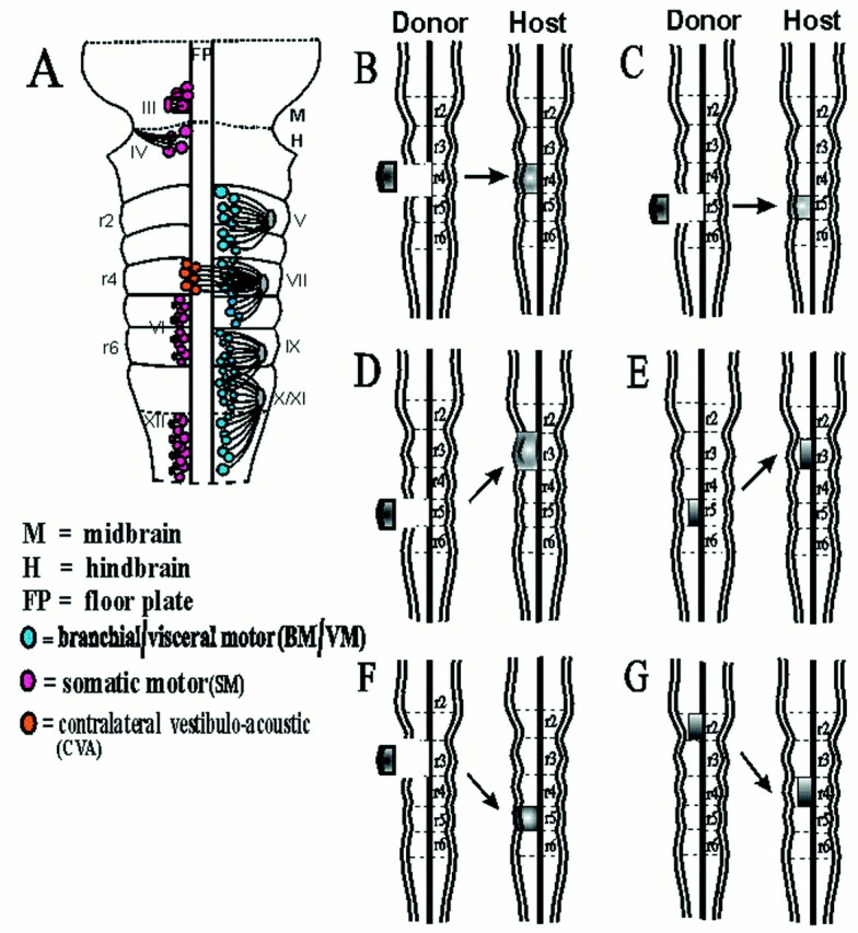
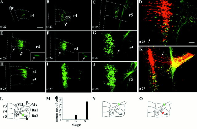
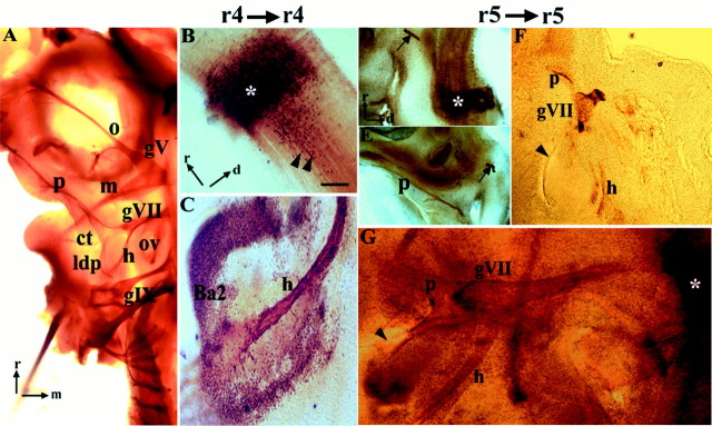
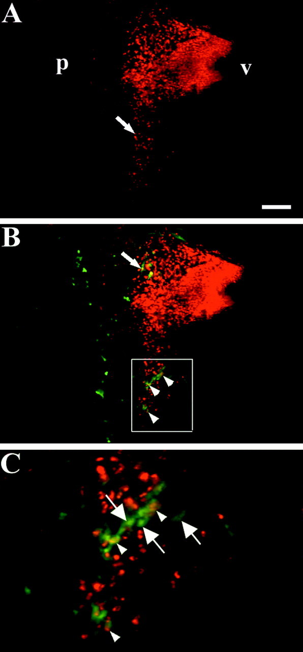
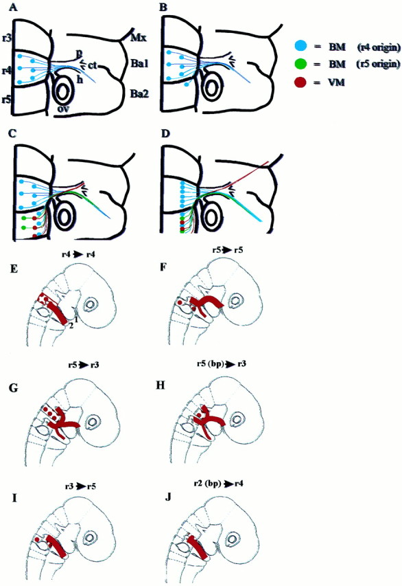
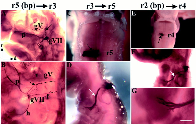
In the chick embryo, facial motor neurons comprise branchiomotor and visceral motor subpopulations, which innervate branchial muscles and parasympathetic ganglia, respectively. Although facial motor neurons are known to develop within hindbrain rhombomere 4 (r4) and r5, the precise origins of branchiomotor and visceral motor neuron subpopulations are unclear. We investigated the organization and axon pathfinding of these motor neurons using axonal tracing and rhombomere transplantation in quail-chick chimeras. Our results show that a large majority of branchiomotor neurons originate in r4 but that a cohort of these neurons undergoes a caudal migration from r4 into r5. By contrast, visceral motor neurons develop exclusively in r5. We found that a striking property of facial visceral motor neurons is the ability of their axons to navigate back to appropriate ganglionic targets in the periphery after heterotopic transplantation. These results complement previous studies in which heterotopic facial branchiomotor neurons sent axons to their correct, branchial arch, target. By contrast, when trigeminal branchiomotor neurons were transplanted heterotopically, we found that they were unable to pathfind correctly, and instead projected to an inappropriate target region. Thus, facial and trigeminal motor neuron populations have different axon pathfinding characteristics.


Figures

Fig. 1.
Fig. 1.
Distribution of motor neurons in the hindbrain and summary of rhombomere transplants. A, Diagram of the ventral aspect of a stage 21 chick embryo showing the motor nuclei of the branchial nerves (V, trigeminal; VII,facial; IX, glossopharyngeal; and X/XI,vagus/cranial accessory) and the somatic motor nuclei (III, oculomotor; IV, trochlear;VI, abducens; and XII, hypoglossal) based on axon tracing (modified after Lumsden, 1990). B–G,Diagrams of orthotopic and heterotopic transplants from donor quail embryos to host chick embryos: r4 orthotopic (B), r5 orthotopic (C), r5 to r3 (D), r5 basal plate to r3 (E), r3 to r5 (F), and r2 basal plate to r4 (G).
Fig. 2.
Fig. 2.
Retrograde labeling of motor neurons in r4 and r5 over a series of developmental stages. A–K, Ventral views of flat-mounted hindbrains showing retrograde labeling of facial BM and/or VM neurons. A, B, E, F, H, I, L, M, Labeling of BM neurons after fluorescein-dextran fills of the hyoid nerve (L, greenbar) at st. 22 (A), st. 23 (B), st. 24 (E, F), st. 25 (H), and st. 27 (I). Between three and five embryos were labeled at each stage. Note the presence of BM neurons in r5 from stage 23 onward. Many of the BM neurons in r5 occupy lateral positions (B, E, arrowheads), whereas others lie closer to the floor plate in a medial position (E, F, arrows). Higher power views of these medially positioned BM neurons are shown in the insets in E andF. C, G, J, Labeling of VM neurons after fluorescein-dextran fills of the palatine nerve (N, green bar) at st. 25 (C), st. 27 (G), and st. 28 (J). Four or five embryos were labeled at each stage. Note the curved trajectory of VM axons (G, arrow). D, K,Double-labeling of BM and VM subpopulations after fluorescein-dextran fills of the palatine nerve (O, green bar) and rhodamine-dextran fills of the hyoid nerve (O, red bar) at st. 25 (D) (n = 3) and st. 27 (K) (n = 5).D, Within r5 a single BM neuron in a medial position has been labeled (arrow), but the majority of facial motor neurons in r5 are located laterally. K, Motor neuron cell bodies at st. 27 occupy the same mediolateral position. The straight or slightly curved axon trajectories of r4 BM neurons are clearly seen (arrow). Presence of apparently double-labeled cells (yellow) in Dand K is an artifact caused by the superposition of labeled neurons at different depths when generating the confocalz-series shown here. Double-labeled cells were never observed in any single focal plane at all depths (data not shown).L, N, O, Schematics of ventral aspect of facial nerve and hindbrain indicating dextran labeling, and quantification of the mean (+SEM) number of BM neurons labeled in r5 at st. 22, 23, and 24 (M). Scale bar (in A):A, B, C, E, F, H, 100 μm. Scale bar (inD): G, I–K, 50 μm. fp,Floor plate; ep, exit point; gVII,geniculate ganglion; ov, otic vesicle; p,palatine nerve; ct, chorda tympani; h,hyoid nerve; Mx, maxilla; Ba1, first branchial arch; Ba2, second branchial arch.Dashed lines in A–C, E, F, andH indicate margins of floor plate and rhombomere boundaries.
Fig. 3.
Fig. 3.
Quail-to-chick orthotopic grafts of r4 and r5.A–G, Anti-neurofilament immunohistochemistry of control (A) and QCPN–QN immunohistochemistry of chimeric (B–G) embryos analyzed at st. 25–30.A, Ventral view of flat-mounted control chick embryo showing the pathways of the branchial nerves V,trigeminal; VII, facial; and IX,glossopharyngeal. Rostral (r) is to thetop, and medial (m) is to theright. B, C, Parasagittal vibratome sections of a chimera with an orthotopic graft of r4. B,Hindbrain with grafted rhombomere (asterisk). Note that many quail cells from the graft have migrated posteriorly (black arrowheads). Rostral and dorsal (d) are indicated by orthogonal arrows. C, Quail axons from motor neurons in r4 innervate the muscle plate of the second branchial arch, which contains quail neural crest cells in the peripheral region. D–G, Parasagittal vibratome sections (D–F) and flat-mount (G) of chimeras with an orthotopic graft of r5. D, Quail graft is visible (asterisk) along with QN+ axons of the ventrally projecting abducens nerve (arrow). Orientation is the same in D–F.E–G, Three examples of the axonal projection patterns of motor neurons in r5. E, In this embryo, motor axons project along the palatine pathway of the facial nerve. An abducens nerve is also visible (arrow). F, This embryo has a palatine nerve and a hyoid projection that terminates abruptly at the level of the first branchial cleft (arrowhead), between the first and second branchial arches. QN+ axons do not reach the core of the second branchial arch (data not shown). G, Ventral view of a st. 25 embryo showing a palatine nerve and a hyoid nerve that terminates proximal to the second branchial arch and caudal to the first branchial cleft (arrowhead). The graft is marked with an asterisk. Scale bar: A, 400 μm;B, C, 100 μm; and D–G, 250 μm. gV, Trigeminal ganglion; gIX,glossopharyngeal ganglion; o, m,ophthalmic and mandibular divisions of trigeminal nerve;ldp, lesser deep petrosal branch of glossopharyngeal nerve.
Fig. 4.
Fig. 4.
Retrograde labeling of the hyoid nerve in a chimera with an orthotopic graft of r4. A–C,Parasagittal vibratome sections through the hindbrain of a st. 26 embryo with an orthotopic r4 graft, after a fluorescein-dextran (green) fill of the hyoid nerve and whole-mount QCPN immunohistochemistry (red). A, Quail cells (red) that have migrated posteriorly from r4 into r5 are clearly seen near the pial surface (arrow).B, QCPN+–dextran-labeled cells are quail BM neurons that are present in r4 (arrow) and r5 (arrowheads). C, Higher power view of theboxed area in B to show quail BM neurons (yellow or redcenter and green surround) in r5 that have migrated posteriorly from r4 (arrowheads). Also visible are QCPN–dextran-labeled endogenous chick BM neurons (arrows) that originate in r5.p, Pial surface; v, ventricular surface. Scale bar: A, B, 100 μm;C, 25 μm.
Fig. 5.
Fig. 5.
Summary diagram of the temporal order of development of motor pathways of the facial nerve in the chick, based on retrograde axon tracing and orthotopic grafts. A, St. 20–22: all BM neurons are confined to r4 and send their axons into the hyoid nerve. B, St. 23: BM neurons are also identified in r5. C, St. 25: BM neurons born in r5 extend axons along the hyoid pathway, and VM neurons in r5 project axons to form the anlage of the palatine nerve. D, St. 27: Cell bodies of BM and VM neurons occupy the same mediolateral position. The palatine nerve has extended to the maxilla to innervate the target ganglia. For abbreviations, see Figure 2 legend. E–J, Motor pathways that result from orthotopic (E, F) and heterotopic (G–J) quail to chick grafts.E, r4 orthotopic grafts: quail axons grow into the second branchial arch. 1 and 2 point out first and second branchial arches. F, r5 orthotopic grafts: quail axons grow into the palatine branch and into the second branchial arch. G, H, r5 to r3 grafts (G,whole rhombomere; H, basal plate graft only): quail axons that exit via the r2 dorsal exit point reroute to join the facial nerve pathway at the geniculate ganglion. Other quail axons exit the hindbrain via the exit point in r4. Axons are distributed in the palatine and hyoid nerves en route to the correct targets. For clarity, axons that traverse the trigeminal nerve in a small minority of chimeras have been omitted. I, r3 to r5 grafts: axons project to an incorrect target, the second branchial arch.J, r2 (basal plate) to r4 grafts: axons project to an incorrect target, the second branchial arch. For abbreviations, see Figure 2 legend.
Fig. 6.
Fig. 6.
BM and VM axon pathfinding after heterotopic quail-to-chick transplants. A–G, QCPN and QN immunohistochemistry in embryos at st. 25–30 after heterotopic grafts of r5 basal plate to r3 (A, B), r3 to r5 (C, D), and r2 basal plate to r4 (E–G). A, B, Ventral views of two flat-mounted embryos showing ectopic QN+ axon tract (A, B, black arrows) projecting from the trigeminal nerve exit point in r2 to the geniculate ganglion. InB, QN+ axons also exit the hindbrain via the exit point in r4 and project to the geniculate ganglion in the facial nerve (f). A palatine nerve is recognizable in both cases and has bifurcated in B to supply both the sphenopalatine and ethmoidal ganglia (data not shown). Two other motor pathways additionally present in B are a hyoid nerve and mandibular projection (arrowhead), which is at the limit of detectability. In B, the connection of the ectopic bundle and facial nerve to the hindbrain was severed during flat-mounting, as indicated by black dashed lines. The grafted rhombomeres (data not shown) resembled the basal plate graft shown in E. C, D, Dorsal (C) and lateral (D) views of whole-mount embryo showing, respectively, the graft (C, black arrow) and axonal projection of trigeminal BM neurons to the second branchial arch (D, white arrow).E–G, Dorsal (E) and lateral (F, G) views of whole-mount embryo showing the grafted basal plate (E, black arrow), the projection of trigeminal BM axons to the second branchial arch (F, white arrow), and, at higher power, the second branchial arch, which is devoid of quail neural crest cells (G). Scale bar (in G): C, G, 200 μm; A, E, 300 μm; B, 500 μm; F, 1200 μm; and D, 1600 μm.

References

    1. Auclair F, Valdes N, Marchand R. Rhombomere-specific origin of branchial and visceral motoneurones of the facial nerve in the rat embryo. J Comp Neurol. 1996;369:451–461. - PubMed
    1. Bell E, Wingate RJ, Lumsden A. Homeotic transformation of rhombomere identity after localized Hoxb1 misexpression. Science. 1999;284:2168–2171. - PubMed
    1. Caton A, Hacker A, Naeem A, Livet J, Maina F, Klein R, Birchmeier C, Guthrie S. The branchial arches and HGF are growth-promoting and chemoattractant for cranial motor axons. Development. 2000;127:1751–1766. - PubMed
    1. D'Amico-Martel A, Norden DM. Contributions of placodal and neural crest cells to avian cranial peripheral ganglia. Am J Anat. 1983;166:445–468. - PubMed
    1. Fritzsch B, Nichols DH. DiI reveals a prenatal arrival of efferents at the differentiating otocyst of mice. Hear Res. 1993;65:51–60. - PubMed

Publication types

MeSH terms

LinkOut - more resources