. 2000 Oct 15;20(20):7760-5.
doi: 10.1523/JNEUROSCI.20-20-07760.2000.

Hypocretin-1 modulates rapid eye movement sleep through activation of locus coeruleus neurons

Affiliations

Hypocretin-1 modulates rapid eye movement sleep through activation of locus coeruleus neurons

P Bourgin et al. J Neurosci. .

Abstract

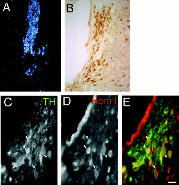
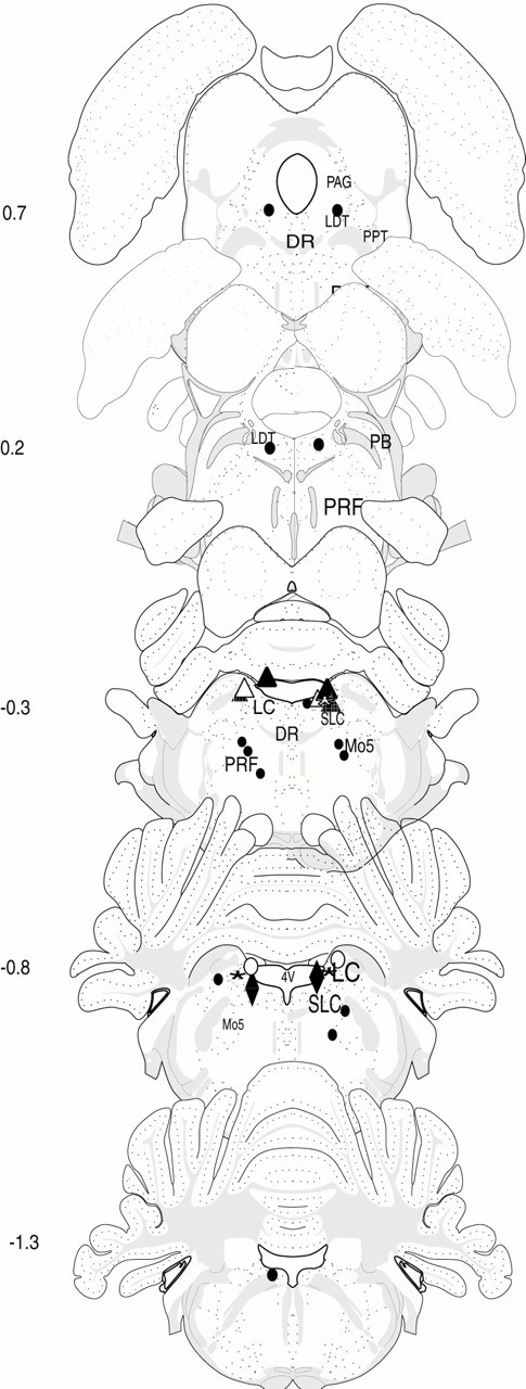
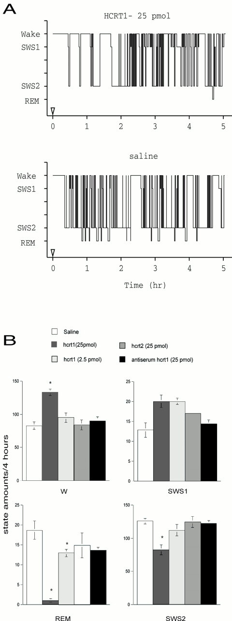
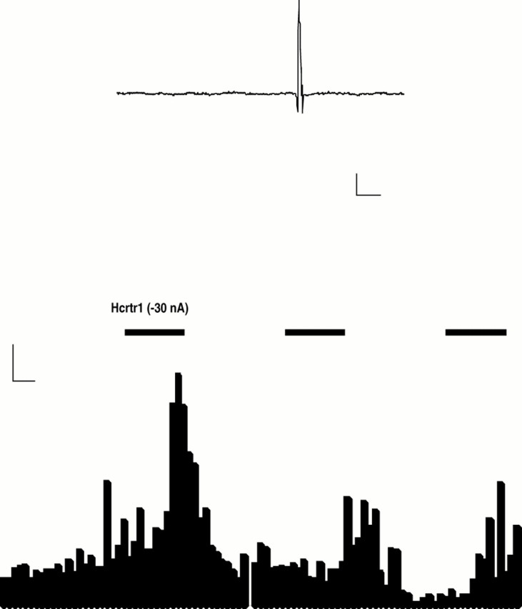
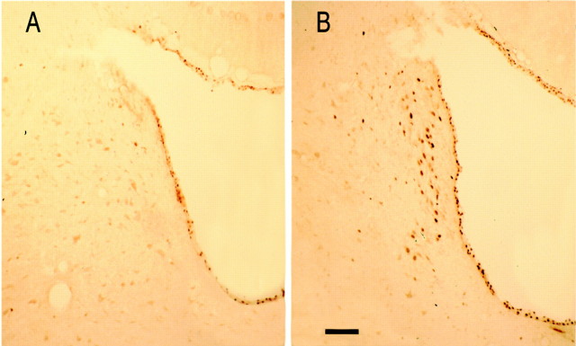
The hypocretins (hcrts), also known as orexins, are two recently identified excitatory neuropeptides that in rat are produced by approximately 1200 neurons whose cell bodies are located in the lateral hypothalamus. The hypocretins/orexins have been implicated in the regulation of rapid eye movement (REM) sleep and the pathophysiology of narcolepsy. In the present study, we investigated whether the locus coeruleus (LC), a structure receiving dense hcrtergic innervation, which is quiescent during REM sleep, might be a target for hcrt to regulate REM sleep. Local administration of hcrt1 but not hcrt2 in the LC suppressed REM sleep in a dose-dependent manner and increased wakefulness at the expense of deep, slow-wave sleep. These effects were blocked with an antibody that neutralizes hcrt binding to hcrt receptor 1. In situ hybridization and immunocytochemistry showed the presence of hcrt receptor 1 but not the presence of hcrt receptor 2 in the LC. Iontophoretic application of hcrt1 enhanced the firing rate of LC neurons in vivo, and local injection of hcrt1 into the LC induced the expression of c-fos in the LC area. We propose that hcrt receptor 1 in the LC is a key target for REM sleep regulation and might be involved in the pathophysiological mechanisms of narcolepsy.


Figures

Fig. 1.
Fig. 1.
Localization of hcrtr1 in the LC.A, Dark-field photomicrograph of the LC hybridized with an hcrtr1 riboprobe. B, Immunocytochemical detection of hcrtr1. Note that most cells in the LC are labeled with the antibody. Scale bar, 50 μm. C–E, Immunofluorescence micrographs showing overlap (yellow, arrows) between hcrtr1-positive (red) and TH-immunoreactive (green) neurons in the LC region. Scale bar, 25 μm.
Fig. 2.
Fig. 2.
Schematic representation of hcrt injection sites. Drawings show coronal sections at five different levels of the brainstem (interaural, −1.3 to +0.7; Swanson, 1992). The black circles represent sites for which no changes in the sleep–wakefulness cycle were observed after injection. Thetriangles and diamonds correspond to the positive sites (at least one site is located in the LC) for which significant changes in W, SWS2, and REM sleep were observed after bilateral infusion of the 25 pmol of hcrt1. For each rat the bilateral sites are represented by a triangle or adiamond and one color. One rat received unilateral injections (red triangle).Mo5, Motor trigeminal nucleus; SLC, subcoeruleus nucleus; PRF, pontine reticular formation;PB, parabrachial nucleus; DR, dorsal raphe; PPT, pedunculopontine tegmentum;PAG, periacqueductal central gray.
Fig. 3.
Fig. 3.
Effects of hcrt1 and hcrt2 on the amounts of W, SWS1, SWS2, and REM sleep after microinjection into the LC.A, Bars represent the amounts of the states of vigilance (mean ± SEM) expressed in minutes (saline,n = 7; hcrt1, 25 pmol, n = 7; hcrt1, 2.5 pmol, n = 5; hcrt2, 25 pmol,n = 4; hcrt1, 25 pmol, + antiserum,n = 4). *Significant difference (p < 0.05, Fisher's exact test) from baseline (rats treated with saline; open bars).B, Representative hypnographs obtained from one rat during 5 hr after the infusion of the vehicle (bottom) and 25 pmol of hcrt1 (top) into the right LC.
Fig. 4.
Fig. 4.
Activation of hypocretin 1 receptors increases the firing rate of locus coeruleus neurons. Top tracing, Filtered recorded signal demonstrating the waveforms of the action potential. Calibration: 100 μV, 5 msec. Bottom, Rate meter record demonstrating that in situmicroelectrophoretic application of hypocretin 1 (1 mg/ml) produced a marked activation of a spontaneously active LC neuron. Calibration: 5 Hz, 5 sec.
Fig. 5.
Fig. 5.
Injection of hcrt1 stimulates c-fosimmunoreactivity in the LC. Photomicrographs show c-fosimmunoreactivity in the LC area after injection of saline (A) or iontophoretic administration of hcrt1 (B). Scale bar, 100 μm.

References

    1. Ahnaou A, Basille M, Gonzalez B, Vaudry H, Hamon M, Adrien J, Bourgin P. Long-term enhancement of REM sleep by the pituitary adenylyl cyclase-activating polypeptide (PACAP) in the pontine reticular formation of the rat. Eur J Neurosci. 1999;11:4051–4058. - PubMed
    1. Aston-Jones G, Bloom FE. Activity of norepinephrine-containing locus coeruleus neurons in behaving rats anticipates fluctuations in the sleep-waking cycle. J Neurosci. 1981;1:876–886. - PMC - PubMed
    1. Berridge CW, Foote SL. Effects of locus coeruleus activation on electroencephalographic activity in neocortex and hippocampus. J Neurosci. 1991;11:3135–3145. - PMC - PubMed
    1. Caballero A, De Andres I. Unilateral lesions in locus coeruleus area enhance paradoxical sleep. Electroencephalogr Clin Neurophysiol. 1986;64:339–346. - PubMed
    1. Chemelli RM, Willie JT, Sinton CM, Elmquist JK, Scammell T, Lee C, Richardson JA, Williams SC, Xiong Y, Kisanuki Y, Fitch TE, Nakazato M, Hammer RE, Saper CB, Yanagisawa M. Narcolepsy in orexin knockout mice: molecular genetics of sleep regulation. Cell. 1999;98:437–451. - PubMed

Publication types

MeSH terms

LinkOut - more resources