. 2000 Nov;20(21):7867-80.
doi: 10.1128/MCB.20.21.7867-7880.2000.

Nckbeta adapter regulates actin polymerization in NIH 3T3 fibroblasts in response to platelet-derived growth factor bb

Affiliations

Nckbeta adapter regulates actin polymerization in NIH 3T3 fibroblasts in response to platelet-derived growth factor bb

M Chen et al. Mol Cell Biol. 2000 Nov.

Abstract

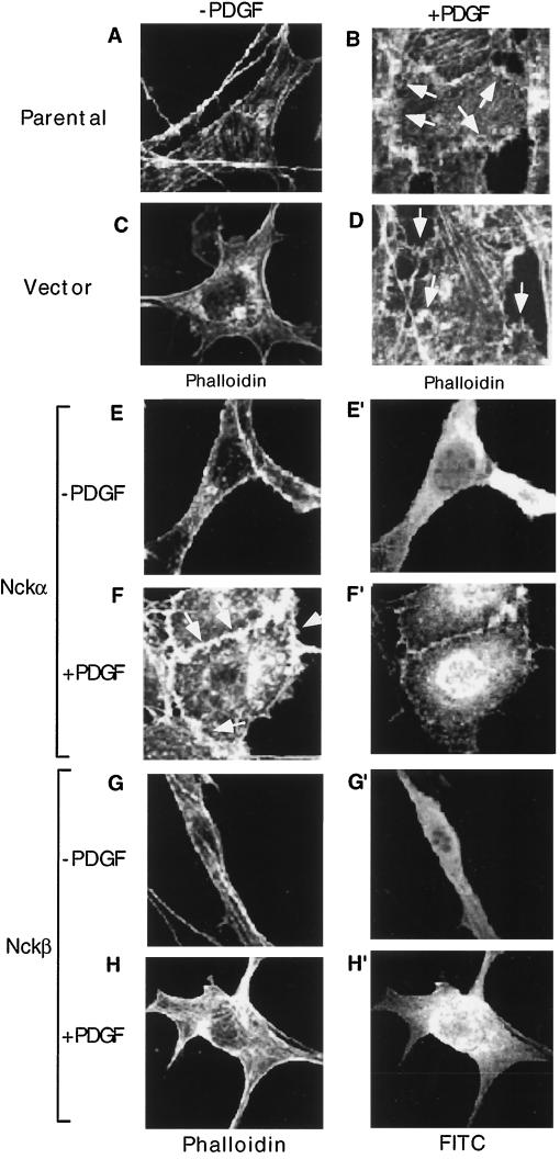
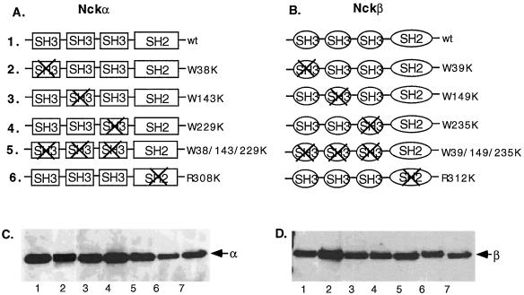
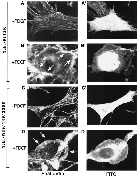
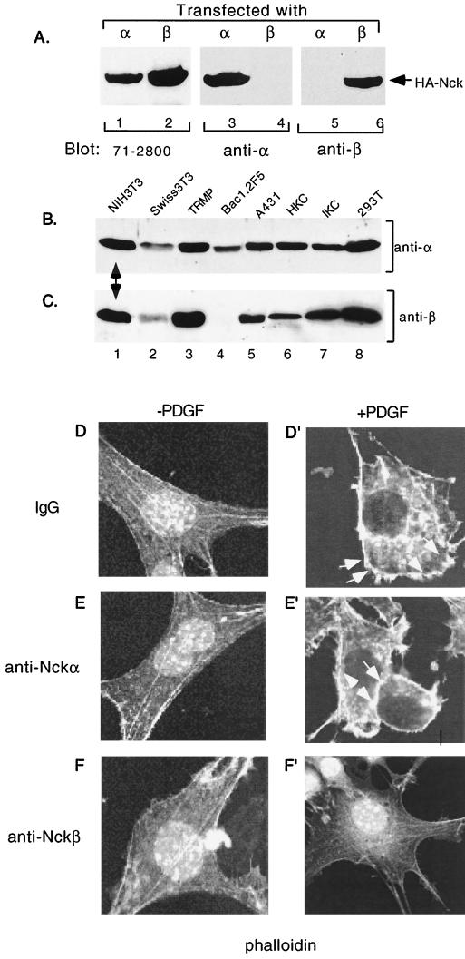
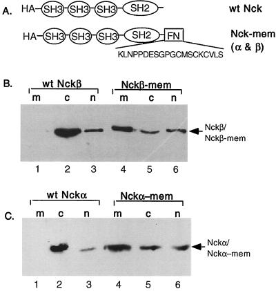
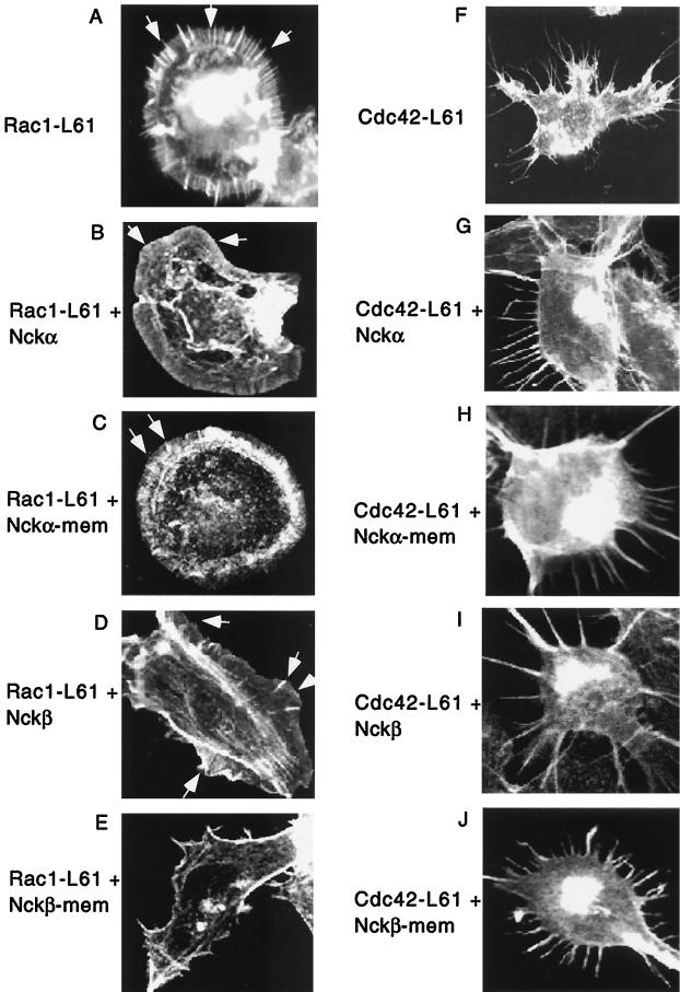
The SH3-SH3-SH3-SH2 adapter Nck represents a two-gene family that includes Nckalpha (Nck) and Nckbeta (Grb4/Nck2), and it links receptor tyrosine kinases to intracellular signaling networks. The function of these mammalian Nck genes has not been established. We report here a specific role for Nckbeta in platelet-derived growth factor (PDGF)-induced actin polymerization in NIH 3T3 cells. Overexpression of Nckbeta but not Nckalpha blocks PDGF-stimulated membrane ruffling and formation of lamellipoda. Mutation in either the SH2 or the middle SH3 domain of Nckbeta abolishes its interfering effect. Nckbeta binds at Tyr-1009 in human PDGF receptor beta (PDGFR-beta) which is different from Nckalpha's binding site, Tyr-751, and does not compete with phosphatidylinositol-3 kinase for binding to PDGFR. Microinjection of an anti-Nckbeta but not an anti-Nckalpha antibody inhibits PDGF-stimulated actin polymerization. Constitutively membrane-bound Nckbeta but not Nckalpha blocks Rac1-L62-induced membrane ruffling and formation of lamellipodia, suggesting that Nckbeta acts in parallel to or downstream of Rac1. This is the first report of Nckbeta's role in receptor tyrosine kinase signaling to the actin cytoskeleton.


Figures

FIG. 1
FIG. 1
Overexpression of Nckβ but not Nckα blocks PDGF-stimulated membrane ruffling. NIH 3T3 cells, cultured in fibronectin-coated (10 μg/ml, 2 h) eight-chamber culture slides, were either untransfected (A and B), transfected with vector alone mixed with a GFP-containing vector (C and D), or transfected with the wild-type Nckα (E to F′) or Nckβ (G to H′) construct at 0.5 μg/well. After 48 h, cells were starved in low-serum medium for an additional 18 h and treated (B, D, F, and H) or not (A, C, E, and G) with PDGF-bb (100 ng/ml) at 37°C for 15 min. Expression of the Nck proteins was monitored by anti-HA antibody blotting, followed by a secondary antibody conjugated with FITC. The actin cytoskeleton was revealed by rhodamine-labeled phalloidin staining. Eighty to 100 cells which showed positive FITC staining were selected and analyzed in each experiment. Vector-transfected cells were identified as GFP positive. Images were recorded with a Zeiss confocal microscope. Magnifications, × 150. This experiment was repeated four times.
FIG. 2
FIG. 2
Nckβ binds at Y1009 in the PDGFR and its overexpression did not affect PI3-K binding to the PDGFR. (A) TRMP cells expressing wild-type (wt) or mutant PDGFR were serum starved and either untreated or treated with PDGF-bb (400 ng/ml) for 5 min. Total lysates (50 μg of protein per lane) of the cells were resolved in an SDS gel, transferred to a nitrocellulose membrane, and blotted with either antiphosphotyrosine (anti-PY) (a) or purified GST-Nckβ (3 μg/ml), followed by anti-GST antibody blotting (b) at 4°C for 2 h. Membranes were washed, and the results were visualized with ECL. (B) Lysates of cells (2 × 10⁶ cells/dish), untransfected (lanes 1 and 2) or transfected with 1.5 μg (lanes 3 and 4) or 5 μg (lanes 5 and 6) of Nckβ cDNA, and either untreated or treated with PDGF (5 min, 37°C), were immunoprecipitated (IP) with an anti-p85 antibody (Z-8; Santa Cruz). The immunoprecipitates were resolved in an SDS gel, transferred to a nitrocellulose membrane, and immunoblotted with either monoclonal anti-PDGFR antibody (61520.11; R&D Systems) (a) or the anti-p85 antibody (b), or the same set of total lysates (30 μg of protein/lane) were directly analyzed by Western blot using anti-HA (12CA5) antibody (c). Results were visualized by ECL. (C) Schematic representation of binding of the two Ncks to human PDGFR-β together with their shared binding partners.
FIG. 3
FIG. 3
Schematic representation of Nckα (A) and Nckβ (B) mutant constructs and their expression. A nucleotide fragment encoding three repeats of the HA peptide YPYDVPDY was linked in frame to the N termini of the Nck genes. Site-directed mutagenesis was carried out to generate point mutations in the previously established conserved sites within the SH2 and SH3 domains (see text). (C and D) Expression of the transgenes in NIH 3T3 cells following transfection is indicated by Western blot analysis (30 μg of total cellular protein/lane) using anti-HA monoclonal antibody 12CA5. The results were visualized by ECL.
FIG. 4
FIG. 4
SH2 and middle SH3 domains of Nckβ are required for the regulatory effect of Nckβ on PDGFR signaling. Cells were transfected with either the SH2 mutant Nckβ-R312K (A, A′, B, and B′), with the triple SH3 mutant Nakβ-W39/149/235K (C, C′, D, and D′), or with the individual SH3 mutants indicated (E to J′). The rest of the experimental procedures were identical to those described for Fig. 1. Three independent experiments were carried out, and they showed similar results.
FIG. 4
FIG. 4
SH2 and middle SH3 domains of Nckβ are required for the regulatory effect of Nckβ on PDGFR signaling. Cells were transfected with either the SH2 mutant Nckβ-R312K (A, A′, B, and B′), with the triple SH3 mutant Nakβ-W39/149/235K (C, C′, D, and D′), or with the individual SH3 mutants indicated (E to J′). The rest of the experimental procedures were identical to those described for Fig. 1. Three independent experiments were carried out, and they showed similar results.
FIG. 5
FIG. 5
Statistical analysis of data shown in Fig. 1 and 4. FITC-staining cells (80 to 100 cells for each of the conditions) were randomly selected and analyzed for membrane ruffling and lamellipodium formation in response to PDGF stimulation.Values are [(number of actin-polymerized cells)/(total number of cells)] × 100. Due to variations in exogenous expression levels of any given HA-tagged Nck construct in different cells, degrees of PDGF-stimulated actin polymerization vary. Nckβ-WTK, Nckβ-W38/143/229K triple mutant.
FIG. 6
FIG. 6
Microinjection of anti-Nckβ but not anti-Nckα antibodies blocks PDGF-stimulated actin polymerization. (A) Lysates of HA-Nckα-transfected (lanes 1, 3, and 5) or HA-Nckβ-transfected (lanes 2, 4, and 6) cells were resolved in an SDS gel, transferred to a nitrocellulose membrane, and blotted with either 71-2800 (Zymed; cross-reacting with α and β) (lanes 1 and 2), anti-Nckα (lanes 3 and 4), or anti-Nckβ (lanes 5 and 6) antibody. Results were visualized by ECL. (B and C) Total lysates of the eight indicated cell lines were resolved in duplicate SDS gels and subjected to Western blotting using either anti-Nckα (B) or anti-Nckβ (C) antibody, followed by ECL. (D to F′) Serum-starved NIH 3T3 cells, cultured in eight-chamber coverslips, were microinjected with either control IgG or antibodies (500 ng/μl), together with FITC-dextran as a marker protein to identify injected cells. Cells were then stimulated with PDGF-bb (100 ng/ml) for 15 min at 37°C. The actin cytoskeleton was revealed by rhodamine-labeled phalloidin staining as described in the text. Images were recorded with a Zeiss confocal microscope (magnification, × 150). For one experiment, 25 to 50 cells were injected with each antibody, and the experiment was repeated three times.
FIG. 7
FIG. 7
Construction of membrane-bound Nckα and Nckβ. The Ras farnesylation sequence, KLNPPDESGPGCMSCKCVLS, was linked in frame to the C termini (immediately following the last amino acid residues) of the Nck genes (A). Wild-type (wt) HA-Nckβ and HA-Nckβ-mem gene constructs (B) or HA-Nckα and HA-Nckα-mem and constructs (C) were transfected into NIH 3T3 cells. After 48 h, cells were subjected to a cellular fractionation procedure (see text) to obtain the membrane (m), cytosol (c), and nuclear (n) factions. Equal portions of each of the fractions were analyzed by Western blot analysis using anti-HA monoclonal antibody 12CA5. The results were visualized by ECL.
FIG. 8
FIG. 8
Nckβ-mem but not Nckα-mem inhibits Rac1-induced membrane ruffling and lamellipodium formation. Cells were transfected with Rac1-L61 (A) or Cdc42-L61 (F) (0.3 mg/well) alone or cotransfected with Rac1-L61 plus wild-type Nckα (B), Rac1-L61 plus Nckα-mem (C), Rac1-L61 plus wild-type Nckβ (D), Rac1-L61 plus Nckβ-mem (E), Cdc42-L61 plus wild-type Nckα. (G), Cdc42-L61 plus Nckα-mem (H), Cdc42-L61 plus wild-type Nckβ (I) or Cdc42-L61 plus wild-type Nckβ-mem (J) (Rac/Cdc42:Nck ratio, 0:3:2.5). To identify the double-transfected Rac1/Cdc42 plus Nck cells, staining with a combination of mouse anti-HA antibody 12CA5 followed by FITC-conjugated rabbit anti-mouse IgG and rabbit anti-Myc antibody followed by AMCA-conjugated goat anti-rabbit IgG was used. Changes in actin polymerization were detected by TRITC-conjugated phalloidin. Statistical analysis of Rac1 (K) and Cdc42 (L) was made from 80 to 100 randomly selected FITC and AMCA double-stained positive cells. Values represent [(number of actin-polymerized cells)/(total number of cells selected)] × 100. Four independent experiments were carried out, and they showed similar results.
FIG. 8
FIG. 8
Nckβ-mem but not Nckα-mem inhibits Rac1-induced membrane ruffling and lamellipodium formation. Cells were transfected with Rac1-L61 (A) or Cdc42-L61 (F) (0.3 mg/well) alone or cotransfected with Rac1-L61 plus wild-type Nckα (B), Rac1-L61 plus Nckα-mem (C), Rac1-L61 plus wild-type Nckβ (D), Rac1-L61 plus Nckβ-mem (E), Cdc42-L61 plus wild-type Nckα. (G), Cdc42-L61 plus Nckα-mem (H), Cdc42-L61 plus wild-type Nckβ (I) or Cdc42-L61 plus wild-type Nckβ-mem (J) (Rac/Cdc42:Nck ratio, 0:3:2.5). To identify the double-transfected Rac1/Cdc42 plus Nck cells, staining with a combination of mouse anti-HA antibody 12CA5 followed by FITC-conjugated rabbit anti-mouse IgG and rabbit anti-Myc antibody followed by AMCA-conjugated goat anti-rabbit IgG was used. Changes in actin polymerization were detected by TRITC-conjugated phalloidin. Statistical analysis of Rac1 (K) and Cdc42 (L) was made from 80 to 100 randomly selected FITC and AMCA double-stained positive cells. Values represent [(number of actin-polymerized cells)/(total number of cells selected)] × 100. Four independent experiments were carried out, and they showed similar results.

References

    1. Becker E, Huynh-Do U, Holland S, Pawson T, Daniel T O, Skolnik E Y. Nck-interacting Ste20 kinase couples Eph receptors to c-Jun N-terminal kinase and integrin activation. Mol Cell Biol. 2000;20:1537–1545. - PMC - PubMed
    1. Birge R B, Knudsen B S, Besser D, Hanafusa H. SH2 and SH3-containing adaptor proteins: redundant or independent mediators of intracellular signal transduction. Genes Cells. 1996;1:595–613. - PubMed
    1. Braverman L E, Quilliam L A. Identification of Grb4/Nck, a src homology 2 and 3 domain-containing adapter protein having similar binding and biological properties to Nck. J Biol Chem. 1999;274:5542–5559. - PubMed
    1. Bubeck Wardenburg J, Pappu R, Bu J Y, Mayer B, Chernoff J, Straus D, Chan A C. Regulation of PAK activation and the T cell cytoskeleton by the linker protein SLP-76. Immunity. 1998;9:607–616. - PubMed
    1. Chen M, She H, Davis E M, Spicer C M, Kim L, Ren R, Le Beau M, Li W. Nck family genes, chromosomal location, expression and signaling specificity. J Biol Chem. 1998;273:25171–25178. - PubMed

Publication types

MeSH terms