. 2000 Nov;20(21):7943-54.
doi: 10.1128/MCB.20.21.7943-7954.2000.

Functional characterization of nuclear localization signals in yeast Sm proteins

Affiliations

Functional characterization of nuclear localization signals in yeast Sm proteins

R Bordonné. Mol Cell Biol. 2000 Nov.

Abstract

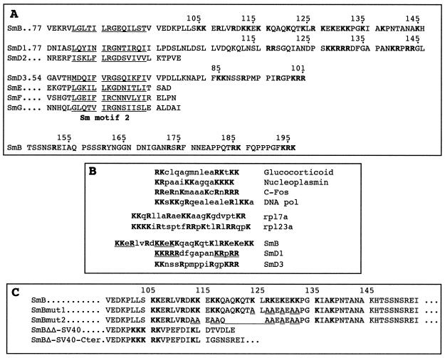
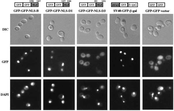
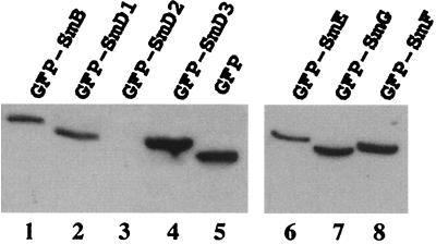
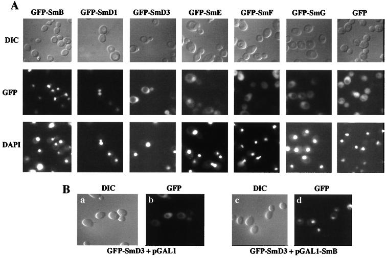
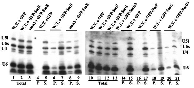
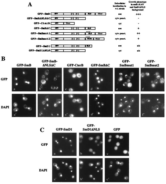
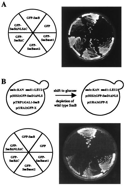
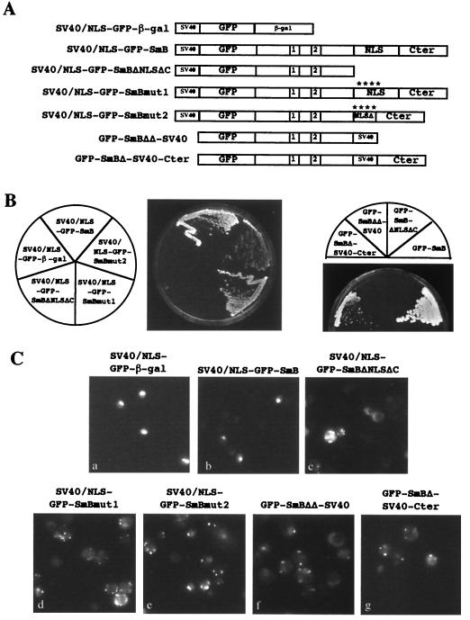
In mammals, nuclear localization of U-snRNP particles requires the snRNA hypermethylated cap structure and the Sm core complex. The nature of the signal located within the Sm core proteins is still unknown, both in humans and yeast. Close examination of the sequences of the yeast SmB, SmD1, and SmD3 carboxyl-terminal domains reveals the presence of basic regions that are reminiscent of nuclear localization signals (NLSs). Fluorescence microscopy studies using green fluorescent protein (GFP)-fusion proteins indicate that both yeast SmB and SmD1 basic amino acid stretches exhibit nuclear localization properties. Accordingly, deletions or mutations in the NLS-like motifs of SmB and SmD1 dramatically reduce nuclear fluorescence of the GFP-Sm mutant fusion alleles. Phenotypic analyses indicate that the NLS-like motifs of SmB and SmD1 are functionally redundant: each NLS-like motif can be deleted without affecting yeast viability whereas a simultaneous deletion of both NLS-like motifs is lethal. Taken together, these findings suggest that, in the doughnut-like structure formed by the Sm core complex, the carboxyl-terminal extensions of Sm proteins may form an evolutionarily conserved basic amino acid-rich protuberance that functions as a nuclear localization determinant.


Figures

FIG. 1
FIG. 1
Amino acid sequence comparison of the C-terminal extensions of yeast Sm proteins. (A) The C-terminal domains of the yeast SmB, SmD1, and SmD3 proteins contain regions rich in basic amino acids (in bold). The evolutionarily conserved Sm motif 2 is underlined. The numbers at the left correspond to the position of the first amino acid shown in the sequence. (B) Comparison of the NLS-like motifs found in the yeast Sm proteins with other nuclear localization motifs. The positions of the basic amino acid-rich regions in the yeast Sm proteins are as follows: SmB, residues 105 to 132; SmD1, residues 128 to 144; and SmD3, residues 85 to 101. The sequences of SmB and SmD1 showing similarities with classical monopartite SV40-type NLSs are underlined. (C) Mutations generated in the SmB NLS-like motif. Basic amino acids are shown in bold. The mutated and deleted positions are underlined in the sequences of the SmBmut1 and SmBmut2 alleles.
FIG. 2
FIG. 2
Localization properties of the putative NLS-like motifs identified in yeast SmB, SmD1, and SmD3 proteins. The NLS-like motif from each indicated Sm protein was fused to the C-terminal domain of a dimeric GFP-GFP reporter protein. The portion of protein sequence used is indicated above each construct. These fusion proteins as well as a fusion carrying the NLS of SV40 Tag at the C terminus of a GFP–β-Gal reporter protein (SV40-GFP-β-Gal) were expressed in wild-type cells. Cells were observed by using differential interference contrast (DIC), and GFP was detected by fluorescence microscopy (GFP). The position of the nuclei was visualized with DAPI (4′,6′-diamidino-2-phenylindole).
FIG. 3
FIG. 3
Western analysis of GFP-Sm fusion proteins. Equivalent amounts of cell extracts prepared from wild-type strains carrying the indicated GFP-Sm fusion proteins were fractionated by SDS-polyacrylamide gel electrophoresis and immunoblotted with anti-GFP antibodies. Both panels represent two independent migrations. The GFP-SmD2 fusion protein (lane 3) was not stably expressed for unknown reasons. Control extract was made from a wild-type strain carrying the GFP vector alone (lane 5). The predicted molecular masses of the proteins are as follows: GFP-SmB, 55 kDa; GFP-SmD1, 45 kDa; GFP-SmD2, 42 kDa; GFP-SmD3, 37 kDa; GFP-SmE, 39 kDa; GFP-SmF, 38 kDa; GFP-SmG, 36 kDa; GFP, 27 kDa.
FIG. 4
FIG. 4
Cellular localizations of yeast GFP-Sm fusion proteins. (A) The indicated GFP-Sm fusion proteins were expressed in a wild-type strain, and living cells were observed using differential interference contrast (DIC) and by fluorescence microscopy (GFP). The position of the nuclei was visualized with DAPI (see text for details). (B) GFP-SmD3 localizes to the nucleus upon SmB overexpression. The YAMB haploid wild-type strain was transformed with the GFP-SmD3 fusion protein and with either the empty pGAL1 vector (a and b) or the pGAL1-SmB plasmid (c and d). Transformants were grown in galactose-containing medium, and the cells were observed using differential interference contrast (DIC) and fluorescence microscopy (GFP).
FIG. 5
FIG. 5
Incorporation of the GFP-Sm fusion proteins into snRNPs. Strains containing the indicated GFP-Sm fusion protein were grown under conditions maintaining the plasmid. Whole-cell extracts were prepared, and snRNPs were immunoprecipitated with anti-GFP antibodies. RNA was extracted from the supernatants (S), the pellets (P), and equivalent aliquots of the total lysates (Total), separated on denaturing polyacrylamide gels, and subjected to Northern analysis. Hybridization was with probes specific for the yeast U4, U5, and U6 snRNAs. W.T., wild-type strain; smeΔ, strain carrying a chromosomal deletion of the SmE gene (4).
FIG. 6
FIG. 6
Intracellular localizations of GFP-SmB and GFP-SmD1 mutant fusion alleles in wild-type cells. (A) Schematic representation of the GFP-Sm mutant alleles. The structures of the mutant fusion constructs are schematically shown: GFP represents the reporter protein, 1 and 2 represent the conserved Sm1 and Sm2 motifs, NLS represents the NLS-like motifs of SmB and SmD1, and Cter represents the SmB C-terminal region (residues 148 to 196). The mutations generated in the GFP-SmBmut1 and GFP-SmBmut2 alleles are detailed in Fig. 1C. A summary of the subcellular localizations in wild-type cells and the growth phenotypes of the mutants in an smb::KAN SmD1ΔNLS context (see Fig. 7B) is shown at the right. nuc, nucleus; cyt. + punct., cytoplasmic staining with fluorescent sites; +++, viable; −, lethal; ++ and +, viable with growth defect; n/a, not applicable. (B) Subcellular localizations of the GFP-SmB mutant alleles. The GFP-SmB fusion alleles were expressed in wild-type cells, and GFP was detected in living cells by fluorescence microscopy. The position of the nuclei was visualized with DAPI. (C) Subcellular localizations of the GFP-SmD1 fusion alleles. The GFP-SmD1 fusion alleles were expressed in wild-type cells, and GFP and DAPI were detected in living cells by fluorescence microscopy.
FIG. 7
FIG. 7
Growth phenotypes of GFP-SmB mutant fusion alleles. (A) An smb::KAN strain carrying a pGAL1-SmB gene was transformed with the indicated GFP-SmB fusion alleles. The different strains, which grow on galactose-based media, were streaked on glucose-based media, which repress the production of the wild-type SmB gene. The phenotypes were observed after 4 days at 30°C. (B) Simultaneous deletion of the SmB- and SmD1-NLS-like motifs gives rise to yeast cell lethality. Above, strain YRB120 carries chromosomal disruptions of the SMB and SMD1 genes. This strain is able to grow on galactose-based media, since it carries the SmB gene under the GAL1 promoter [p(TRP1)GAL1-SmB plasmid] and the GFP-SmD1ΔNLS mutant allele [p(HIS3)GFP-SmD1ΔNLS plasmid]. This strain was transformed with different constructs of GFP-SmB mutant alleles carried by a URA3 plasmid [p(URA3)GFP-X plasmid]. When placed on glucose-based media, the wild-type SmB gene is repressed, allowing the determination of the growth phenotype of the GFP-SmB mutant alleles. Below, growth assay. The growth phenotypes of the indicated GFP-SmB mutants were determined after 4 days at 30°C.
FIG. 8
FIG. 8
Growth phenotypes and intracellular localizations of SV40 NLS containing GFP-SmB mutant alleles. (A) Schematic representation of the GFP-SmB mutant alleles carrying the NLS motif of SV40 Tag. The structures of the mutant fusion constructs are as described in the legend to Fig. 6A. (B) Growth assay. The constructs were transformed in the YRB120 strain, and their phenotypes were determined as described in the legend to Fig. 7B. (C) Subcellular localizations of the SV40 NLS containing GFP-SmB fusion alleles in wild-type cells as detected by fluorescence microscopy.

Similar articles

Cited by

References

    1. Achsel T, Brahms H, Kastner B, Bachi A, Wilm M, Lührmann R. A doughnut-shaped heteromer of human Sm-like proteins binds to the 3′-end of U6 snRNA, thereby facilitating U4/U6 duplex formation in vitro. EMBO J. 1999;18:5789–5802. - PMC - PubMed
    1. Blum S, Mueller M, Schmid S R, Linder P, Trachsel H. Translation in Saccharomyces cerevisiae: initiation factor 4A-dependent cell-free system. Proc Natl Acad Sci USA. 1989;86:6043–6046. - PMC - PubMed
    1. Bordonné R, Banroques J, Abelson J, Guthrie C. Domains of yeast U4 spliceosomal RNA required for PRP4 protein binding, snRNP-snRNP interactions, and pre-mRNA splicing in vivo. Genes Dev. 1990;4:1185–1196. - PubMed
    1. Bordonné R, Tarassov I. The yeast SME1 gene encodes the homologue of the human E core protein. Gene. 1996;176:111–117. - PubMed
    1. Camasses A, Bragado-Nilsson E, Martin R, Séraphin B, Bordonné R. Interactions within the yeast Sm core complex: from proteins to amino acids. Mol Cell Biol. 1998;18:1956–1966. - PMC - PubMed

Publication types

MeSH terms