. 2000 Nov;20(21):8244-53.
doi: 10.1128/MCB.20.21.8244-8253.2000.

Human scribble (Vartul) is targeted for ubiquitin-mediated degradation by the high-risk papillomavirus E6 proteins and the E6AP ubiquitin-protein ligase

Affiliations

Human scribble (Vartul) is targeted for ubiquitin-mediated degradation by the high-risk papillomavirus E6 proteins and the E6AP ubiquitin-protein ligase

S Nakagawa et al. Mol Cell Biol. 2000 Nov.

Abstract

The high-risk human papillomavirus (HPV) E6 proteins stimulate the ubiquitination and degradation of p53, dependent on the E6AP ubiquitin-protein ligase. Other proteins have also been shown to be targeted for degradation by E6, including hDlg, the human homolog of the Drosophila melanogaster Discs large (Dlg) tumor suppressor. We show here that the human homolog of the Drosophila Scribble (Vartul) (hScrib) tumor suppressor protein is also targeted for ubiquitination by the E6-E6AP complex in vitro and that expression of E6 induces degradation of hScrib in vivo. Characterization of the E6AP-E6-hScrib complex indicated that hScrib binds directly to E6 and that the binding is mediated by the PDZ domains of hScrib and a carboxyl-terminal epitope conserved among the high-risk HPV E6 proteins. Green fluorescent protein-hScrib was localized to the periphery of MDCK cells, where it colocalized with ZO-1, a component of tight junctions. E6 expression resulted in loss of integrity of tight junctions, as measured by ZO-1 localization, and this effect was dependent on the PDZ binding epitope of E6. Thus, the high-risk HPV E6 proteins induce the degradation of the human homologs of two Drosophila PDZ domain-containing tumor suppressor proteins, hDlg and hScrib, both of which are associated with cell junction complexes. The fact that Scrib/Vart and Dlg appear to cooperate in a pathway that controls Drosophila epithelial cell growth suggests that the combined targeting of hScrib and hDlg is an important component of the biologic activity of high-risk HPV E6 proteins.


Figures

FIG. 1
FIG. 1
Biochemical screen for E6-dependent E6AP binding proteins. (A) GST-E6AP on glutathione-Sepharose beads was incubated with 35S-labeled whole-cell extract from C-33A cells, without (−) or with (+) partially purified baculovirus-expressed HPV39 E6 protein. Bound proteins were detected by SDS-PAGE and autoradiography. Molecular weight markers are indicated, as well as positions of p220 and proteins that likely correspond to p53 and E6AP. (B) The identities of the bands shown in panel A as p53 and E6AP were confirmed by performing binding experiments in duplicate, releasing the bound proteins in SDS-PAGE loading buffer, and then either running the proteins directly (lanes 1 and 2) or immunoprecipitating (ip) then with anti-p53 or anti-E6AP antibody (lanes 3 and 4). Lane 5, direct immunoprecipitation of the proteins from the labeled extract. (C) Binding experiment as in panel A with GST-E6AP (wild-type [WT]; lanes 1 and 2) or GST-E6APΔ378-395, which had the E6 binding domain deleted (lanes 3 and 4).
FIG. 2
FIG. 2
Schematic of human Scrib protein relative to Drosophila Scrib, human KIAA1225, rat densin-180, and human Dlg (upper) and schematic representation of Blast2 comparisons of hScrib to Drosophila Scrib, densin-180, and hDlg (lower). LRRs and PDZ domains are indicated.
FIG. 3
FIG. 3
hScrib has characteristics of an E6-dependent substrate of E6AP. (A) Binding of rabbit reticulocyte lysate-translated 35S-labeled p53 and hScrib to GST-E6AP was performed under conditions described in the legend for Fig. 1 in the absence or presence of HPV39 E6 (lanes 1 to 4). Lanes 5 to 8, effect of the addition of HPV39 E6 to in vitro p53 and hScrib under conditions that support E6AP-dependent ubiquitination. The high-molecular-weight material (ubn substrate) represents the multiubiquitination of p53 and hScrib (lanes 6 and 8, respectively). The same amounts of translation product were used for binding (lanes 1 to 4) and ubiquitination reactions (lanes 5 to 8). (B) Wheat germ extract-translated hScrib was incubated with or without HPV16 E6 and purified E6AP, as indicated, in the presence of added E1 and E2 (UbcH7) protein, ubiquitin, and ATP. hScrib was ubiquitinated only in the presence of both E6 and E6AP (lane 3).
FIG. 4
FIG. 4
(A) Schematic of hScrib regions that were expressed as GST fusion proteins. (B) GST-hScrib fusion proteins were assayed for binding to HPV16 E6 protein synthesized in a wheat germ extract translation system. (C) Full-length GST-hScrib (amino acids 1 to 1551) and the GST fusion to the PDZ domain region (amino acids 655 to 1126) were assayed for binding to E6AP in the absence and presence of HPV16 E6 protein. The amount of translation product used in the binding experiment is indicated (in.).
FIG. 5
FIG. 5
(A) The carboxyl-terminal 16 residues of HPV16 E6, from the last C-x-x-C sequence to amino acid 151, are shown on the top line, and the corresponding region of HPV11 E6 is shown on the bottom line. Mutations of HPV16 E6 substituted the indicated amino acids for those present at analogous positions of HPV11 E6, relative to the carboxyl termini of the two proteins. In the HPV16 E6 Δ151 mutant the last amino acid was deleted without replacement. (B) HPV11 E6, HPV16 E6, and the mutants described for panel A were synthesized in a wheat germ extract translation system and assayed for binding to GST-hScrib (amino acids 1 to 1551). The relative input amounts are shown. (C) In vitro-translated (rabbit reticulocyte lysate) p53 and hScrib were incubated without E6 (−) or with wild-type (wt) HPV16 E6 or the HPV16 E6 SAT8–10 mutant (SAT). p53 was ubiquitinated in the presence of wt HPV16 E6, while hScrib was ubiquitinated in the presence of either the wt or mutant protein. (D) Binding of wt HPV16 E6, the SAT8–10 mutant, and HPV11 E6 to GST-E6AP (wt) or GST-E6APΔ378-395 (ΔE6), confirming that the SAT8–10 mutant binds to E6AP similarly to the wt protein. Amounts of input proteins were similar (not shown).
FIG. 6
FIG. 6
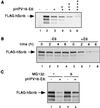
HPV16 E6 expression affects steady-state level and half-life of hScrib in cells. (A) 293-T cells were transfected with a FLAG-hScrib-expressing plasmid without (lane 2) or with increasing amounts of HPV16 E6 expression plasmid (lanes 3 to 6; 0.5, 1.0, 1.5, and 2.0 μg, respectively). Cell extracts were made 48 h posttransfection and analyzed for FLAG-hScrib levels by immunoblotting with an anti-FLAG antibody. Lane 1, mock-transfected cells. (B) 293-T cells were transfected with a FLAG-hScrib-expressing plasmid without (lanes 1 to 4) or with an E6 expression plasmid (lanes 5 to 8; equivalent to lane 3 in panel A). Cycloheximide was added 24 h posttransfection, and cell extracts were made at the indicated times after addition, followed by immunoblotting analysis as for panel A. (C) Cells were transfected with a plasmid expressing FLAG-hScrib without or with an HPV16 E6-expressing plasmid, and levels of FLAG-hScrib without (lanes 1 and 2) or with incubation of MG132 for 3 h (lanes 3 and 4) were compared. Cell extracts were made and analyzed as for panel A.
FIG. 7
FIG. 7
GFP-hScrib-transfected MDCK cells were probed with an anti-ZO-1 antibody and rhodamine-conjugated secondary antibody, and cells were analyzed by confocal laser scanning microscopy.
FIG. 8
FIG. 8
(A) MDCK cells were transfected with a GFP-hScrib-expressing plasmid alone (top) or with a plasmid expressing the HPV16 E6 Δ151 mutant (bottom), and the fixed cells were probed with anti-ZO-1 antibody and rhodamine-conjugated secondary antibody. Cells were observed by fluorescence microscopy. (B) MDCK cells were transfected with pEGFP-C1 vector alone (top) or pEGFP-C1 vector with pCDNA-HPV16 E6 plasmid (bottom). The GFP signal served as a marker for transfected cells, and the ZO-1 antibody was detected with a rhodamine-conjugated secondary antibody.

Similar articles

Cited by

References

    1. Bilder D, Perrimon N. Localization of apical determinants by the basolateral PDZ protein Scribble. Nature. 2000;403:676–680. - PubMed
    1. Bilder D, Li M, Perrimon N. Cooperative regulation of cell polarity and growth by Drosophila tumor suppressors. Science. 2000;289:113–116. - PubMed
    1. Bilder D, Birnbaum D, Borg J-P, Bryant P, Huibregtse J M, Jansen E, Kennedy M B, Labouesse M, Legouis R, Mechler B, Perrimon N, Petit M, Sinha P. Collective nomenclature for LAP proteins. Nat Cell Biol. 2000;2:E114. - PubMed
    1. Chen J J, Hong Y, Androphy E J. Mutational analysis of transcriptional activation by the bovine papillomavirus type 1 E6. Virology. 1997;236:30–36. - PubMed
    1. Crook T, Tidy J A, Vousden K H. Degradation of p53 can be targeted by HPV E6 sequences distinct from those required for p53 binding and trans-activation. Cell. 1991;67:547–556. - PubMed

Publication types

MeSH terms

Substances