. 2000 Nov;20(21):8283-9.
doi: 10.1128/MCB.20.21.8283-8289.2000.

DNA replication is required To elicit cellular responses to psoralen-induced DNA interstrand cross-links

Affiliations

DNA replication is required To elicit cellular responses to psoralen-induced DNA interstrand cross-links

Y M Akkari et al. Mol Cell Biol. 2000 Nov.

Abstract

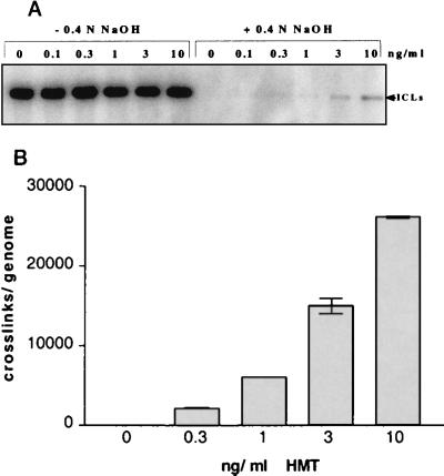
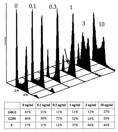
Following introduction of DNA interstrand cross-links (ICLs), mammalian cells display chromosome breakage or cell cycle delay with a 4N DNA content. To further understand the nature of the delay, previously described as a G(2)/M arrest, we developed a protocol to generate ICLs during specific intervals of the cell cycle. Synchronous populations of G(1), S, and G(2) cells were treated with photoactivated 4'-hydroxymethyl-4,5',8-trimethylpsoralen (HMT) and scored for normal passage into mitosis. In contrast to what was found for ionizing radiation, ICLs introduced during G(2) did not result in a G(2)/M arrest, mitotic arrest, or chromosome breakage. Rather, subsequent passage through S phase was required to trigger both chromosome breakage and arrest in the next cell cycle. Similarly, ICLs introduced during G(1) did not cause a G(1)/S arrest. We conclude that DNA replication is required to elicit the cellular responses of cell cycle arrest and genomic instability after psoralen-induced ICLs. In primary human fibroblasts, the 4N DNA content cell cycle arrest triggered by ICLs was long lasting but reversible. Kinetic analysis suggested that these cells could remove up to approximately 2,500 ICLs/genome at an average rate of 11 ICLs/genome/h.


Figures

FIG. 1
FIG. 1
(A) Detection of ICLs in the 17-kb 28S rRNA gene after treatment with 0.4 N NaOH. The covalent bond in cross-linked DNA made it resistant to alkaline denaturation and hence visible as a double-stranded DNA band. Increasing doses of HMT gave increasing levels of ICLs. (B) The numbers of ICLs/genome were calculated (see Materials and Methods and references therein) and plotted for each HMT dose. The graph shows a nearly linear dose response.
FIG. 2
FIG. 2
(A) Effect of HMT plus UVA on the growth of PD743.F as a function of time. At 0.3 ng of HMT/ml, the cells were arrested for ∼8 days and then resumed entry into the cell cycle. At 1 to 3 ng of HMT/ml, cells remained arrested for the duration of the experiment. (B) Following treatment, cells were incubated with BrdU and labeled with an anti-BrdU antibody at various time points. As cells not treated with HMT (0 ng/ml) reached confluency, BrdU-positive cells decreased. Cells treated with 0.3 ng of HMT/ml were completely negative on day 7, followed by an abrupt increase in labeling at the time of recovery. At 3 ng/ml, cells remained unlabeled after they arrested. (C) Clonogenic survival assay of cells treated with 0, 0.15, 0.3, and 3 ng of HMT/ml and allowed to recover and form clones. With the exception of 3 ng of HMT/ml, all doses allowed 60% or more of cells to recover.
FIG. 2
FIG. 2
(A) Effect of HMT plus UVA on the growth of PD743.F as a function of time. At 0.3 ng of HMT/ml, the cells were arrested for ∼8 days and then resumed entry into the cell cycle. At 1 to 3 ng of HMT/ml, cells remained arrested for the duration of the experiment. (B) Following treatment, cells were incubated with BrdU and labeled with an anti-BrdU antibody at various time points. As cells not treated with HMT (0 ng/ml) reached confluency, BrdU-positive cells decreased. Cells treated with 0.3 ng of HMT/ml were completely negative on day 7, followed by an abrupt increase in labeling at the time of recovery. At 3 ng/ml, cells remained unlabeled after they arrested. (C) Clonogenic survival assay of cells treated with 0, 0.15, 0.3, and 3 ng of HMT/ml and allowed to recover and form clones. With the exception of 3 ng of HMT/ml, all doses allowed 60% or more of cells to recover.
FIG. 3
FIG. 3
Flow cytometry analysis of PD743.F 24 h after treatment with HMT plus UVA. At 0.1 to 1 ng of HMT/ml, cells were arrested with a near-4N DNA content. A shoulder of unreplicated DNA was, however, apparent (arrow). At 3 and 10 ng of HMT/ml, cells were clearly arrested with an intermediate DNA content, i.e., in S phase. The table below shows the corresponding percentages of cells in G1, G2, and S phase.
FIG. 4
FIG. 4
Synchronization and treatment of PD743.F cells in G2. (Left) cells were synchronized in G2 and treated with 3 ng of HMT/ml (generating 16,000 ICLs/genome). (Right) Seven hours after treatment, most cells have divided and entered G1. The table below shows the corresponding percentages of cells in G1, G2, and S phases.
FIG. 5
FIG. 5
(A) FACS analysis of PD743.F cells treated in G1 after serum starvation with either HMT plus UVA or ionizing radiation. Following treatment, cells were released into the cell cycle by serum addition and analyzed at three different time points. Untreated cells progress into the cell cycle as observed after 24 h and 40 h. Cells treated with HMT at 0.3 ng/ml (generating ∼2,500 ICLs/genome) also showed progress into S phase after 24 h, and by 40 h cells were arrested with a 4N DNA content. In contrast, cells treated with 5 and 10 Gy did not enter the cell cycle within these time points. (B) Comparison of cell viability in response to HMT-plus-UVA treatment of cycling cells (a) to that for cells retained in G1 by serum starvation (b). Approximately 2 weeks following treatment with 3 ng of HMT/ml (generating ∼16,000 ICLs/genome), cycling cells show a higher degree of cell death than cells retained in G1 by serum starvation.
FIG. 5
FIG. 5
(A) FACS analysis of PD743.F cells treated in G1 after serum starvation with either HMT plus UVA or ionizing radiation. Following treatment, cells were released into the cell cycle by serum addition and analyzed at three different time points. Untreated cells progress into the cell cycle as observed after 24 h and 40 h. Cells treated with HMT at 0.3 ng/ml (generating ∼2,500 ICLs/genome) also showed progress into S phase after 24 h, and by 40 h cells were arrested with a 4N DNA content. In contrast, cells treated with 5 and 10 Gy did not enter the cell cycle within these time points. (B) Comparison of cell viability in response to HMT-plus-UVA treatment of cycling cells (a) to that for cells retained in G1 by serum starvation (b). Approximately 2 weeks following treatment with 3 ng of HMT/ml (generating ∼16,000 ICLs/genome), cycling cells show a higher degree of cell death than cells retained in G1 by serum starvation.
FIG. 6
FIG. 6
A model for the cellular response to ICLs. When ICLs are introduced prior to replication, DNA synthesis stalls at the lesion. This unreplicated DNA triggers a cell cycle arrest, whereby cells do not enter mitosis unless caffeine is added to the cells. As a result of an aberrant mitosis, chromosome breaks form. Conversely, if ICLs are introduced in G2 (post-replication DNA cross-link), the cells are unable to recognize the lesion, and can enter a normal mitosis. Cell cycle arrest will only result when the cells undergo DNA replication again.

References

    1. Auerbach A D, Wolman S R. Susceptibility of Fanconi's anaemia fibroblasts to chromosome damage by carcinogens. Nature. 1976;261:494–496. - PubMed
    1. Bempong M A, Trower E C. Sensitivity of rat testes to inhibitors of nucleic acid synthesis. III. The inheritance of mitomycin C-induced structural rearrangements of chromosomes. J Hered. 1975;66:285–289. - PubMed
    1. Bessho T, Mu D, Sancar A. Initiation of DNA interstrand cross-link repair in humans: the nucleotide excision repair system makes dual incisions 5′ to the cross-linked base and removes a 22- to 28-nucleotide-long damage-free strand. Mol Cell Biol. 1997;17:6822–6830. - PMC - PubMed
    1. Cheng S, Van Houten B, Gamper H B, Sancar A, Hearst J E. Use of psoralen-modified oligonucleotides to trap three-stranded RecA-DNA complexes and repair of these cross-linked complexes by ABC excinuclease. J Biol Chem. 1988;263:15110–15117. - PubMed
    1. de Andrade H H, Marques E K, Schenberg A C, Henriques J A. The PSO4 gene is responsible for an error-prone recombinational DNA repair pathway in Saccharomyces cerevisiae. Mol Gen Genet. 1989;217:419–426. - PubMed

Publication types

MeSH terms

LinkOut - more resources