

Sulindac inhibits neointimal formation after arterial injury in wild-type and apolipoprotein E-deficient mice
- PMID: 11027305
- PMCID: PMC18838
- DOI: 10.1073/pnas.210394497
Sulindac inhibits neointimal formation after arterial injury in wild-type and apolipoprotein E-deficient mice
Abstract
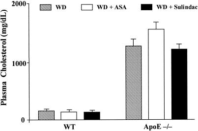
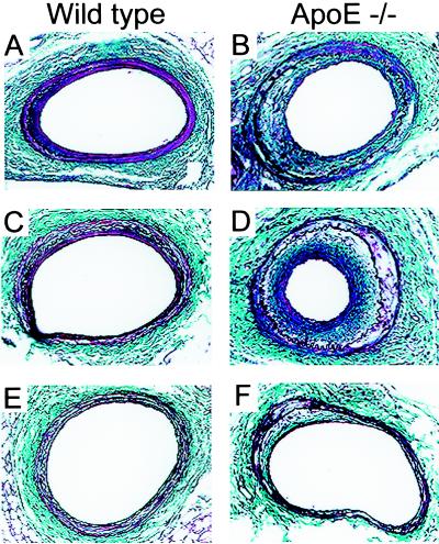
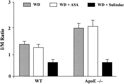
Neointimal hyperplasia is a critical component of restenosis, a major complication of angioplasty and related therapeutic procedures. We studied the effects of hyperlipidemia and the nonsteroidal anti-inflammatory drugs, aspirin (acetyl-salicylic acid; ASA), and sulindac, on neointimal formation in a mouse femoral arterial injury model. At 2 months of age, normolipidemic, wild-type (WT), and hyperlipidemic, apolipoprotein E-deficient (apoE-/-) mice were divided into three treatment groups: Western-type diet (WD), WD + ASA (200 mg/kg food), and WD + sulindac (300 mg/kg food). After 1 week, mice underwent arterial injury and treatments were maintained for 4 weeks. Histomorphometry of the injured arteries showed striking effects of plasma cholesterol levels and drug treatment on neointimal hyperplasia. In the WD or WD + ASA groups, apoE-/- mice had twice the neointimal area than WT mice ( approximately 30,000 vs. 13,000 microm(2) per section; P < 0.0001). Compared with ASA or WD alone, sulindac treatment resulted in approximately 70% (P = 0.0001) and 50% (P = 0.01) reductions in the neointimal area in apoE-/- and WT mice, respectively. ASA, at a dose sufficient to inhibit platelet aggregation, did not affect neointimal formation in mice of either genotype. Evidence of macrophages was noted in the lesions of apoE-/- mice in the WD and WD + ASA groups, but remarkably, none was detectable with sulindac treatment, despite hyperlipidemia, suggesting early steps in the response to injury were abrogated. These results demonstrate sulindac reduces neointimal formation in both normolipidemic and hyperlipidemic settings and raise the possibility that similar benefits may be obtained in patients undergoing angioplasty and related procedures.
Figures

Comment in
-
Anti-inflammatories for cardiovascular disease.Proc Natl Acad Sci U S A. 2000 Nov 7;97(23):12400-1. doi: 10.1073/pnas.240459597. Proc Natl Acad Sci U S A. 2000. PMID: 11058172 Free PMC article. Review. No abstract available.
References
-
- Bittl J A. N Engl J Med. 1996;24:1290–1302. - PubMed
-
- Knight C J, Curzen N P, Groves P H, Patel D J, Goodall A H, Wright C, Clarke D, Oldershaw P J, Fox K M. Eur Heart J. 1999;20:1783–1790. - PubMed
-
- Ross R. N Engl J Med. 1999;340:115–126. - PubMed
-
- Popma J, Califf R, Topol E. Circulation. 1991;84:1426–1436. - PubMed
-
- Liu M W, Roubin G S, King S B., III Circulation. 1989;79:1374–1387. - PubMed
Publication types
MeSH terms
Substances
Grants and funding
LinkOut - more resources
Full Text Sources
Other Literature Sources
Molecular Biology Databases
Miscellaneous
