

Functional switching of GABAergic synapses by ryanodine receptor activation
- PMID: 11027306
- PMCID: PMC17336
- DOI: 10.1073/pnas.210396697
Functional switching of GABAergic synapses by ryanodine receptor activation
Abstract
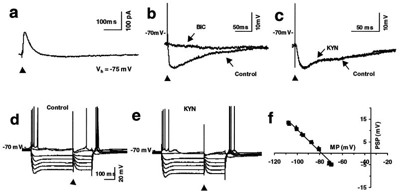
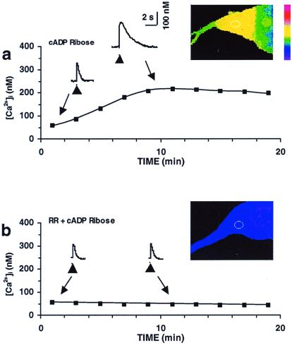
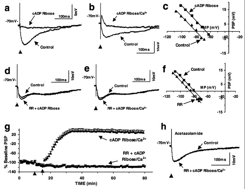
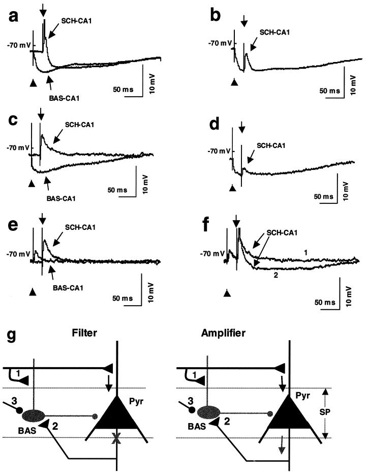
The role of the ryanodine receptor (RyR) in modifiability of synapses made by the basket interneurons onto the hippocampal CA1 pyramidal cells was examined in rats. Associating single-cell RyR activation with postsynaptic depolarization increased intracellular free Ca(2+) concentrations and reversed the basket interneuron-CA1 inhibitory postsynaptic potential into an excitatory postsynaptic potential. This synaptic transformation was accompanied by a shift of the reversal potential from that of chloride toward that of bicarbonate. This inhibitory postsynaptic potential-excitatory postsynaptic potential transformation was prevented by blocking RyR or carbonic anhydrase. Associated postsynaptic depolarization and RyR activation, therefore, changes GABAergic synapses from excitation filters to amplifier and, thereby, shapes information flow through the hippocampal network.
Figures
Similar articles
-
Calexcitin transformation of GABAergic synapses: from excitation filter to amplifier.Proc Natl Acad Sci U S A. 1999 Jun 8;96(12):7023-8. doi: 10.1073/pnas.96.12.7023. Proc Natl Acad Sci U S A. 1999. PMID: 10359832 Free PMC article.
-
Theta rhythm of hippocampal CA1 neuron activity: gating by GABAergic synaptic depolarization.J Neurophysiol. 2001 Jan;85(1):269-79. doi: 10.1152/jn.2001.85.1.269. J Neurophysiol. 2001. PMID: 11152726
-
Monosynaptic GABA-mediated inhibitory postsynaptic potentials in CA1 pyramidal cells of hyperexcitable hippocampal slices from kainic acid-treated rats.Neuroscience. 1993 Feb;52(3):541-54. doi: 10.1016/0306-4522(93)90404-4. Neuroscience. 1993. PMID: 8095707
-
A kainate receptor increases the efficacy of GABAergic synapses.Neuron. 2001 May;30(2):503-13. doi: 10.1016/s0896-6273(01)00298-7. Neuron. 2001. PMID: 11395010
-
Reduction of GABA-mediated inhibitory postsynaptic potentials in hippocampal CA1 pyramidal neurons following oral flurazepam administration.Neuroscience. 1995 May;66(1):87-99. doi: 10.1016/0306-4522(94)00558-m. Neuroscience. 1995. PMID: 7637878
Cited by
-
The role of carbonic anhydrases in extinction of contextual fear memory.Proc Natl Acad Sci U S A. 2020 Jul 7;117(27):16000-16008. doi: 10.1073/pnas.1910690117. Epub 2020 Jun 22. Proc Natl Acad Sci U S A. 2020. PMID: 32571910 Free PMC article.
-
Transcranial magnetic stimulation (TMS) of the human frontal cortex: implications for repetitive TMS treatment of depression.J Psychiatry Neurosci. 2004 Jul;29(4):268-79. J Psychiatry Neurosci. 2004. PMID: 15309043 Free PMC article. Review.
-
Molecular pharmacodynamics, clinical therapeutics, and pharmacokinetics of topiramate.CNS Neurosci Ther. 2008 Summer;14(2):120-42. doi: 10.1111/j.1527-3458.2008.00041.x. CNS Neurosci Ther. 2008. PMID: 18482025 Free PMC article. Review.
-
Memory-specific temporal profiles of gene expression in the hippocampus.Proc Natl Acad Sci U S A. 2002 Dec 10;99(25):16279-84. doi: 10.1073/pnas.242597199. Epub 2002 Dec 2. Proc Natl Acad Sci U S A. 2002. PMID: 12461180 Free PMC article.
-
GABAB receptor- and metabotropic glutamate receptor-dependent cooperative long-term potentiation of rat hippocampal GABAA synaptic transmission.J Physiol. 2003 Nov 15;553(Pt 1):155-67. doi: 10.1113/jphysiol.2003.049015. Epub 2003 Sep 8. J Physiol. 2003. PMID: 12963794 Free PMC article.
References
MeSH terms
Substances
LinkOut - more resources
Full Text Sources
Other Literature Sources
Miscellaneous
