. 2000 Oct 24;97(22):12289-94.
doi: 10.1073/pnas.220398297.

The Ebola virus VP35 protein functions as a type I IFN antagonist

Affiliations

The Ebola virus VP35 protein functions as a type I IFN antagonist

C F Basler et al. Proc Natl Acad Sci U S A. .

Abstract

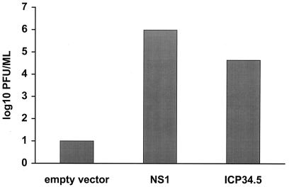
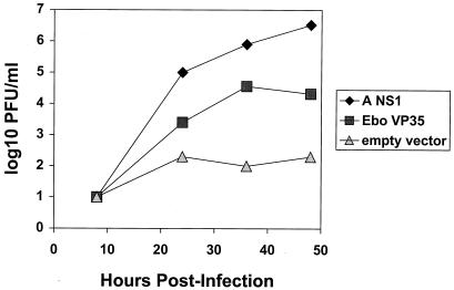
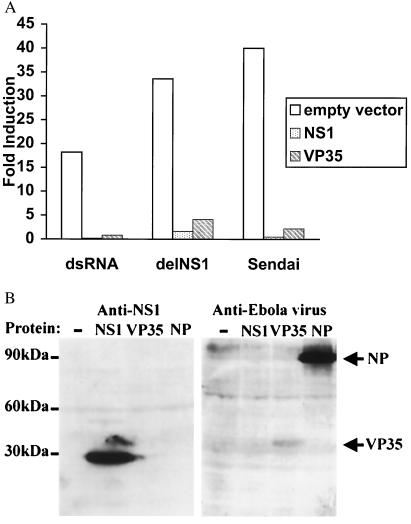
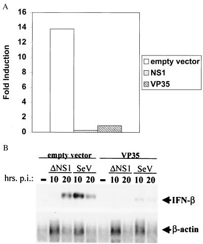
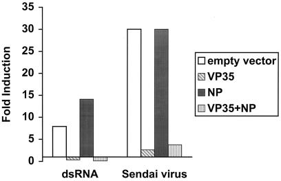
An assay has been developed that allows the identification of molecules that function as type I IFN antagonists. Using this assay, we have identified an Ebola virus-encoded inhibitor of the type I IFN response, the Ebola virus VP35 protein. The assay relies on the properties of an influenza virus mutant, influenza delNS1 virus, which lacks the NS1 ORF and, therefore, does not produce the NS1 protein. When cells are infected with influenza delNS1 virus, large amounts of type I IFN are produced. As a consequence, influenza delNS1 virus replicates poorly. However, high-efficiency transient transfection of a plasmid encoding a protein that interferes with type I IFN-induced antiviral functions, such as the influenza A virus NS1 protein or the herpes simplex virus protein ICP34.5, rescues growth of influenza delNS1 virus. When plasmids expressing individual Ebola virus proteins were transfected into Madin Darby canine kidney cells, the Ebola virus VP35 protein enhanced influenza delNS1 virus growth more than 100-fold. VP35 subsequently was shown to block double-stranded RNA- and virus-mediated induction of an IFN-stimulated response element reporter gene and to block double-stranded RNA- and virus-mediated induction of the IFN-beta promoter. The Ebola virus VP35 therefore is likely to inhibit induction of type I IFN in Ebola virus-infected cells and may be an important determinant of Ebola virus virulence in vivo.


Figures

Figure 1
Figure 1
Growth of the influenza delNS1 virus is complemented by transient transfection of an influenza A NS1 protein or an HSV ICP34.5 expression plasmid. MDCK cells were transfected with 4 μg of empty expression plasmid (pCAGGS), pCAGGS-PR8 NS1 SAM (23), or pCAGGS-γ134.5. Twenty-four hours later, the cells were infected with influenza delNS1 virus (moi = 0.001). Forty-eight hours posttransfection, viral titers were determined by plaque assay. The results are the average of two independent experiments.
Figure 2
Figure 2
Growth of the influenza delNS1 virus is complemented by the Ebola virus VP35 protein. MDCK cells were transfected with 4 μg of empty expression plasmid (pcDNA3), NS1 expression plasmid, or Ebola virus VP35 expression plasmid. Twenty-four hours later, the cells were infected with influenza delNS1 virus (moi = 0.001). Viral titers were determined by plaque assay at the indicated times.
Figure 3
Figure 3
Expression of Ebola virus VP35 protein inhibits dsRNA- or virus-mediated induction of an ISRE. (A) Fold induction of an ISRE promoter–CAT reporter gene in the presence of empty vector, NS1 expression plasmid, or VP35 expression plasmid. 293 cells were transfected with 4 μg of the indicated expression plasmid plus 0.3 μg each of the reporter plasmids pHISG-54-CAT and pGL2-Control. Twenty-four hours posttransfection, the cells were mock-treated or treated with the indicated IFN inducer. The CAT activities were normalized to the corresponding luciferase activities to determine fold induction. (B) Western blot showing NS1, VP35, and Ebola virus NP expression. 293 cells were transfected with 4 μg of the indicated plasmids. Forty-eight hours later, cell lysates were prepared and Western blots were performed by using the indicated antiserum.
Figure 4
Figure 4
The VP35 protein of Ebola virus inhibits induction of the IFN-β promoter. (A) Inhibition of induction of the mouse IFN-β promoter. 293 cells were transfected with 4 μg of the indicated expression plasmid plus 0.3 μg each of the reporter plasmids pIFN-β-CAT and pGL2-Control. Twenty-four hours posttransfection, the cells were mock-transfected or transfected with 40 μg of polyI:polyC. (B) Northern blot showing VP35-mediated inhibition of endogenous IFN-β induction. 293 cells were transfected with either empty vector or VP35 expression plasmid. Twenty-four hours later, the cells were mock-infected (−) or infected with influenza delNS1 virus (delNS1) or Sendai virus (SeV) (moi = 1). Total RNA was prepared from cells at 10 h or 20 h posttransfection. Mock-transfected cell RNA was prepared at the same time as the 20-h postinfection samples. Northern blots were performed to detect IFN-β or β-actin mRNAs. Note that less total RNA was obtained when cells, including the mock-infected cells, were lysed at the 20-h postinfection time point.
Figure 5
Figure 5
The Ebola virus VP35 protein inhibits type I IFN induction when coexpressed with Ebola virus NP. Fold induction of the IFN-inducible ISRE-driven reporter in the presence of empty vector, VP35, NP, or VP35 plus NP. 293 cells were transfected with a total of 4 μg of expression plasmid, including 2 μg of a plasmid encoding an individual protein and 2 μg of a second plasmid (either empty vector or a second expression plasmid) plus 0.3 μg each of the reporter plasmids pHISG-54-CAT and pGL2-Control. Twenty-four hours posttransfection, the cells were mock-treated or treated with the indicated IFN inducer. Twenty-four hours postinduction, CAT and luciferase assays were performed. The CAT activities were normalized to the corresponding luciferase activities to determine fold induction.

References

    1. Klenk H-D, Slenczka W, Feldmann H. In: Encyclopedia of Virology. Webster R G, Granoff A, editors. Vol. 2. New York: Academic; 1994. pp. 827–831.
    1. Peters C J, Khan A S. Curr Top Microbiol Immunol. 1999;235:85–95. - PubMed
    1. Villinger F, Rollin P E, Brar S S, Chikkala N F, Winter J, Sundstrom J B, Zaki S R, Swanepoel R, Ansari A A, Peters C J. J Infect Dis. 1999;179, Suppl. 1:S188–S191. - PubMed
    1. Yang Z, Duckers H J, Sullivan N J, Sanchez A, Nabel E G, Nabel G J. Nat Med. 2000;6:886–889. - PubMed
    1. Yang Z, Delgado R, Xu L, Todd R F, Nabel E G, Sanchez A, Nabel G J. Science. 1998;279:1034–1037. - PubMed

Publication types