. 2000 Oct 24;97(22):11893-8.
doi: 10.1073/pnas.220413297.

Cytoplasmic catalytic subunit of protein kinase A mediates cross-repression by NF-kappa B and the glucocorticoid receptor

Affiliations

Cytoplasmic catalytic subunit of protein kinase A mediates cross-repression by NF-kappa B and the glucocorticoid receptor

V Doucas et al. Proc Natl Acad Sci U S A. .

Abstract

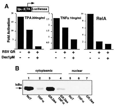
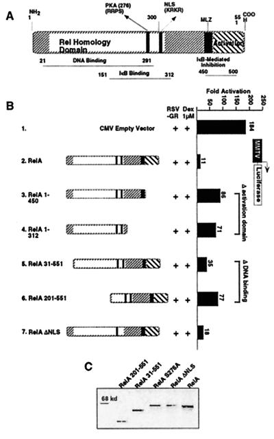
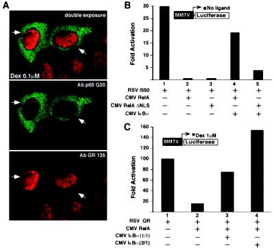
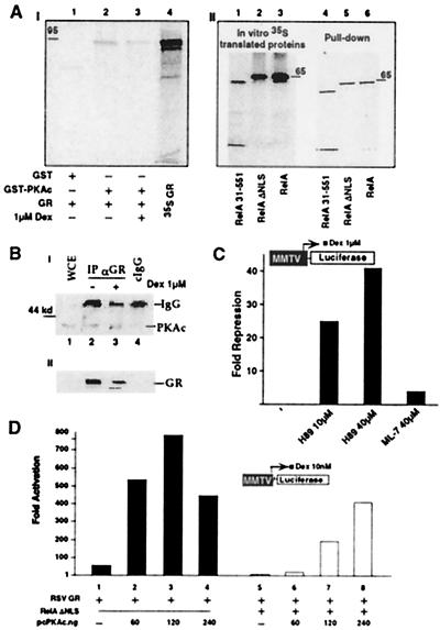
Negative transcriptional regulation or cross-coupling between NF-kappa B (RelA) and the glucocorticoid receptor (GR) is proposed to play a regulatory role in human physiology and disease. Despite previous advances, the biochemical basis of this phenomenon remains a subject of controversy. We show here that the inhibition of GR activity by RelA does not require the RelA DNA binding, transactivation, or nuclear localization domains. Surprisingly, RelA repression of GR is abolished by mutation of the conserved protein kinase A (PKA) site at amino acid residue 276 of RelA. We show that GR associates in vivo and in vitro with the catalytic subunit of PKA (PKAc) in a ligand-independent manner and that GR transcription depends on PKA signaling. Indeed, we demonstrated that GR-mediated inhibition of NF-kappa B transactivation is PKAc-dependent. In contrast to previous models, we suggest that the cross-coupling requires a cytoplasmic step and is regulated by a PKAc-associated signaling.


Figures

Figure 1
Figure 1
GR mediates trans-repression of TNF-α and TPA-activated RelA-dependent signaling. (A) GR represses NF-κB-dependent transcription. CV1 cells (48-well plates) were transiently cotransfected with 120 ng of Igk3-Luc reporter construct, 75 ng of CMXβgal, and 75 ng of RSVGR or RSV empty expression vectors. Cells were costimulated with TPA or TNF-α in the absence or presence of Dex for 4 h before the assay, as indicated. The y axis shows activation as measured by Igk3-driven Luc activity. The histogram is representative of three independent experiments. (B) Protein levels of IkBα are not affected by GR.Dex treatment in 293 cells. Western blot analysis of IkBα in cytoplasmic or nuclear extracts in cells without treatment (Not), treated with TNF-α, overexpression of GR and Dex treatment (GR.Dex), or in combination of TNF-α and GR.Dex.
Figure 2
Figure 2
Mapping the domain of RelA involved in GR repression. (A) Schematic of RelA. (B) RelA cross-coupling domains. CV1 cells (48-well plates) were transiently cotransfected with 120 ng of MMTV-Luc reporter construct, 75 ng of CMXβgal, 37.5 ng of RSVGR, and CMV empty vector or CMVRelA mutants at 3:1 M ratio with GR, as indicated. Cells were stimulated with Dex at 1 μM for 10 h before the assay. The y axis shows activation as measured by MMTV-driven Luc activity conducted in quadruplicate assays. (C) Expression levels of RelA proteins. Western blot analysis of RelA wild-type and mutant proteins.
Figure 3
Figure 3
Cytoplasmic form of RelA controls GR transcription. (A) Immunohistochemistry detecting relative cellular distribution of Dex-activated GR and RelAΔNLS proteins. Confocal image represents a double-exposure photograph. Primary antibodies are used as indicated. Green corresponds to the RelA labeling revealed with the FITC-conjugated second antibody, and red corresponds to the GR labeling revealed with the Texas red conjugated second antibody. HeLa cells were transiently cotransfected with the CMVRelAΔNLS expression vector and the MMTV-Luc reporter, as indicated in Fig. 2B. Cells were treated with 0.1 μM Dex for 3 h before fixation. The filled arrowheads point to the transfected cells. Similar results were obtained in CV1 cells by using ectopically expressed GR and RelAΔNLS proteins (data not shown). (B) IκBα-free RelA wild type and RelAΔNLS equally repress a constitutively active GR mutant (I550). The transient transfection assay was performed as in Fig. 2B. The RSVI550 was used at 37.5 ng, and the CMVIκBα was transfected at 1:1 M ratio with the RelA expression vector. All of the transfections were equalized for the total amount of expression vectors, i.e., CMV empty vector. (C) IκBα sequesters RelA and blocks RelA-mediated GR repression. The experiment was performed as above but cells were stimulated with Dex at 1 μM for 10 h before the assay. The CMVIκBα was used at 1:1 or 1:2 M ratio with RelA expression vector, as indicated. The y axis shows activation as measured by MMTV-driven Luc activity conducted in quadruplicate assays.
Figure 4
Figure 4
Point mutations of RelA PKA phosphorylation site abolish RelA-mediated GR repression. A conserved PKA phosphorylation site of RelA, Ser-276, was mutated to Ala (S276A) or Gly (S276G). Transfection was performed as in Fig. 2B. CMV RelA mutants were assayed as indicated. The y axis shows activation as measured by MMTV-driven Luc activity conducted in quadruplicate assays.
Figure 5
Figure 5
PKAc is associated with GR and potentiates GR activity. (A) In vitro association of PKAc subunit and GR (95 kDa). (AI) The 35S-labeled GR protein was incubated with GST-PKAc or GST alone on glutathione-agarose beads, in the absence or presence of 1 μM Dex as indicated. The bound proteins were analyzed by SDS/PAGE and fluorography (lanes 1–3). Input was included in lane 4. (AII) The same conditions as in AI but using 35S-labeled RelA wild-type and mutants. Input proteins (lanes 1–3) and the pull-down proteins (lanes 4–6) are indicated. (B) In vivo association of PKAc and GR wild-type protein. (BI) Human 293 cells were cotransfected with CMXGR and pcPKAc expression vectors. Cells were mock-treated (lane 2) or activated with 1 μM Dex for 12 h (lane 3) and harvested 48 h after transfection. Cell lysates were immunoprecipitated with 5 μl of GR-135 polyclonal antibody (lanes 2 and 3) or with the same amount of normal rabbit IgG as a control (lane 4). Immunoprecipitates then were analyzed by Western blot with PKAc-specific antibody. WCE were loaded in parallel with the immunoprecipitates to show comigration (lane 1). (BII) An aliquot of the WCE was analyzed directly with Western blot by using GR-specific antibody. (C) H-89, a PKAc-specific inhibitor represses GR activity. CV-1 cells were transfected with MMTV-Luc, CMXβgal, and RSVGR as in Fig. 2B. After transfection cells were stimulated for 4 h with 1 μM Dex, with or without H-89 or ML-7, at the indicated concentrations. Cellular extracts were used to measure Luc activity. The y axis shows arbitrary unit of repression as measured by MMTV-driven Luc activity. The histogram is representative of at least three independent experiments. (D) PKAc potentiates GR transcription and reverses RelA repression of GR. CV1 cells were cotransfected with 120 ng of MMTV-Luc reporter construct, 75 ng of CMXβgal, 33 ng of RSVGR, CMV empty vector or CMVRelA ΔNLS at 120 ng, and increasing concentration of PKAc expression vector, as indicated. Cells were stimulated with Dex at 10 nM for 10 h before the assay. All of the transfections were normalized for the total amount of expression vectors, i.e., CMV empty vector. The y axis shows arbitrary unit of activation as measured by MMTV-driven Luc activity.
Figure 6
Figure 6
GR competes with RelA for PKAc association in vivo. (A) GR blocks RelA:PKAc protein association in vivo. Human 293T cells were transfected with 5 μg of CMVRelA, 5 μg of pcPKAc, and 5 μg of CMXGR or empty vector, as indicated. Cells were mock-treated or activated with 1 μM Dex for 12 h and harvested 48 h after transfection. (AI) Cell lysates were immunoprecipitated with 5 μl (1 μg) of RelA sc-109 rabbit polyclonal antibody (lanes 3–6) or with the same amount of normal rabbit IgG as control (lane 2). Immunoprecipitates were analyzed by Western blot with PKAc-specific antibody. WCE (lane 1) were loaded in parallel with the immunoprecipitates to show comigration. (AII) Quantitation of PKAc coprecipitated by RelA-specific antibody. The y axis shows the ratio of PKAc to IgG. Quantitation is performed with NIH image program. (AIII) Similar amount of WCE from cells corresponding to lanes 3–6 in AI was immunostained with GR-specific antibody (sc-1002). (AIV) The same blot shown in AIII was stripped and reprobed with RelA-specific antibody. (B) GR represses PKAc-dependent NF-κB transactivation. CV1 cells were cotransfected with 120 ng of Igk3-Luc reporter construct, 75 ng of CMXβgal, 33 ng of CMV RelA, and increasing amount of pcPKAc expression vector, as indicated. The RSVGR and RSVGRΔ589–697 were used at 3:1 M ratio with RelA. All of the transfection points were equalized for the total amount of expression vectors. The y axis shows fold activation as measured by Igk3-driven Luc activity. The histogram is representative of a triplicate experiment.

References

    1. Karin M, Hunter T. Curr Biol. 1995;5:747–757. - PubMed
    1. Evans R M. Science. 1988;240:889–895. - PMC - PubMed
    1. Mangelsdorf D J, Thummel C, Beato M, Herrlich P, Schutz G, Umesono K, Blumberg B, Kastner P, Mark M, Chambon P, et al. Cell. 1995;83:835–839. - PMC - PubMed
    1. Mangelsdorf D J, Evans R M. Cell. 1995;83:841–850. - PubMed
    1. Beato M, Herrlich P, Schutz G. Cell. 1995;83:851–857. - PubMed

Publication types

MeSH terms

Substances