. 2000 Oct 10;97(21):11472-7.
doi: 10.1073/pnas.97.21.11472.

Specific Th2 cells accumulate in the central nervous system of mice protected against experimental autoimmune encephalomyelitis by copolymer 1

Affiliations

Specific Th2 cells accumulate in the central nervous system of mice protected against experimental autoimmune encephalomyelitis by copolymer 1

R Aharoni et al. Proc Natl Acad Sci U S A. .

Abstract

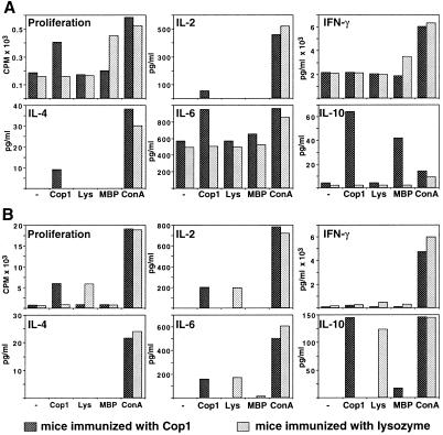
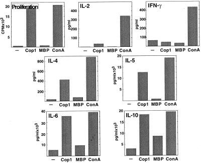
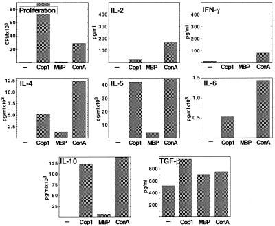
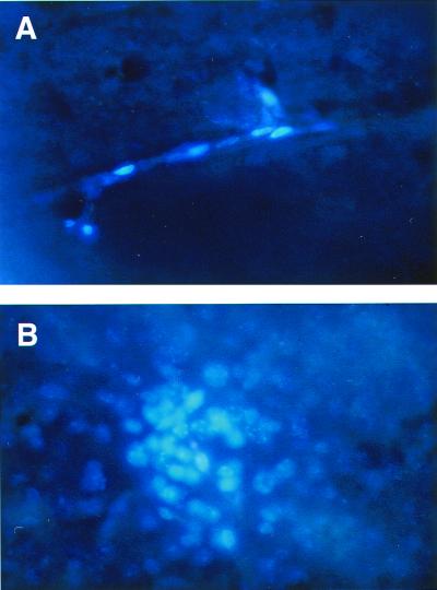
This study addresses the issue of the effect of immunomodulating therapies in the target organ-the central nervous system (CNS)-in the case of multiple sclerosis. Copolymer 1 (Cop 1, Copaxone, glatiramer acetate), an approved drug for the treatment of multiple sclerosis, is a potent inducer of Th2 regulatory cells in both mice and humans. Highly reactive Cop 1-specific T cell lines that secrete IL-4, IL-5, IL-6, IL-10, and transforming growth factor-beta in response to Cop 1 and crossreact with myelin basic protein (MBP) at the level of Th2 cytokine secretion were established from both brains and spinal cords of Cop 1-treated mice. In contrast, no reactivity to the control antigen lysozyme could be obtained in lymphocytes isolated from CNS of mice injected with lysozyme. Adoptively transferred labeled Cop 1-specific suppressor cells were found in brain sections 7 and 10 days after their injection to the periphery, whereas lysozyme-specific cells were absent in the CNS. Hence, Cop 1-induced Th2 cells cross the blood-brain barrier and accumulate in the CNS, where they can be stimulated in situ by MBP and thereby exert therapeutic effects in the diseased organ. This therapeutic effect was manifested, in brains of experimental autoimmune encephalomyelitis-induced mice, by a decrease in the inflammatory cytokine interferon-gamma and by secretion of the anti-inflammatory cytokine IL-10 in response to the autoantigen MBP.


Figures

Figure 1
Figure 1
Proliferation and cytokine secretion by cells isolated from EAE-induced mice pretreated with Cop 1 (dark bars) or lysozyme (light bars). Reactivity of whole lymphocyte populations isolated from brains (A) and spleens (B) is demonstrated. Mice rendered resistant to EAE by injection of Cop 1 (10 mg per mouse in incomplete Freund's adjuvant) and control mice similarly injected with lysozyme were challenged for disease with spinal cord homogenate. One month later, the mice were perfused, and whole lymphocyte population isolated from brains and spleens was evaluated for their reactivity. Proliferation was measured by thymidine incorporation (cpm) into triplicate cultures and cytokine secretion (pg/ml) by quantitative ELISA in duplicates. The response to medium alone (−), Cop 1 (50 μg/ml), MBP (100 μg/ml), and Con A (5 μg/ml) was tested. Standard deviations were under 20% of the mean. Results represent one of four independent experiments.
Figure 2
Figure 2
Proliferation and cytokine secretion by the short-term line Cop-Br-1 originated from brains of EAE-induced mice pretreated with Cop 1, after one in vitro stimulation with Cop 1. Proliferation (cpm) into triplicate cultures and cytokine secretion (pg/ml) in duplicates were measured in response to medium alone (−), Cop 1 (50 μg/ml), MBP (100 μg/ml), and Con A (5 μg/ml). Standard deviations were under 20% of the mean.
Figure 3
Figure 3
Proliferation and cytokine secretion by the long-term T cell line Cop-Br-1, originated from brains of EAE-induced mice pretreated with Cop 1, after three in vitro stimulations with Cop 1. Proliferation (cpm) into triplicate cultures and cytokine secretion (pg/ml) in duplicates were measured in response to medium alone (−), Cop 1 (50 μg/ml), MBP (100 μg/ml), and Con A (5 μg/ml). Standard deviations were under 20% of the mean.
Figure 4
Figure 4
Brain sections of mouse adoptively transferred with Cop 1-specific T cells. Activated T cells of the Cop 1-specific line Ts-D were labeled with the fluorescent dye Hoechst and injected intraperitoneally to normal mice with subsequent injection of pertussis toxin. After 7 days, brains were removed, fixed in paraformaldehyde, and sectioned (40 μm). Cells in blood vessels (A) and a cluster of cells in the brain tissue (B) are demonstrated. (×400.)

References

    1. Wekerle H, Linington C, Lassmann H, Meyermann R. Trends Neurosci. 1986;9:271–277.
    1. Cross A H, O'Mara T, Raine C S. Neurology. 1993;43:1028–1033. - PubMed
    1. Wekerle H. Lab Invest. 1984;51:199–205. - PubMed
    1. Lehmann P V. Am J Pathol. 1998;153:677–680. - PMC - PubMed
    1. Teitelbaum D, Aharoni R, Fridkis-Hareli M, Arnon R, Sela M. In: The Decade in Autoimmunity. Shoenfeld Y, editor. New York: Elsevier; 1998. pp. 183–188.

Publication types

LinkOut - more resources