. 2000 Oct 10;97(21):11516-21.
doi: 10.1073/pnas.97.21.11516.

Invasion in vitro of mosquito midgut cells by the malaria parasite proceeds by a conserved mechanism and results in death of the invaded midgut cells

Affiliations

Invasion in vitro of mosquito midgut cells by the malaria parasite proceeds by a conserved mechanism and results in death of the invaded midgut cells

H Zieler et al. Proc Natl Acad Sci U S A. .

Abstract

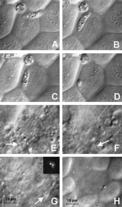
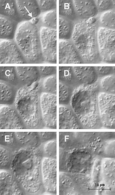
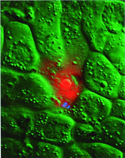
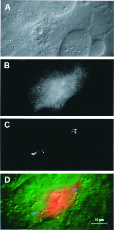
Using an in vitro culture system, we observed the migration of malaria ookinetes on the surface of the mosquito midgut and invasion of the midgut epithelium. Ookinetes display constrictions during migration to the midgut surface and a gliding motion once on the luminal midgut surface. Invasion of a midgut cell always occurs at its lateral apical surface. Invasion is rapid and is often followed by invasion of a neighboring midgut cell by the ookinete. The morphology of the invaded cells changes dramatically after invasion, and invaded cells die rapidly. Midgut cell death is accompanied by activation of a caspase-3-like protease, suggesting cell death is apoptotic. The events occurring during invasion were identical for two different species of Plasmodium and two different genera of mosquitoes; they probably represent a universal mechanism of mosquito midgut penetration by the malaria parasite.


Figures

Figure 1
Figure 1
DIC images of the invasion of a P. gallinaceum ookinete into an A. aegypti midgut cell. The times are given relative to the start of invasion. The scale bar in G is for images A–G; image H is reduced in magnification to show the difference in refractive index of invaded cells relative to neighboring cells. (A) T = −48 sec. (B) T = −15 sec. The ookinete is gliding on the surface of the cell to be invaded. (C) T = 0 sec. Invasion into the lateral apical membrane of the midgut cell has begun and the anterior end of the ookinete has disappeared beneath the cell surface. (D) T = 7 sec. Invasion is near completion, and only the posterior end of the ookinete is visible on the midgut cell surface. (E) T = 2.8 min. The ookinete (arrow) is barely visible inside the invaded cell. (F) T = 3.4 min. The ookinete (arrow) invades a neighboring cell. (G) T = 5.0 min. The ookinete (arrow) is entirely within the neighboring cell. (Inset) Polarized light microscopy unambiguously identifies the position of the ookinete on the basis of the birefringent hemozoin pigment granules. (H) T = 11.3 min. The change in surface morphology and decrease in refractive index of invaded cells are hallmarks of the invasion process.
Figure 2
Figure 2
Rapid morphological change and death of a midgut cell during invasion. The magnification for all six images is identical. The focal plane for images A–D is identical; the focal plane for image E is slightly lower and for image F slightly higher than for images A–D. (A) T = 0. An ookinete is positioned between the apical free margins of two midgut cells with only the posterior end visible (arrow). The focal plane of the image is slightly below the cell surface. (B) T = 8 sec. The midgut cell is beginning to disintegrate. (C) T = 17 sec. The cell continues to disintegrate and cell contents are being discharged. The cell's nucleus has become more prominent. (D) T = 32 sec. The ookinete is completely interiorized within the midgut cell and has disappeared from view. (E) T = 57 sec. The midgut cell's morphology is grossly altered. (F) T = 1.4 min. A surface view of the invaded cell showing the marked decrease in refractive index of the cell.
Figure 3
Figure 3
Composite color image of an ookinete inside a midgut cell showing ethidium homodimer staining of the invaded cell. The DIC image of the cell is shown in green, the ethidium homodimer fluorescence is shown in red, and the hemozoin pigment granules of the ookinete are shown in blue.
Figure 4
Figure 4
Images of an invaded cell stained with PhiPhiLux-G1D2. (A) DIC image of the invaded cell. (B) Increased fluorescence of the invaded cell compared with its neighbors. (C) Polarized light microscope image of hemozoin pigment granules of two ookinetes in the invaded cell. (D) Composite color image with DIC in green, fluorescence in red, and ookinete position in blue, showing the overall relationship between these three independent parameters.
Figure 5
Figure 5
Schematic diagram of ookinete movement and midgut invasion. The ookinete first contacts the MN, through which it must navigate to reach the midgut surface. In the process of migrating through the MN, the ookinete develops constrictions along its length. Once on the surface of the midgut epithelium (ME), the parasite glides rapidly and tends to stay in the crevices between cells. Invasion occurs at the lateral apical membrane of a midgut cell. During its descent toward the basement membrane (BM), the ookinete often crosses to a neighboring cell. The refractive index of the invaded cells decreases, and their nuclei become more visible.

References

    1. Shortt H E. Trans R Soc Trop Med Hyg. 1948;42:227–230. - PubMed
    1. Stohler H. Acta Trop. 1957;14:302–352. - PubMed
    1. Garnham P C C, Bird R G, Baker J R. Trans R Soc Trop Med Hyg. 1962;56:116–120. - PubMed
    1. Mehlhorn H, Peters W, Haberkorn A. Protistologica. 1980;16:135–154.
    1. Meis J F, Ponnudurai T. Parasitol Res. 1987;73:500–506. - PubMed

Publication types