. 2000 Oct 10;97(21):11545-50.
doi: 10.1073/pnas.97.21.11545.

Stimulation of glutamate receptor protein synthesis and membrane insertion within isolated neuronal dendrites

Affiliations

Stimulation of glutamate receptor protein synthesis and membrane insertion within isolated neuronal dendrites

J E Kacharmina et al. Proc Natl Acad Sci U S A. .

Abstract

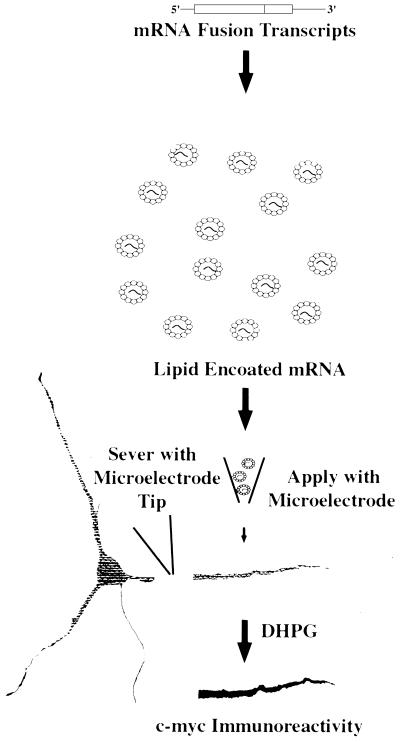
The selective subcellular localization of mRNAs to dendrites and the recent demonstration of local protein synthesis have highlighted the potential role of postsynaptic sites in modulation of cell-cell communication. We show that epitope-tagged subunit 2 of the ionotopic glutamate receptor, GluR2, mRNA transfected into isolated hippocampal neuronal dendrites is translated in response to pharmacologic stimulation. Further, confocal imaging of N-terminally labeled GluR2 reveals that the newly synthesized GluR2 protein can integrate into the dendritic membrane with the N terminus externally localized. These data demonstrate that integral membrane proteins can be synthesized in dendrites and can locally integrate into the cell membrane.


Figures

Figure 1
Figure 1
Single dendrite transfection protocol. This schematic shows the experimental procedure for mRNA transfection of isolated dendrites. This assay was used to generate the data in the subsequent figures.
Figure 2
Figure 2
Epitope-tagged GluR2–cMyc mRNA transfection into cells and single dendrites. A, C, and E contain phase contrast photographs of cells in B, D, and F, respectively. (B) Immunohistochemical background staining of hippocampal neurons that have not been transfected with the fusion mRNA construct but have been taken through the immunohistochemical procedure. (D) This photomicrograph shows the immunohistochemcial background staining of cells that had been transfected with the fusion construct followed by immunohistochemical staining in the absence of cMyc primary antibody. (F) Immunohistochemical proof of local protein synthesis of the transfected reporter construct mRNA in single, isolated dendrites and whole cells. Also visible in this field of cells are some cells that were not transfected. All of the cultures were treated with DHPG. *, transfected cell body; large arrow, untransfected cell; small arrow, transfected dendrite.
Figure 3
Figure 3
Metabotrobic glutamate receptor activation of GluR2–cMyc protein translation. A, C, E, and G are phase contrast photographs of cell fields that correspond to B, D, F, and H, respectively. (B) These cells show that transfection of GluR2–cMyc fusion construct mRNA into cells, in the absence of DHPG stimulation, results in minimal translated protein. (D) This nontransfected dendritic process is stained with the cMyc antibody and shows no immunoreactivity. (F) This nontransfected dendrite is stained with an ATF-2 antibody and exhibits no immunostaining. The Inset in the upper left corner shows a stained neuron from the same culture, thus showing that ATF-2 is present in the neuronal cell body but is absent from dendrites. (H) GluR2–cMyc immunoreactivity generated after transfection of the GluR2–cMyc fusion construct mRNA that had been stimulated by the addition of 500 μM DHPG into the media. Arrow points to the individual isolated dendrite described herein.
Figure 4
Figure 4
Membrane localization of dendritically synthesized c-Myc–GluR2. A, C, and E show immunofluorescence of MAP-2, whereas B, D, and F show the double-labeled anti-cMyc immunofluorescence in the same dendrites. G shows a phase contrast photomicrograph of the dendrite shown in E and F. (A and B) Dendrites that have been permeabilized and not transfected with reporter RNA. (C and D) Dendrites that have been permeabilized and transfected. (E–G) Intact cells that have been transfected with reporter RNA.

References

    1. Petralia R S, Esteban J, Wang Y-X, Partridge J, Zhao H-M, Wenthold R. Nature Neurosci. 1999;2:31–36. - PubMed
    1. Asztely F, Erdemli G, Kullmann D M. Neuron. 1997;18:281–293. - PubMed
    1. Rusakov D A, Kullmann D M. J Neurosci. 1998;18:3158–3170. - PMC - PubMed
    1. He Y, Janssen W G, Morrison J H. J Neurosci Res. 1998;54:444–449. - PubMed
    1. Nusser C, Lujan R, Laube G, Roberts J D, Molnar E, Somogyi P. Neuron. 1998;21:545–559. - PubMed

Publication types

LinkOut - more resources