. 2000 Oct 10;97(21):11609-13.
doi: 10.1073/pnas.97.21.11609.

Nitric oxide prevents cardiovascular disease and determines survival in polyglobulic mice overexpressing erythropoietin

Affiliations

Nitric oxide prevents cardiovascular disease and determines survival in polyglobulic mice overexpressing erythropoietin

F T Ruschitzka et al. Proc Natl Acad Sci U S A. .

Abstract

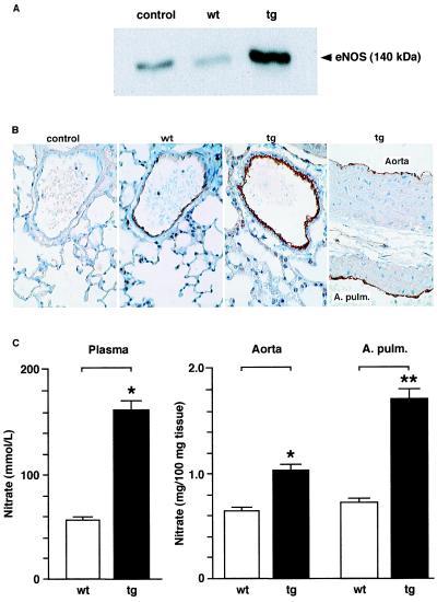
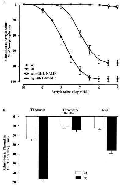
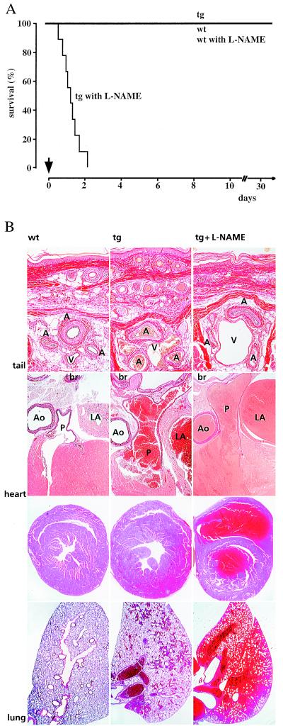
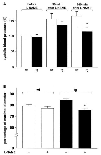
Nitric oxide (NO) induces vasodilatatory, antiaggregatory, and antiproliferative effects in vitro. To delineate potential beneficial effects of NO in preventing vascular disease in vivo, we generated transgenic mice overexpressing human erythropoietin. These animals induce polyglobulia known to be associated with a high incidence of vascular disease. Despite hematocrit levels of 80%, adult transgenic mice did not develop hypertension or thromboembolism. Endothelial NO synthase levels, NO-mediated endothelium-dependent relaxation and circulating and vascular tissue NO levels were markedly increased. Administration of the NO synthase inhibitor N(G)-nitro-L-arginine methyl ester (L-NAME) led to vasoconstriction of peripheral resistance vessels, hypertension, and death of transgenic mice, whereas wild-type siblings developed hypertension but did not show increased mortality. L-NAME-treated polyglobulic mice revealed acute left ventricular dilatation and vascular engorgement associated with pulmonary congestion and hemorrhage. In conclusion, we here unequivocally demonstrate that endothelial NO maintains normotension, prevents cardiovascular dysfunction, and critically determines survival in vivo under conditions of increased hematocrit.


Figures

Figure 1
Figure 1
eNOS expression and NO tissue levels in wild-type (wt) and transgenic (tg) arteries. (A) Representative Western blot of eNOS protein levels in the endothelium scraped from thoracic aortas. The rabbit polyclonal antibody NOS3 (St. Cruz) was used at a 1:1000 dilution. Lysate derived from a human endothelial cell line (TransLab) was loaded as control following the supplier‘s instructions. (B) Immunochemical analysis of eNOS expression in the thoracic aorta and pulmonary arteries (A. Pulm.) of wild-type and transgenic mice using the anti-eNOS antibody described above. Note that eNOS expression is restricted to the endothelium and is markedly increased in the transgenic tissue. In the negative control, the primary antibody was omitted. (C) Quantitation of NO levels in the circulation (Left, n = 4) and in vascular tissue (Right, n = 6–7) of wild-type and transgenic mice (*, P < 0.05, **, P = 0.007).
Figure 2
Figure 2
Endothelium-dependent relaxation of aortic rings is mediated by endothelium-derived NO. (A) Aortic rings were prepared from mice 30 ± 2 weeks old. Acetylcholine (10−10 to 3 × 10⁻⁵ M) was added in the presence or absence of the NOS inhibitor L-NAME (10−7 M) to aortic rings previously contracted with norepinephrine (70% of the response obtained with 0.1 M KCl; precontraction did not differ between wild-type and transgenic group). Compared with control littermates, endothelium-dependent relaxation of aortic rings is augmented in transgenic mice (mean ± SEM, wild type versus transgenic). Half-maximal response to acetylcholine was expressed as negative logarithm (pD2): 7.43 ± 0.1 vs. 8.36 ± 0.1 (P < 0.0001); area under the curve: 153 ± 10 vs. 283 ± 18 (P < 0.0001); maximal relaxations: −76.8 ± 3.4 vs. −97.5 ± 3.3 (P = 0.001). Note that the tissue weight of the wild-type (5.4 ± 1.7 mg) and transgenic (6.2 ± 1.9 mg) aortic rings did not differ significantly between both groups. (B) Relaxation of precontracted aortic rings upon addition of thrombin (3 × 10⁻⁸ M) alone, a combination of thrombin and hirudin (5 units/ml; note that 1 unit of hirudin neutralizes 3 × 10⁻⁸ M thrombin), or thrombin receptor agonist peptide (TRAP) (10−5 M) to the bath solution (n = 6–7).
Figure 3
Figure 3
Treatment of conscious wild-type and transgenic mice with L-NAME. (A) Drinking water supplemented or not with L-NAME at a final concentration of 0.5 g/liter was applied ad libitum to 4 to 5-month-old male transgenic mice and wild-type control littermates (n = 9–10), resulting in a calculated dosage of approximately 40 mg of L-NAME per kg body weight and day. All nine transgenic animals died within 52 h after L-NAME application, whereas mortality did not increase in wild-type siblings and nonexposed transgenic mice. The arrow indicates the start of L-NAME application. (B) Morphological analysis of tail, heart, and lung from wild-type and transgenic animals exposed or not to L-NAME. Compared with wild-type controls, transgenic mice showed enlargement and congestion of muscular arteries and great veins. Addition of L-NAME to the drinking water resulted in vasoconstriction of tail muscular arteries and further venous congestion (top row). Elastin-van Gieson staining; A, artery; V, vein (magnification, ×125). Transgenic mice showed increased diameters of pulmonary artery, pulmonary vein, and left atrium. Ingestion of L-NAME resulted in acute left ventricular dysfunction (middle rows). Elastin-van Gieson staining (second row); Ao, aorta; P, pulmonary trunc; LA, left atrium; br, broncus (×20). Hematoxylin–eosin staining (third row, ×15). Lungs of transgenic mice showed focal pulmonary hemorrhage. Exposure to L-NAME resulted in severe pulmonary congestion and massive hemorrhage (bottom row). Hematoxylin–eosin staining (×15).
Figure 4
Figure 4
Effect of L-NAME on systolic blood pressure and arteriolar diameters in anesthetized wild-type and transgenic mice. (A) Anesthetized male mice 4–5 months old (n = 6–8) received 50 mg of L-NAME per kg body weight intravenously within 15 min. Subsequently, L-NAME was infused at a dosage of 30 mg per kg per h. Three of eight transgenic mice died within 4 h after application of the NOS inhibitor was started, whereas all wild-type mice survived this procedure (*, P < 0.05). (B) Intravital microscopy of arterioles (40–100 μm; n = 14–15) was performed in anesthetized male mice 4–5 months old (n = 3 each genotype). Arteriolar diameters are depicted as a fraction of maximal diameter as determined at the end of the experiment. L-NAME (3 × 10⁻⁵ M) was applied locally to the cremaster muscle, which did not affect arterial pressure, and diameters were recorded before and 30 min after start of treatment (*, P < 0.05 vs. untreated controls).

References

    1. Vallance P, Collier J, Moncada S. Lancet. 1989;2:997–1000. - PubMed
    1. Pohl U, Holtz J, Busse R, Bassenge E. Hypertension. 1986;8:37–44. - PubMed
    1. Lüscher T F, Vanhoutte P M. The Endothelium: Modulator of Cardiovascular Function. Boca Raton, FL: CRC Press; 1990.
    1. Furchgott R F, Zawadzki J V. Nature (London) 1980;299:373–376. - PubMed
    1. Rapoport R M, Draznin M B, Murad F. Nature (London) 1983;306:174–176. - PubMed

Publication types

MeSH terms

LinkOut - more resources