. 2000 Dec 8;275(49):38640-4.
doi: 10.1074/jbc.C000587200.

The lipids C2- and C16-ceramide form large stable channels. Implications for apoptosis

Affiliations

The lipids C2- and C16-ceramide form large stable channels. Implications for apoptosis

L J Siskind et al. J Biol Chem. .

Abstract

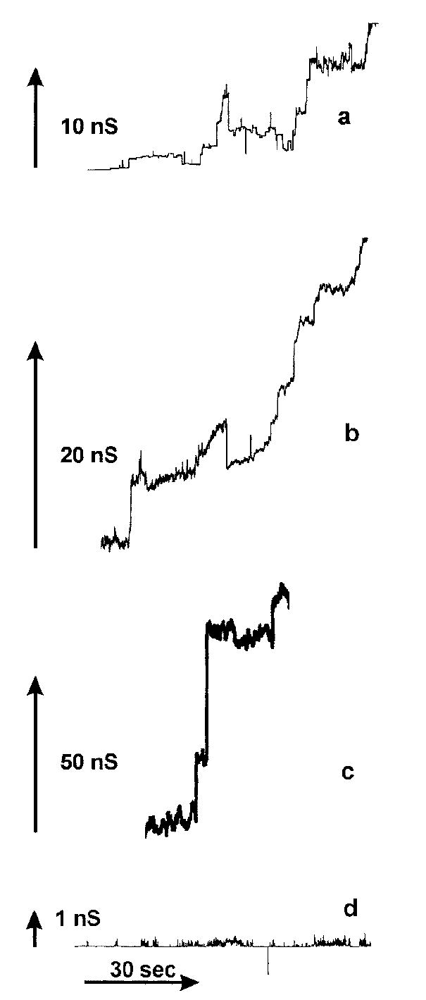
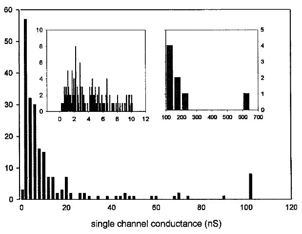
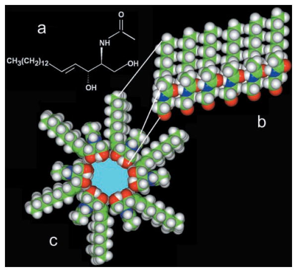
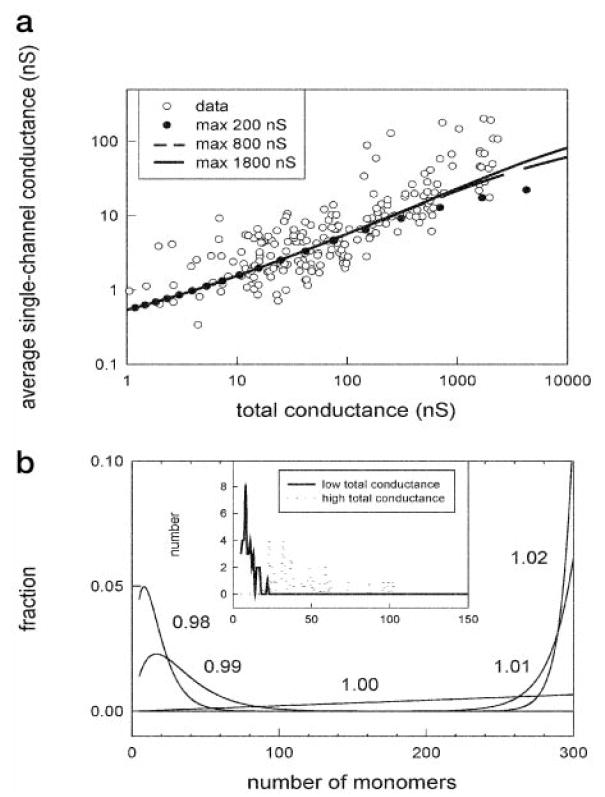
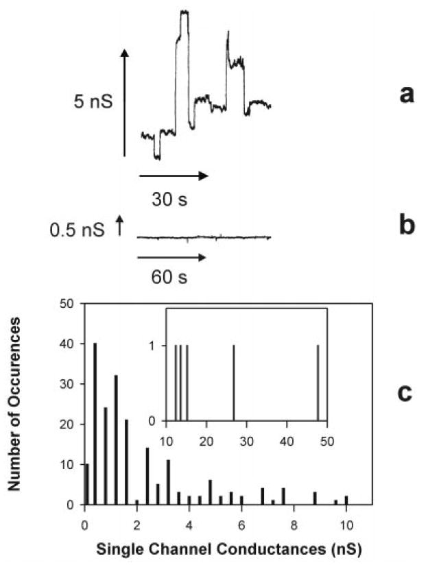
We report that physiological concentrations of both short- and long-chain ceramides, despite being lipids, form large stable pores in membranes. Some of these pores should be large enough to allow cytochrome c to permeate. Dihydroceramide differs from ceramide by the reduction of one double bond, and yet both its apoptogenic and channel-forming activities are greatly reduced. A structural model provides insight into how ceramides might form pores. According to a mathematical model, both the individual conductance of the channels and the overall membrane conductance are directly related to the overall concentration of ceramide in the membrane. Slight changes in concentration have dramatic effects on the size of the channels formed, providing an easy way for rapidly altering membrane permeability by changing the activity of local synthetic and catabolic enzymes. A possible role for these channels in apoptosis is discussed.


Figures

Fig. 1
Fig. 1. C2-ceramide forms channels in planar phospholipid membranes
a–c, continuous current recordings of channel formation induced by the addition of 5 μm C2-ceramide to the aqueous solution, which consisted of 1 m KCl, 1 mm MgCl2, and 5 mm MES (pH 6). The applied voltage was clamped at 9 mV. d, current recordings in the presence of 120 μm C2-dihydroceramide. Conditions were as in a–c.
Fig. 2
Fig. 2. Distribution of single channel conductances produced by 5 μm C2-ceramide
Data were compiled from four separate experiments using the conditions stated for Fig. 1, a–c. Only distinct vertical current increments were measured. The left and right insets are detailed distributions of channels with single channel conductances below 10 nS and greater than 100 nS, respectively.
Fig. 3
Fig. 3. Structural model for ceramide pore formation
a, structure of a C2-ceramide molecule. b, a column of ceramide residues held together by intermolecular hydrogen bonds between amide and carbonyl groups. This column would span the hydrophobic portion of the membrane and, in association with other columns, would form pores of various sizes. c, cross-sectional view of a ceramide channel consisting of 6 columns of ceramide molecules. The columns are held together via intermolecular hydrogen bonds between hydroxyl groups along the channel lumen. The ceramide molecules form an annulus that encloses a polar inner space lined with hydroxyl groups. Note that the hydrocarbon chain of the sphingoid base backbone has been shortened to increase the image size of the polar region.
Fig. 4
Fig. 4. The average size of ceramide channels increased with increasing ceramide content in the membrane
a, the average single channel conductance increased as the total conductance increases. The dots are individual experimental observations, and Equation 4 was used to generate the fitted lines. The size of the largest possible channel was chosen to be 200, 800, or 1800 nS. b, the main figure is a theoretical distribution of single-channel sizes as a function of ceramide monomer concentration in the membrane (numbers next to curves). The inset is a plot of the number of channels observed that would correspond to channels with the indicated number of monomers. The solid line represents the channels formed at low membrane conductance (bottom fifth), and the dotted line represents the channels formed at high conductance (top fifth).
Fig. 5
Fig. 5. C16-Ceramide forms channels in planar phospholipid membranes
a, continuous current recording of channel formation induced by membranes made in the presence of 5 mol % C16-ceramide. The aqueous solution was identical to that used in C2-ceramide experiments. b, typical current recording from a membrane formed in the presence of 5 mol % C18-dihydroceramide. c, distribution of single channel conductances produced by membranes made in the presence of 5 mol % C16-ceramide.

Similar articles

Cited by

  • Sphingosine, a product of ceramide hydrolysis, influences the formation of ceramide channels.
    Elrick MJ, Fluss S, Colombini M. Elrick MJ, et al. Biophys J. 2006 Sep 1;91(5):1749-56. doi: 10.1529/biophysj.106.088443. Epub 2006 Jun 16. Biophys J. 2006. PMID: 16782799 Free PMC article.
  • Molecular mechanisms of cell death: recommendations of the Nomenclature Committee on Cell Death 2018.
    Galluzzi L, Vitale I, Aaronson SA, Abrams JM, Adam D, Agostinis P, Alnemri ES, Altucci L, Amelio I, Andrews DW, Annicchiarico-Petruzzelli M, Antonov AV, Arama E, Baehrecke EH, Barlev NA, Bazan NG, Bernassola F, Bertrand MJM, Bianchi K, Blagosklonny MV, Blomgren K, Borner C, Boya P, Brenner C, Campanella M, Candi E, Carmona-Gutierrez D, Cecconi F, Chan FK, Chandel NS, Cheng EH, Chipuk JE, Cidlowski JA, Ciechanover A, Cohen GM, Conrad M, Cubillos-Ruiz JR, Czabotar PE, D'Angiolella V, Dawson TM, Dawson VL, De Laurenzi V, De Maria R, Debatin KM, DeBerardinis RJ, Deshmukh M, Di Daniele N, Di Virgilio F, Dixit VM, Dixon SJ, Duckett CS, Dynlacht BD, El-Deiry WS, Elrod JW, Fimia GM, Fulda S, García-Sáez AJ, Garg AD, Garrido C, Gavathiotis E, Golstein P, Gottlieb E, Green DR, Greene LA, Gronemeyer H, Gross A, Hajnoczky G, Hardwick JM, Harris IS, Hengartner MO, Hetz C, Ichijo H, Jäättelä M, Joseph B, Jost PJ, Juin PP, Kaiser WJ, Karin M, Kaufmann T, Kepp O, Kimchi A, Kitsis RN, Klionsky DJ, Knight RA, Kumar S, Lee SW, Lemasters JJ, Levine B, Linkermann A, Lipton SA, Lockshin RA, López-Otín C, Lowe SW, Luedde T, Lugli E, MacFarlane M, Madeo F, Malewicz M, Malorni W, Manic G, Marine JC, Marti… See abstract for full author list ➔ Galluzzi L, et al. Cell Death Differ. 2018 Mar;25(3):486-541. doi: 10.1038/s41418-017-0012-4. Epub 2018 Jan 23. Cell Death Differ. 2018. PMID: 29362479 Free PMC article. Review.
  • Ceramide microdomains: the major influencers of the sphingolipid media platform.
    Weber-Stout M, Nicholson RJ, Dumaguit CDC, Holland WL, Summers SA. Weber-Stout M, et al. Biochem Soc Trans. 2024 Aug 28;52(4):1765-1776. doi: 10.1042/BST20231395. Biochem Soc Trans. 2024. PMID: 39082976 Free PMC article. Review.
  • Ceramides and Acute Kidney Injury.
    Nicholson RJ, Holland WL, Summers SA. Nicholson RJ, et al. Semin Nephrol. 2022 May;42(3):151281. doi: 10.1016/j.semnephrol.2022.10.007. Epub 2022 Nov 18. Semin Nephrol. 2022. PMID: 36404212 Free PMC article. Review.
  • The role of sphingolipids and ceramide in pulmonary inflammation in cystic fibrosis.
    Becker KA, Riethmüller J, Zhang Y, Gulbins E. Becker KA, et al. Open Respir Med J. 2010 Mar 30;4:39-47. doi: 10.2174/1874306401004020039. Open Respir Med J. 2010. PMID: 20556203 Free PMC article.

References

    1. Kolesnick RN, Krönke M. Annu Rev Physiol. 1998;60:643–665. - PubMed
    1. Hannun YA. Science. 1996;274:1855–1859. - PubMed
    1. Ariga T, Jarvis WD, Yu RK. J Lipid Res. 1998;39:1–16. - PubMed
    1. Bernardi P, Scorrano L, Colonna R, Petronilli V, Di Lisa F. Eur J Biochem. 1999;264:687–701. - PubMed
    1. Crompton M. Biochem J. 1999;342:233–249. - PMC - PubMed