

Ethylene induces epidermal cell death at the site of adventitious root emergence in rice
- PMID: 11027711
- PMCID: PMC59167
- DOI: 10.1104/pp.124.2.609
Ethylene induces epidermal cell death at the site of adventitious root emergence in rice
Abstract
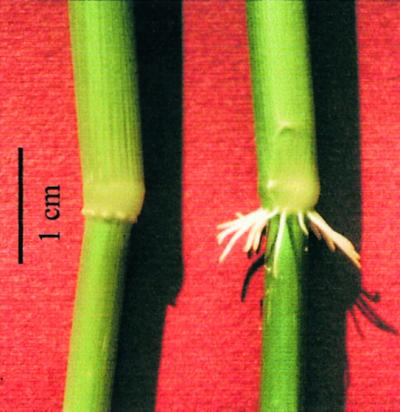
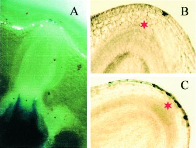
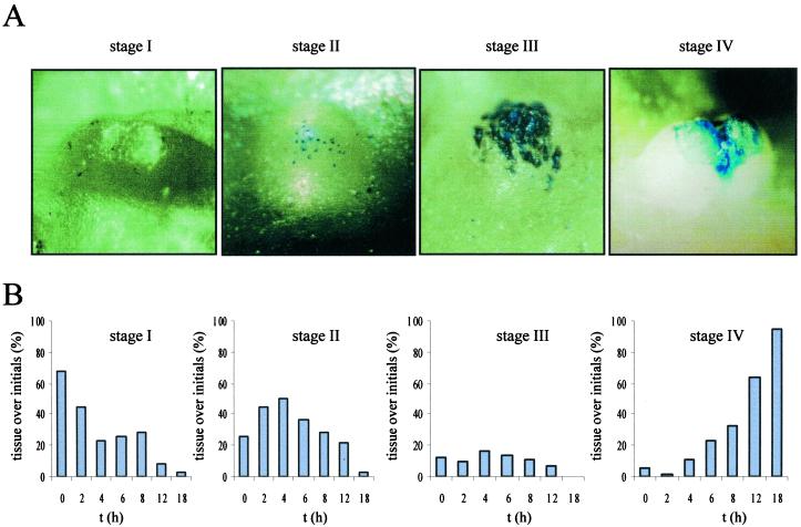
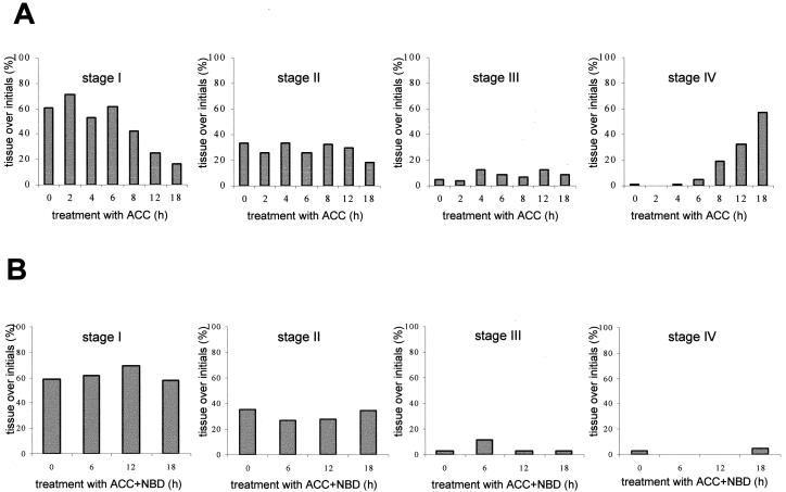
In deepwater rice (Oryza sativa), adventitious root primordia initiate at the nodes as part of normal development. Emergence of the roots is dependent on flooding of the plant and is mediated by ethylene action. Root growth was preceded by the induced death of epidermal cells of the node external to the tip of the root primordium. Cell death proceeded until the epidermis split open. Through this crack the root eventually emerged. Induced death was confined to nodal epidermal cells covering the tip of the primordia. Our results suggest that this process facilitates adventitious root emergence and prevents injury to the growing root. Cell death was inducible not only by submergence but also by application of 1-aminocyclopropane-1-carboxylic acid, the natural precursor of ethylene and it was suppressed in the presence of 2,5-norbornadiene (bicyclo[2.2.1]hepta-2,5-diene), an inhibitor of ethylene action. Adventitious root growth and epidermal cell death are therefore linked to the ethylene signaling pathway, which is activated in response to low oxygen stress.
Figures
Similar articles
-
Interactions between ethylene, gibberellin and abscisic acid regulate emergence and growth rate of adventitious roots in deepwater rice.Planta. 2006 Feb;223(3):604-12. doi: 10.1007/s00425-005-0111-1. Epub 2005 Sep 14. Planta. 2006. PMID: 16160845
-
Epidermal cell death in rice is regulated by ethylene, gibberellin, and abscisic acid.Plant Physiol. 2005 Oct;139(2):713-21. doi: 10.1104/pp.105.064469. Epub 2005 Sep 16. Plant Physiol. 2005. PMID: 16169967 Free PMC article.
-
Heterotrimeric G protein signaling is required for epidermal cell death in rice.Plant Physiol. 2009 Oct;151(2):732-40. doi: 10.1104/pp.109.142133. Epub 2009 Aug 5. Plant Physiol. 2009. PMID: 19656904 Free PMC article.
-
Regulation of submergence-induced enhanced shoot elongation in Oryza sativa L.Ann Bot. 2003 Jan;91 Spec No(2):263-70. doi: 10.1093/aob/mcf121. Ann Bot. 2003. PMID: 12509346 Free PMC article. Review.
-
G proteins as regulators in ethylene-mediated hypoxia signaling.Plant Signal Behav. 2010 Apr;5(4):375-8. doi: 10.4161/psb.5.4.10910. Plant Signal Behav. 2010. PMID: 20948297 Free PMC article. Review.
Cited by
-
Comparative RNA-seq based transcriptome profiling of waterlogging response in cucumber hypocotyls reveals novel insights into the de novo adventitious root primordia initiation.BMC Plant Biol. 2017 Jul 26;17(1):129. doi: 10.1186/s12870-017-1081-8. BMC Plant Biol. 2017. PMID: 28747176 Free PMC article.
-
The Receptor-Like Kinase SIT1 Mediates Salt Sensitivity by Activating MAPK3/6 and Regulating Ethylene Homeostasis in Rice.Plant Cell. 2014 Jun;26(6):2538-2553. doi: 10.1105/tpc.114.125187. Epub 2014 Jun 6. Plant Cell. 2014. PMID: 24907341 Free PMC article.
-
Novel Role of JAC1 in Influencing Photosynthesis, Stomatal Conductance, and Photooxidative Stress Signalling Pathway in Arabidopsis thaliana.Front Plant Sci. 2020 Jul 29;11:1124. doi: 10.3389/fpls.2020.01124. eCollection 2020. Front Plant Sci. 2020. PMID: 32849690 Free PMC article.
-
Environmental stress alters genes expression and induces ovule abortion: reactive oxygen species appear as ovules commit to abort.Planta. 2005 Nov;222(4):632-42. doi: 10.1007/s00425-005-0010-5. Epub 2005 Nov 4. Planta. 2005. PMID: 16133218
-
Genome-wide expression of transcriptomes and their co-expression pattern in subtropical maize (Zea mays L.) under waterlogging stress.PLoS One. 2013 Aug 6;8(8):e70433. doi: 10.1371/journal.pone.0070433. Print 2013. PLoS One. 2013. PMID: 23936429 Free PMC article.
References
-
- Armstrong W, Brändle R, Jackson MB. Mechanisms of flood tolerance in plants. Acta Bot Neerl. 1994;43:307–358.
-
- Bell JK, McCully ME. A histological study of lateral root initiation and development in Zea mays. Protoplasma. 1970;70:179–205.
-
- Bleecker AB, Schuette JL, Kende H. Anatomical analysis of growth and developmental patterns in the internode of deepwater rice. Planta. 1986;169:490–497. - PubMed
-
- Buckner B, Janick-Buckner D, Gray J, Johal GS. Cell-death mechanisms in maize. Trends Plant Sci. 1998;3:218–223.
Publication types
MeSH terms
Substances
LinkOut - more resources
Full Text Sources
