. 2000 Oct;124(2):665-80.
doi: 10.1104/pp.124.2.665.

Development of peltate glandular trichomes of peppermint

Affiliations

Development of peltate glandular trichomes of peppermint

G W Turner et al. Plant Physiol. 2000 Oct.

Abstract

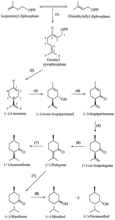
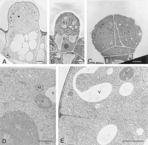
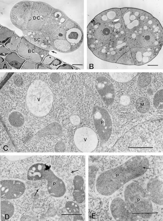
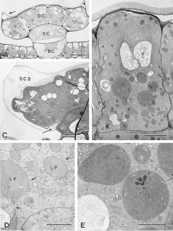
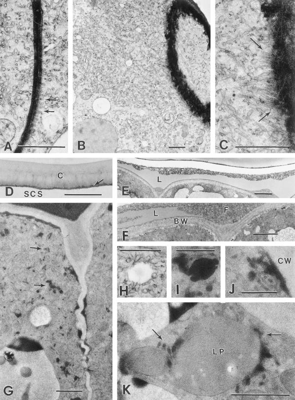
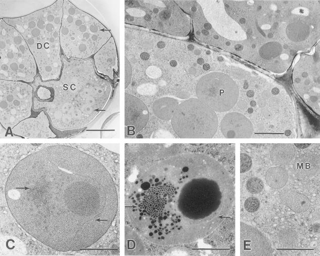
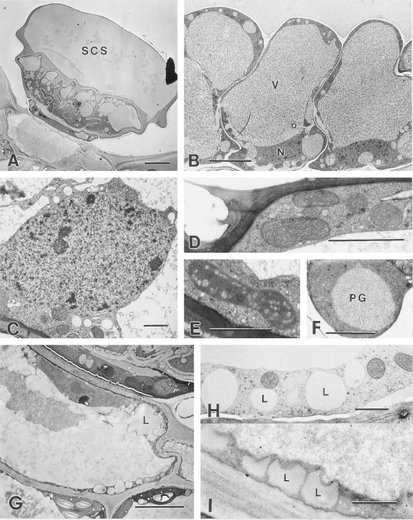
Cryofixation and conventional chemical fixation methods were employed to examine the ultrastructure of developing peltate glandular trichomes of peppermint (Mentha x piperita). Our results are discussed in relation to monoterpene production and the mechanism of essential oil secretion. Peltate glands arise as epidermal protuberances (initials) that divide asymmetrically to produce a vacuolate basal cell, a stalk cell, and a cytoplasmically dense apical cell. Further divisions of the apical cell produce a peltate trichome with one basal cell, one stalk cell, and eight glandular (secretory) disc cells. Presecretory gland cells resemble meristematic cells because they contain proplastids, small vacuoles, and large nuclei. The secretory phase coincides with the separation and filling of the sub-cuticular oil storage space, the maturation of glandular disc cell leucoplasts in which monoterpene biosynthesis is known to be initiated, and the formation of extensive smooth endoplasmic reticulum at which hydroxylation steps of the monoterpene biosynthetic pathway occur. The smooth endoplasmic reticulum of the secretory cells appears to form associations with both the leucoplasts and the plasma membrane bordering the sub-cuticular oil storage cavity, often contains densely staining material, and may be involved with the transport of the monoterpene-rich secretion product. Associated changes in the ultrastructure of the secretory stage stalk cell are also described, as is the ultrastructure of the fragile post-secretory gland for which cryofixation methods are particularly well suited for the preservation of organizational integrity.


Figures

Figure 1
Figure 1
The principal pathway for monoterpene biosynthesis in peppermint. The responsible enzymes are: geranyl diphosphate synthase (1), (4S)-(−)-limonene synthase (2), cytochrome P450 (−)-limonene-3-hydroxylase (3), (−)-trans-isopiperitenol dehydrogenase (4), (−)-isopiperitenone reductase (5), (+)-cis-isopulegone isomerase (6), (+)-pulegone reductase (7), and (−)-menthone reductase (8).
Figure 2
Figure 2
Early presecretory stage. A, Cryofixed glandular trichome initial consisting of a single cell with a vacuolate basal region and an apical region containing the nucleus, numerous mitochondria, and plastids, but few vacuoles. Bar = 2 μm. B, Chemically fixed glandular trichome initial after periclinal cell divisions, with a vacuolate basal cell (BC), narrow stalk cell (SC), and the apical disc initial cell (AI). Bar = 5 μm. C, Chemically fixed developing peltate glandular trichome with four apical disc cells (DC). Bar = 4 μm. D, Cryofixed apical initial of a developing glandular trichome with a single apical cell. The nucleus and nucleolus are large and the ribosomes are abundant but ER is relatively sparse. P, Proplastid; M, mitochondrion; V, vacuole. Bar = 1 μm. E, Periclinal section through a cryofixed, two-celled, apical disc. P, Proplastid; V, vacuole. Bar = 1 μm.
Figure 3
Figure 3
Middle- to late-presecretory stage. A, Transverse section through a chemically fixed presecretory peltate gland with an eight-celled apical disc. BC, Basal cell; SC, stalk cell; DC, disc cell. Arrow indicates a thickening stalk cell lateral wall. Bar = 5 mm. B, Periclinal section through a cryofixed apical disc of a presecretory stage gland. Bar = 5 μm. C, Higher magnification of the specimen shown in (B). The disc cells appear typical of developing plant cells with numerous ribosomes, small vacuoles (V), abundant Golgi, mitochondria (M), and small proplastids (P). Bar = 1 μm. D, Cryofixed apical disc cells of a late-presecretory gland. SER (arrows) is relatively abundant near the enlarging plastids (P). Bar = 1 μm. E, Cryofixed stalk cell of a late-presecretory stage gland. Plastids (P) remain relatively narrow and contain numerous tubular membranes (arrow). Bar = 1 μm.
Figure 4
Figure 4
Secretory stage. A, Cryofixed early-secretory stage peltate gland. DC, Apical disc cell; SC, stalk cell; BC, basal cell. Arrow indicates the lateral rim of raised cuticle. Bar = 10 μm. B, Cryofixed apical disc cell of a secretory stage peltate gland. Large leucoplasts (LP) occur mainly in the basal one-half of the cell. An extensive SER occurs throughout the cell, except near vacuoles (V). Lateral disc cell walls remain thin and have become densely staining. Bar = 3 μm. C, Chemically fixed secretory stage peltate gland. Arrow indicates the lowermost extension of the sub-cuticular oil storage space (SCS) at the juncture of the stalk and disc cells. Bar = 5 μm. D, Portion of a cryofixed disc cell showing leucoplasts (LP) in close contact with SER. Upper arrows indicate apparent contact between SER and plastid membranes. The structure indicated by the lower arrow is a narrow portion (near the edge) of a leucoplast branch. Bar = 1 μm. E, Cryofixed leucoplasts (LP) of a secretory stage gland in close contact with SER. Bar = 1 μm.
Figure 5
Figure 5
Secretory stage. A, SER adjacent to a densely staining radial wall of a cryofixed disc cell showing the apparent alignment of the SER along the plasma membrane (arrows). Bar = 1 μm. B, Extensively developed SER in a cryofixed secretory stage disc cell, near a glancing section through the boundary wall bordering the sub-cuticular storage space (upper right). Bar = 1 μm. C, Higher magnification of (B) showing the alignment of SER adjacent to the plasma membrane along the boundary wall (arrows). Bar = 1 μm. D, Elevated cuticle (C) at the apex of a cryofixed secretory stage gland. Arrow indicates a thin layer of residual cell wall. Bar = 1 μm. E, Sub-cuticular storage space in the apical region of a chemically fixed early secretory stage peltate gland containing lipid-like material (L) and fibrillar material (F). Bar = 2 μm. F, Higher magnification of a sub-cuticular region of a chemically fixed peltate gland showing layers of lipid-like material (L) within the fibrillar matrix (F) near the disc cell boundary cell wall (BW). Bar = 1 μm. G, Microwave-fixed glandular disc cells of a secretory phase peltate gland. Note the dark lipid-like deposits within the SER (arrows) and along the lateral cell walls. Bar = 1 μm. H, Vesicle-like structure in close contact with SER of a cryofixed glandular disc cell. Equivalent structures in microwave-fixed tissues consistently contain osmiophilic material. Compare with (I). Bar = 0.5 μm. I, Lipid-containing vesicle in close contact with SER of a microwave-fixed glandular disc cell. Bar = 0.5 μm. J, Enlargement of (G) showing a lipid-filled region of SER in close contact with the plasma membrane along the lateral cell wall (CW). Bar = 0.5 μm. K, Leucoplast (LP) from a microwave-fixed glandular disc cell in close contact with lipid-filled periplastic SER (arrows). Bar = 1 μm.
Figure 6
Figure 6
A, Peridermal section through glandular disc cells (DC) and the stalk cell (SC) of a cryofixed secretory stage peltate gland. Leucoplasts (upper arrow) of disc cells stain darkly and have few plastoglobuli, whereas stalk cell plastids (lower arrow) stain lightly and have large plastoglobuli. Bar = 10 μm. B, Transverse section through a cryofixed secretory stage gland. The stalk cell contains large plastids (P), abundant SER, numerous darkly staining mitochondria, and numerous lightly stained microbodies. Bar = 2 μm. C, Typical stalk cell plastid from a cryofixed gland. The left arrow indicates a small prolamellar body-like region of crystaloid plastid membranes. The right arrow indicates an isolated membrane forming a pocket around the large plastoglobule. Bar = 1 μm. D, Typical glandular stalk cell plastid from a chemically fixed peltate gland showing a similar prolamellar body-like region (arrow), and an isolated membrane enclosing a large plastoglobule. Compare with (C). Bar = 1 μm. E, Microbodies (MB) surrounded by an extensive SER in the stalk cell of a cryofixed secretory stage gland. Bar = 1 μm.
Figure 7
Figure 7
Post-secretory stage. A, Chemically fixed post-secretory stage peltate gland showing a large SCS containing lipid above the fibrillar material coating the glandular disc cells. Bar = 10 μm. B, Cryofixed glandular disc cells from a post-secretory phase peltate gland. The cell walls show less distortion than those of post-secretory stage glands prepared by chemical fixation. The cells contain nuclei (N), mitochondria, and large central vacuoles (V). Bar = 5 μm. C, Typical nucleus from a cryofixed glandular disc cell containing heterochromatin and lacking well-developed nucleoli. Bar = 1 μm. D, Mitochondria adjacent to the sub-cuticular space of a cryofixed post-secretory glandular disc cell. Bar = 1 μm. E, Typical plastid from a cryofixed post-secretory glandular disc cell that is smaller than the large leucoplasts of secretory stage glands. Bar = 1 μm. F, Cryofixed stalk cell plastid with large plastoglobule (PG). Bar = 1 μm. G, Chemically fixed basal cell of a post-secretory stage peltate gland. The peripheral cytoplasm contains numerous lipid spherosomes (L). Bar = 5 μm. H, Cryofixed peripheral cytoplasm of a post-secretory gland basal cell showing vacuole-like structures (L) that correspond to lipid spherosomes of chemically fixed specimens. Compare with (I). Bar = 1 μm. I, Peripheral cytoplasm showing lipid spherosomes (L) of a chemically fixed post-secretory gland basal cell. Compare with (H). Bar = 1 μm.

References

    1. Amelunxen F. Elektronenmikroskopische Untersuchungen an den Drüsenschuppen von Mentha piperita L. Plant Med. 1965;13:457–473.
    1. Ascensão L, Marques N, Pais MS. Peltate glandular trichomes of Leonotis leonurus leaves: ultrastructure and histochemical characterization of secretions. Int J Plant Sci. 1997;158:249–258.
    1. Bisio A, Corallo A, Gastaldo P, Romussi G, Ciarallo G, Fontana N, De Tommasi N, Profumo P. Glandular hairs and secreted material in Salvia blepharophylla Brandegee ex Epling grown in Italy. Ann Bot. 1999;83:441–452.
    1. Black SM, Harikrishna JA, Szklarz GD, Miller WL. The mitochondrial environment is required for the activity of the cholesterol side-chain cleavage enzyme, cytochrome P450scc. Proc Natl Acad Sci USA. 1994;91:7247–7251. - PMC - PubMed
    1. Bosabalidis A, Gabrieli C, Niopas I. Flavone aglycones in glandular hairs of Origanum × intercedens. Phytochemistry. 1998;49:1549–1553. - PubMed

Publication types

LinkOut - more resources