. 2000 Oct;11(10):3277-88.
doi: 10.1091/mbc.11.10.3277.

The diabetes autoantigen ICA69 and its Caenorhabditis elegans homologue, ric-19, are conserved regulators of neuroendocrine secretion

Affiliations
Free PMC article

The diabetes autoantigen ICA69 and its Caenorhabditis elegans homologue, ric-19, are conserved regulators of neuroendocrine secretion

M Pilon et al. Mol Biol Cell. 2000 Oct.
Free PMC article

Abstract

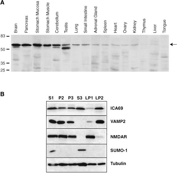
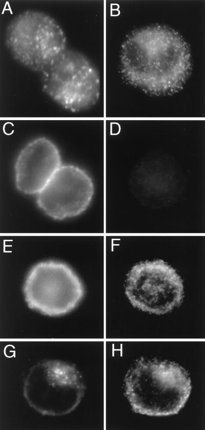
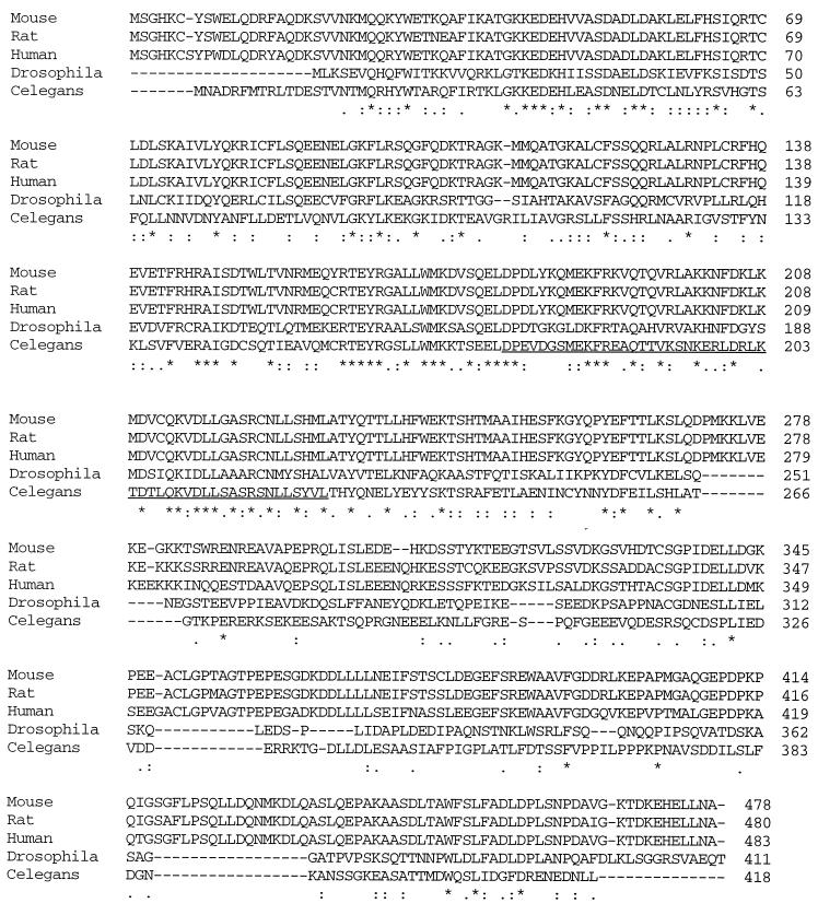
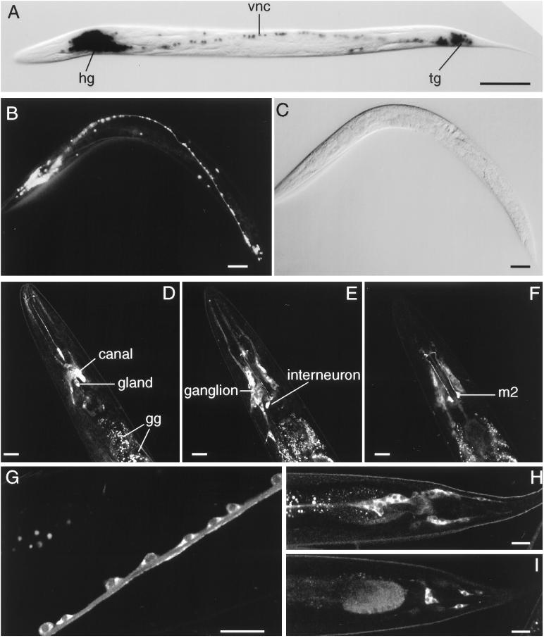
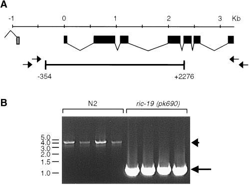
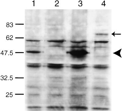
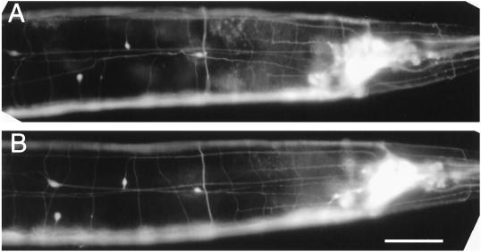
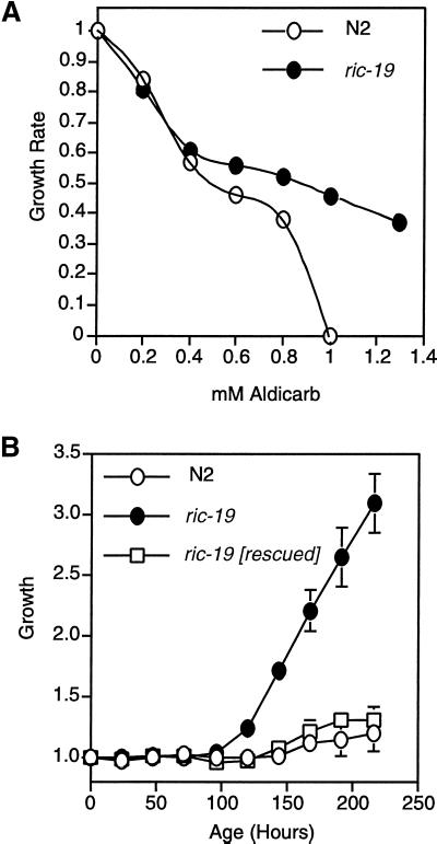
ICA69 is a diabetes autoantigen with no homologue of known function. Given that most diabetes autoantigens are associated with neuroendocrine secretory vesicles, we sought to determine if this is also the case for ICA69 and whether this protein participates in the process of neuroendocrine secretion. Western blot analysis of ICA69 tissue distribution in the mouse revealed a correlation between expression levels and secretory activity, with the highest expression levels in brain, pancreas, and stomach mucosa. Subcellular fractionation of mouse brain revealed that although most of the ICA69 pool is cytosolic and soluble, a subpopulation is membrane-bound and coenriched with synaptic vesicles. We used immunostaining in the HIT insulin-secreting beta-cell line to show that ICA69 localizes in a punctate manner distinct from the insulin granules, suggesting an association with the synaptic-like microvesicles found in these cells. To pursue functional studies on ICA69, we chose to use the model organism Caenorhabditis elegans, for which a homologue of ICA69 exists. We show that the promoter of the C. elegans ICA69 homologue is specifically expressed in all neurons and specialized secretory cells. A deletion mutant was isolated and found to exhibit resistance to the drug aldicarb (an inhibitor of acetylcholinesterase), suggesting defective neurotransmitter secretion in the mutant. On the basis of the aldicarb resistance phenotype, we named the gene ric-19 (resistance to inhibitors of cholinesterase-19). The resistance to aldicarb was rescued by introducing a ric-19 transgene into the ric-19 mutant background. This is the first study aimed at dissecting ICA69 function, and our results are consistent with the interpretation that ICA69/RIC-19 is an evolutionarily conserved cytosolic protein participating in the process of neuroendocrine secretion via association with certain secretory vesicles.


Figures

Figure 1
Figure 1
(A) Tissue distribution of mouse ICA69. A total of 40 μg of protein from the various tissues was loaded in each lane. The predominant ICA69 band is indicated by the arrow. (B) Subcellular fractionation of mouse brain and Western blot detection of ICA69, VAMP-2, NMDAR, SUMO-1, and β-tubulin is shown. The nature of the different fractions is as follows: S1, postnuclear supernatant; P2, crude synaptosomes; P3, microsomes; S3, cytosolic soluble; LP1, plasma membranes of synaptosomes; LP2, synaptic vesicles. A total of 40 μg of protein was loaded in each lane.
Figure 2
Figure 2
Immunodetection of ICA69, SNAP-25, and insulin in HIT-1 cells. (A) Two cells stained with anti-ICA69. Note the punctate distribution of the staining. (B) One cell stained with anti-insulin. Again note the punctate staining pattern. (C) Two cells stained with anti-SNAP-25. Note that the signal is localized to the plasma membrane. (D) One cell stained with a control guinea pig antibody. Note the absence of staining. (E and F) One cell costained for SNAP-25 (E) and insulin (F). Note the absence of colocalization. (G and H) One cell costained for ICA69 (G) and insulin (H). Note that although both antigens exhibit a punctate distribution, the signals do not exhibit colocalization (compare G and H).
Figure 3
Figure 3
Alignment of the ICA69 homologues. Below the sequences, stars, colons, and periods indicate that amino acid positions are identical, highly conserved, or weakly conserved, respectively. The region predicted to encode a coiled-coil motif is underlined in the C. elegans sequence.
Figure 4
Figure 4
(A–C) In vivo ric-19 promoter expression visualized with the use of the NLS–GFP or NLS–lacZ reporter genes. (A) NLS–lacZ staining in an animal transgenic for the array Ex[pCelicaDP-lacZ; pRF4]. (B) NLS–GFP expression in a young adult transgenic animal carrying the array Ex[pCelicaDP-GFP; pRF4]. (C) Nomarski view of panel b. hg, head ganglia; vnc, ventral nerve cord; tg, tail ganglia. Bars in A–C, 50 μm. (D–I) Confocal microscopy of transgenic animals carrying the array Ex[pRIC19::GFP; pRF4]. (D–F) Three confocal planes of the head region. In D, the excretory canal cell and gland cells are indicated; the autofluorescent gut granules (gg) are also indicated. Note the expression of the RIC-19::GFP protein in neurons of the head ganglion (most visible in E) as well as the high levels of expression in a pharyngeal interneuron (E) and m2 neurons (F). (G) A portion of the ventral nerve cord; note that the RIC-19::GFP fusion protein is expressed in the cytoplasm of each neuron shown. (H and I) Two sections of the tail region; note the expression of the RIC-19::GFP protein in the neurons of the tail ganglia. Bars in D–I, 20 μm.
Figure 5
Figure 5
(A) Structure of the ric-19 gene as well as the approximate positions of the primers used to screen and isolate a deletion mutation spanning positions −354 to 2276. (B) Example of single-worm PCR reactions with the use of the primers AR1 and AL1 on N2 worms (lanes 1–4) or worms homozygous for the ric-19(pk690) allele (lanes 5–8). The arrowhead and arrow in B indicate the amplified wild-type and pk690 alleles of ric-19, respectively.
Figure 6
Figure 6
Western blot detection of the RIC-19 protein. Five micrograms of total worm protein was loaded in each lane and probed with a rabbit antiserum directed against a peptide corresponding to the C-terminal 20 amino acids of the RIC-19 protein. Lane 1, N2; lane 2, AS241; lane 3, AS241 transgenic for the array Ex[pRIC-19; pRF4]; lane 4, AS241 transgenic for the array Ex[pRIC19::GFP; pRF4]. The arrowhead indicates the position of the RIC-19 protein, and the arrow indicates the position of the RIC-19::GFP fusion protein.
Figure 7
Figure 7
ric-19(pk690) animals exhibit no obvious abnormality in neuroarchitecture. Expression of the integrated panneuronal GFP array (evIs11) in wild-type (A) or ric-19(pk690) mutant background (B). Note that the variation in the positions of the four isolated nuclei visible in the left half of each panel is well within the norm. Bar, 20 μm.
Figure 8
Figure 8
The ric-19(pk690) mutant exhibits resistance to aldicarb. (A) Relative growth rate assay. (B) Growth rate assay on NGM plates containing 1 mM aldicarb (see MATERIALS AND METHODS). ric-19 [rescued] is a ric-19(pk690) mutant strain that carries an integrated array harboring the pRIC-19 expression plasmid. Error bars show the SD among five separate plates.

Similar articles

Cited by

References

    1. Aanstoot HJ, et al. Identification and characterization of glima 38, a glycosylated islet cell membrane antigen, which together with GAD65 and IA2 marks the early phases of autoimmune response in type I diabetes. J Clin Invest. 1996;97:2772–2783. - PMC - PubMed
    1. Anderson GL. Responses of dauer larvae of Caenorhabditis elegans (Nematoda:Rhabditidae) to thermal stress and oxygen deprivation. Can J Zool. 1978;56:1786–1791.
    1. Avery L. Motor neuron M3 controls pharyngeal muscle relaxation timing in Caenorhabditis elegans. J Exp Biol. 1993;175:283–297. - PubMed
    1. Baekkeskov S, Aanstoot H, Christgau S, Reetz A, Solimena M, Cascalho M, Folli F, Richter-Olsen H, De Camilli P. Identification of the 64K autoantigen in insulin dependent diabetes as the GABA-synthesizing enzyme glutamic acid decarboxylase. Nature. 1990;347:151–156. - PubMed
    1. Bargmann CI, Harwieg E, Horvitz HR. Odorant-selective genes and neurons mediate olfaction in C. elegans. Cell. 1993;74:515–527. - PubMed

Publication types

MeSH terms