. 2000 Oct;11(10):3289-98.
doi: 10.1091/mbc.11.10.3289.

The R-SNARE endobrevin/VAMP-8 mediates homotypic fusion of early endosomes and late endosomes

Affiliations
Free PMC article

The R-SNARE endobrevin/VAMP-8 mediates homotypic fusion of early endosomes and late endosomes

W Antonin et al. Mol Biol Cell. 2000 Oct.
Free PMC article

Abstract

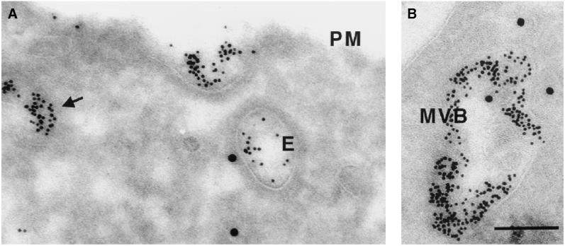
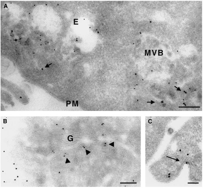
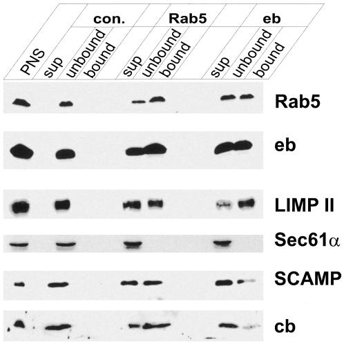
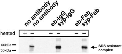
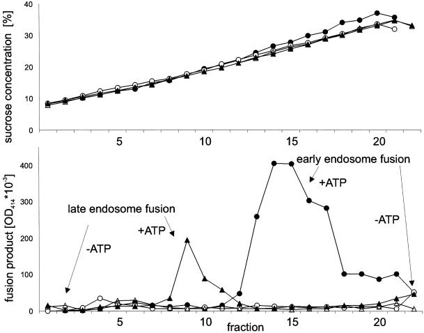
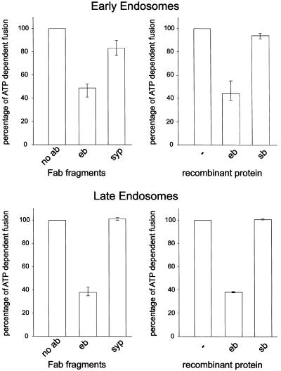
Endobrevin/VAMP-8 is an R-SNARE localized to endosomes, but it is unknown in which intracellular fusion step it operates. Using subcellular fractionation and quantitative immunogold electron microscopy, we found that endobrevin/VAMP-8 is present on all membranes known to communicate with early endosomes, including the plasma membrane, clathrin-coated pits, late endosomes, and membranes of the trans-Golgi network. Affinity-purified antibodies that block the ability of endobrevin/VAMP-8 to form SNARE core complexes potently inhibit homotypic fusion of both early and late endosomes in vitro. Fab fragments were as active as intact immunoglobulin Gs. Recombinant endobrevin/VAMP-8 inhibited both fusion reactions with similar potency. We conclude that endobrevin/VAMP-8 operates as an R-SNARE in the homotypic fusion of early and late endosomes.


Figures

Figure 1
Figure 1
Ultrastructural analysis of endobrevin localization. NRK cells were incubated for 15 min with BSA-gold (5 nm) before fixation and immunolabeling of endobrevin with the use of an affinity-purified rabbit serum and 15 nm protein A–gold (see MATERIALS AND METHODS). PM, plasma membrane; E, endosome of vacuolar type; MVB, multivesicular body. Note the association of endobrevin with tubulovesicular structures (A, triangle), with endosomal structures of vacuolar type (A, and with endosomes appearing as multivesicular bodies (B). These endobrevin-positive compartments are also positive for the endocytic tracer. Bar, 100 nm.
Figure 2
Figure 2
Immunogold localization of endobrevin and LIMP II. Ultrathin cryosections of NRK cells were immunolabeled for endobrevin (15-nm protein A–gold) and either LIMP II (10-nm protein A–gold; A) or the endogenous MPR46 (10-nm protein A–gold; B and C). Endobrevin is detectable in tubulovesicular structures (arrows in A), underneath the plasma membrane (PM), and on endosomal vacuoles (E) and endosomes that appear as multivesicular bodies (MVB), where it often colocalizes with the lysosomal membrane protein LIMP II. We also noted a colocalization of endobrevin with MPR46 in TGN-associated structures (B, arrowheads) and occasionally the appearance in coated pits/vesicles at the plasma membrane (C, arrow). Bars, 100 nm.
Figure 3
Figure 3
Characterization of organelles immunoisolated with microbeads containing antibodies specific for endobrevin and Rab5. A PNS from rat liver was incubated with small amounts of immunobeads containing covalently bound antibodies specific for Rab5 and endobrevin (eb). Glycine-inactivated beads (con.) served as control for nonspecific binding. The protein composition of the bead-bound membranes (bound) was compared with that of unbound membranes that were sedimented by ultracentrifugation (unbound). Equal proportions of all fractions were analyzed by immunoblotting. PNS, postnuclear supernatant used as starting material; sup, membrane-free supernatant obtained after ultracentrifugation and concentrated by precipitation according to Wessel and Flügge (1984); cb, cellubrevin. PNS, supernatant, and unbound fractions, 10 μg of protein/lane; bead-bound material, ∼6 μl of beads. Note that the membrane protein content of the beads is at least 5- to 10-fold lower than of the unbound fraction. Also, note that to saturate the beads with organelles, an excess of PNS was used, explaining why none of the antigens was depleted.
Figure 4
Figure 4
Antibodies specific for endobrevin prevent formation of SDS-resistant SNARE complexes with SNAP-25 and syntaxin 1A. Endobrevin was preincubated with affinity-purified antibodies (IgG) or Fab fragments specific for endobrevin (eb) or synaptophysin (syp) followed by the addition of preassembled SNAP-25/syntaxin 1 complexes. Formation of SDS-resistant complexes was monitored by immunoblotting with the use of an antibody specific for syntaxin 1. SDS-resistant core complexes are dissociated by boiling (left lane; cf. Hayashi et al., 1994).
Figure 5
Figure 5
Separation of early and late endosomes by continuous sucrose density gradient centrifugation after completion of the homotypic fusion reaction. Fusion reactions were carried out for early (circles) and late (triangles) endosomes in the presence (closed symbols) or absence (open symbols) of ATP and then loaded in parallel on top of 10–40% continuous sucrose gradients (see MATERIALS AND METHODS). At the end of the run, fractions of 0.5 ml were collected manually and analyzed by immunoprecipitation for the presence of fusion product. Each immunoprecipitate was assayed for HRP activity with the use of a photometric assay (bottom). The top panel shows the sucrose concentration of each gradient as determined by refractometry (data were obtained from a single experiment).
Figure 6
Figure 6
Fusion of early and late endosomes is inhibited by Fab fragments specific for endobrevin or by recombinant endobrevin. Fusion of early and late endosomes derived from PC12 cells was monitored in vitro with the use of a content-mixing assay (see MATERIALS AND METHODS). For preincubation of PNS, 1.2 μM Fab fragments specific for endobrevin (eb) or synaptophysin (syp) (left panels) or 30 μM soluble recombinant endobrevin or synaptobrevin (sb) (right panels) was used. ATP-dependent fusion activity in the absence of Fab fragments or recombinant protein is defined as 100%. Values are given as means of four independent experiments, and bars indicate the range. Fusion was initiated by mixing donor and acceptor fractions while simultaneously adding ATP and cytosol, followed by 30 min of incubation.

References

    1. Advani RJ, Bae HR, Bock JB, Chao DS, Doung YC, Prekeris R, Yoo JS, Scheller RH. Seven novel mammalian SNARE proteins localize to distinct membrane compartments. J Biol Chem. 1998;273:10317–10324. - PubMed
    1. Advani RJ, Yang B, Prekeris R, Lee KC, Klumperman J, Scheller RH. VAMP-7 mediates vesicular transport from endosomes to lysosomes. J Cell Biol. 1999;146:765–776. - PMC - PubMed
    1. Aniento F, Emans N, Griffiths G, Gruenberg J. Cytoplasmic dynein-dependent vesicular transport from early to late endosomes. J Cell Biol. 1993;123:1373–1387. - PMC - PubMed
    1. Annaert WG, Becker B, Kistner U, Reth M, Jahn R. Export of cellubrevin from the endoplasmic reticulum is controlled by BAP31. J Cell Biol. 1997;139:1397–1410. - PMC - PubMed
    1. Barnstable CJ, Hofstein R, Akagawa K. A marker of early amacrine cell development in rat retina. Brain Res. 1985;352:286–290. - PubMed

Publication types

MeSH terms

LinkOut - more resources