. 2000 Oct;11(10):3315-27.
doi: 10.1091/mbc.11.10.3315.

Forced expression of keratin 16 alters the adhesion, differentiation, and migration of mouse skin keratinocytes

Affiliations
Free PMC article

Forced expression of keratin 16 alters the adhesion, differentiation, and migration of mouse skin keratinocytes

M Wawersik et al. Mol Biol Cell. 2000 Oct.
Free PMC article

Abstract

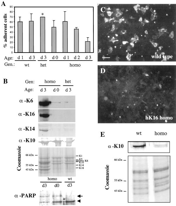
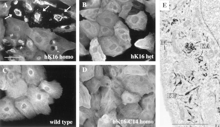
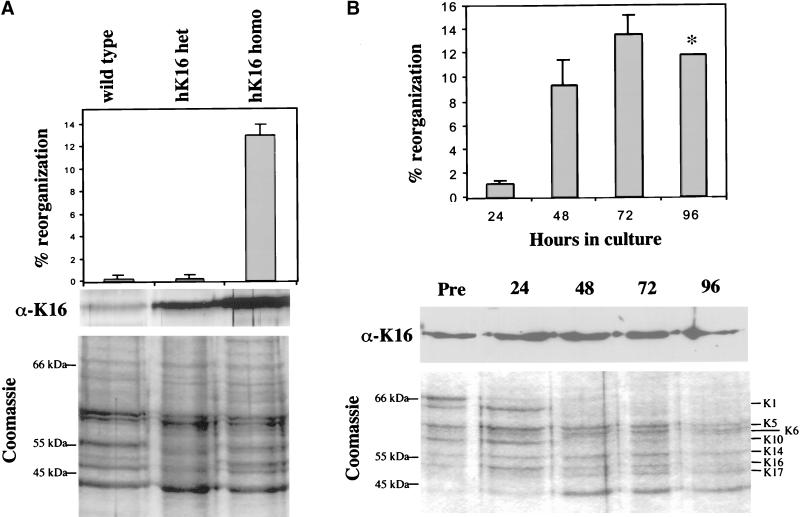
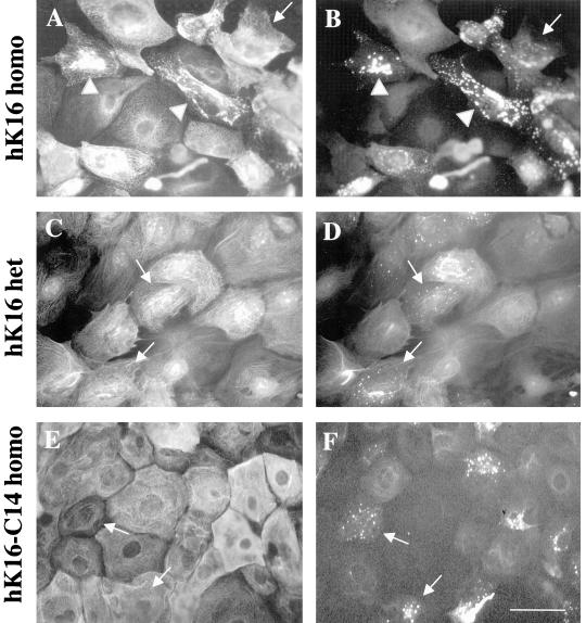
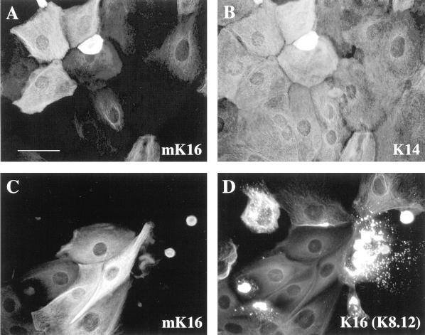
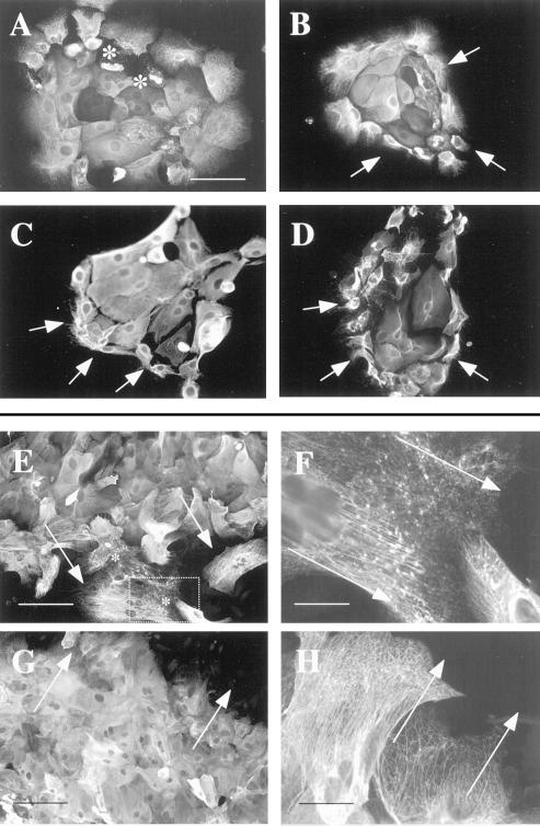
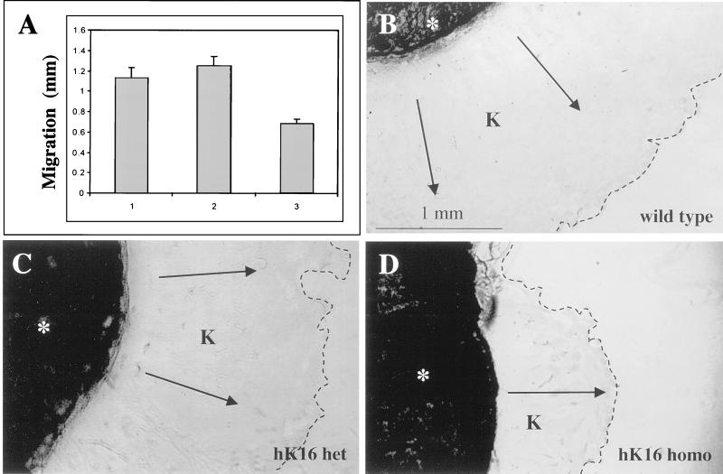
Injury to the skin results in an induction of keratins K6, K16, and K17 concomitant with activation of keratinocytes for reepithelialization. Forced expression of human K16 in skin epithelia of transgenic mice causes a phenotype that mimics several aspects of keratinocyte activation. Two types of transgenic keratinocytes, with forced expression of either human K16 or a K16-C14 chimeric cDNA, were analyzed in primary culture to assess the impact of K16 expression at a cellular level. High K16-C14-expressing and low K16-expressing transgenic keratinocytes behave similar to wild type in all aspects tested. In contrast, high K16-expressing transgenic keratinocytes show alterations in plating efficiency and calcium-induced differentiation, but proliferate normally. Migration of keratinocytes is reduced in K16 transgenic skin explants compared with controls. Finally, a subset of high K16-expressing transgenic keratinocytes develops major changes in the organization of keratin filaments in a time- and calcium concentration-dependent manner. These changes coincide with alterations in keratin content while the steady-state levels of K16 protein remain stable. We conclude that forced expression of K16 in progenitor skin keratinocytes directly impacts properties such as adhesion, differentiation, and migration, and that these effects depend upon determinants contained within its carboxy terminus.


Figures

Figure 1
Figure 1
Plating efficiency and differentiation of K16 ectopic keratinocytes. (A) Plating efficiency of 0–3-d old (d 0, d 1, d 2, or d 3) keratinocytes isolated from wild-type (wt), heterozygous (het), or homozygous (homo) K16 ectopic mice was determined after 6 h in standard calcium (0.2 mM) medium conditions. Adherent and nonadherent keratinocytes were collected, counted, and plating efficiency was determined as a ratio (mean ± SE). With the exception of d 3 heterozygous keratinocytes (∗), plating efficiency was quantitated two or more times. (B) Total protein extracts prepared from nonadherent cells were electrophoresed and revealed by Coomassie blue staining or Western blotting. When probed with antibodies for K6, K14, or K16 (1275), extracts prepared from 3-d-old homozygous K16 keratinocytes (d 3, homo) showed increased amounts of these antigens compared with 0-d-old K16 homozygous (d 0; homo) or 3-d-old K16 heterozygous (d 3; het) keratinocytes. Levels of K10 antigen are similar in all three types of keratinocytes. Coomassie staining of duplicate gels show the relative amounts of keratins present in these samples. When reacted with antibodies directed against PARP (α-PARP), d 3 homozygous K16 keratinocyte (d 3; homo) proteins show similar amounts of uncleaved (arrow) and cleaved (arrowhead) antigen, whereas d 0 homozygous and d 3 wild-type keratinocytes display a slight increase in the 85-kDa cleavage product. (C and D) To assess differentiation, equal numbers of keratinocytes were plated and grown to confluency under low-calcium (0.05 mM) conditions for 24 h and then shifted to high calcium (2 mM) for 24 h. Differentiation of wild-type (C) and homozygous K16 keratinocytes (D) was assessed by immunostaining for K10. Bar, 15 μm. (E) Protein extracts from keratinocytes grown under high-calcium conditions were subjected to SDS-PAGE, transfer onto nitrocellulose, and Western blot analysis. There is significantly less K10 antigen in homozygous K16 keratinocytes (homo) compared with wild-type (see Western blot and Coomassie-stained gel), confirming the immunoflorescence data.
Figure 2
Figure 2
Organization of keratin filaments in primary keratinocyte cultures. (A–D) Keratinocytes from wild-type, homozygous K16-C14 ectopic, and heterozygous and homozygous K16 ectopic keratinocytes were isolated and grown in culture for 72 h in standard calcium (0.2 mM) medium conditions and then immunostained to reveal keratin filament organization. (A) K16 homozygous cultures stained with the anti-K16 (1275) polyclonal antibody show striking alterations in filament organization in a subset of cells (arrows). No filament reorganization is observed in heterozygous K16 (B) cultures immunostained with anti-K16 anti-K16 (1275), or in wild-type (C) and homozygous K16-C14 (D) cultures immunostained for K17. Bar, 20 μm. (E) Similar homozygous K16 cultures were processed for electron microscopy studies. A subset of keratinocytes shows large electron-dense aggregates near the nucleus that are consistent with keratin aggregates (KA). Adjacent to these aggregates are short keratin filaments (KF). The mitochondria (M) and nuclei (N) in these cells are intact. Bar, 2 μm.
Figure 3
Figure 3
Dose and time dependency of keratin filament reorganization. (A) The percentage of keratinocytes showing severe alterations in keratin filament organization, as assessed by immunostaining, was determined in wild-type, heterozygous K16, and homozygous K16 cultures grown for 72 h in 0.2 mM calcium-containing medium. Bar graph: homozygous K16 keratinocytes show 13 ± 0.9% filament reorganization, whereas wild-type and heterozygous K16 cultures show <1%. To relate the degree of filament reorganization to transgene protein dose, total protein extracts were prepared and analyzed. Quantitation of K16 levels in these extracts through Western blotting indicates that homozygous K16 keratinocytes contain 2-fold more K16 than in heterozygous K16 cells. Levels of endogenous mouse K16 observed in wild-type keratinocyte extracts are much lower in comparison. Coomassie staining shows that equal amounts of protein were loaded from these extracts. (B) Homozygous K16 ectopic cultures were grown in 0.2 mM calcium for 24–96 h and then either immunostained to assess filament reorganization, or processed for protein extraction and analysis. The percentage of keratinocytes showing filament reorganization is 1.3 ± 0.2% at 24 h, increases to 9.4 ± 2.1% after 48 h, peaks at 13 ± 1.6% after 72 h, and is 11.9% after 96 h. With the exception of the 96-h time point (∗), these values were determined through two or more separate experiments. The levels of K16 detected through Western blotting do not vary between total cell extracts before placement in culture (pre) and after 24–96 h in culture (see α-K16 Western). Levels of K16 assessed by Western blotting were normalized to duplicate Coomassie-stained gels (see Coomassie).
Figure 4
Figure 4
Detection of nonfilamentous keratins: primary keratinocytes were grown for 72 h at 0.2 mM calcium, fixed with 100% methanol, and then coimmunostained with the K8.12 antibody and another anti-keratin antiserum. Homozygous K16 keratinocytes show K8.12 staining (B) as small punctae (arrow) or larger aggregates (arrowheads). Double-immunofluorescence by using the anti-K16 (1275) polyclonal antiserum (A) reveals that the large K8.12 aggregates (B) colocalize with reorganized keratin filaments (compare A and B). Similar analyses in heterozygous K16 (D) and homozygous K16-C14 (F) keratinocyte cultures reveal the presence of K8.12 immunostaining as small punctae only (arrows). Double-immunofluorescence by using the anti-K16 (1275) polyclonal antibody (C) or with anti-K17 antiserum (E) illustrates that keratinocytes showing reactivity with K8.12 (arrows) feature an intact keratin filament network (compare C and D; E and F). Bar, 20 μm.
Figure 5
Figure 5
Mutual exclusion of mK16 staining and keratin filament reorganization. Keratinocytes were grown for 72 h in 0.2 mM calcium, fixed, and immunostained. (A and B) Coimmunostaining of wild-type keratinocytes with an anti-mouse K16-specific antibody (A) and a monoclonal anti-K14 (LL001) antibody (B) shows that the mouse K16 antigen is expressed in a subset of keratinocytes in primary cultures. (C and D) Coimmunostaining of homozygous K16 keratinocytes with the anti-mouse K16-specific antibody (C) and monoclonal antibody K8.12 (D) as a marker of keratin aggregates shows that mouse K16 antigen cannot be detected in from K8.12-positive keratinocytes. Bar, 20 μm.
Figure 6
Figure 6
Localization of cells with altered keratin filament networks in heat-shocked and explant cultures. (A–D) Heat–shock experiments. Cultures were grown for 72 h in standard (0.2 mM) calcium medium conditions at 37°C, and then heat shocked for up to 90 min at 43°C, fixed, and analyzed. Wild-type and homozygous K16 cultures were stained with the anti-K17 and anti-K16 (1275) antisera, respectively. (A) Homozygous K16 keratinocyte colony before heat shock. Keratinocytes with reorganized keratin networks are randomly distributed (∗). (B) Homozygous K16 keratinocyte colony at 45 min after heat shock. Several keratinocytes located at the edge show retracted keratin IF networks (arrows). This is not seen in wild-type cultures (our unpublished observations). (C) Wild-type keratinocyte colony at 90 min after heat shock. Many cells located at the edge of colonies in wild-type cultures now show a retraction of keratin filaments toward the nucleus. (D) Homozygous K16 cultures at 90 min after heat shock. Keratin IF retraction is more severe and affect virtually all keratinocytes located at the edge(see arrows). Bar, 25 μm. (E–H) Skin explant cultures. Full-thickness skin punch biopsies of wild-type and homozygous K16 keratinocytes were placed into culture for 7–8 d. Cells migrating out of the explant were fixed and analyzed. (E and F) Homozygous K16 ectopic cultures were immunostained using anti-K16 (1275) antiserum. (E) Filament reorganization, depicted by ∗, occurs specifically at the leading edge of migrating keratinocytes. (F) The cell boxed in E is shown at higher magnification, illustrating filament morphology in leading edge cells. (G and H) Wild-type keratinocyte cultures double-immunostained using the anti-K17 polyclonal antiserum and the LL001 anti-K14 monoclonal antibody. Anti-K17 staining is shown. No keratin punctae or aberrant filament organization is seen in leading edge keratinocytes under low or high magnification. Bar, 25 μm (E and G) or 6.5 μm (F and H).
Figure 7
Figure 7
Keratinocyte migration in cultured skin explants. (A) Average distance of keratinocyte migration out of skin explants isolated from wild-type (wt), heterozygous (het), or homozygous (homo) K16 ectopic mice was determined. In each instance, the value reported (mean ± SE) was calculated from a pool of 25–30 explants (see MATERIALS AND METHODS). (B–D) Representative examples of skin explants from wild-type (B), heterozygous K16 (C), and homozygous K16 (D) mice after 8 d of culture. The explants (∗) were immunostained for K17 to reveal migrating keratinocytes (K). The keratinocyte migrating front is depicted by a dashed line.

Similar articles

Cited by

References

    1. Carter WG, Kaur P, Gil SG, Gahr PJ, Wayner EA. Distinct functions for integrins α3β1 in focal adhesions and α6β4/bullous pemphigoid antigen in a new stable anchoring contact (S.A.C) of keratinocytes: relation to hemidesmosomes. J Cell Biol. 1990;111:3141–3154. - PMC - PubMed
    1. Carter WG, Ryan MC, Gahr PJ. Epiligrin, a new cell adhesion ligand for integrin α3-β1 in epithelial basements membranes. Cell. 1991;65:599–610. - PubMed
    1. Casciola-Rosen LA, Anhalt GJ, Rosen A. DNA-dependent protein kinase is one of a subset of autoantigens specifically cleaved early during apoptosis. J Exp Med. 1995;182:1625–1634. - PMC - PubMed
    1. Choi Y, Fuchs E. TGF-β and retinoic acid: regulators of growth and modifiers of differentiation in human epidermal cells. Cell Regul. 1990;1:791–809. - PMC - PubMed
    1. Clark RAF. Mechanisms of cutaneous wound repair. In: Fitzpatrick TB, Eisen AZ, Wolff K, Freedberg IM, Austen, M.D MD, editors. Dermatology in General Medicine. I. New York: McGraw-Hill Book Company; 1993. pp. 473–486.

Publication types

MeSH terms